
Abstract
Ascites and renal dysfunction are frequent complications experienced by patients with cirrhosis of the liver. Ascites is the pathologic accumulation of fluid in the peritoneal cavity, and is one of the cardinal signs of portal hypertension. The diagnostic evaluation of ascites involves assessment of its granulocyte count and protein concentration to exclude complications such as infection or malignoma and to allow risk stratification for the development of spontaneous peritonitis. Although sodium restriction and diuretics remain the cornerstone of the management of ascites, many patients require additional therapy when they become refractory to this treatment. In this situation, the treatment of choice is repeated large-volume paracentesis. Alteration in splanchnic hemodynamics is one of the most important changes underlying the development of ascites. Further splanchnic dilation leads to changes in systemic hemodynamics, activating vasopressor agents and leading to decreased renal perfusion. Small alterations in renal function influence the prognosis, which depends on the cause of renal failure. Prerenal failure is evident in about 70% of patients, whereas in about 30% of patients the cause is hepatorenal syndrome (HRS), which is associated with a worse prognosis. Therefore, effective therapy is of great clinical importance. Recent data indicate that use of the new definition of acute kidney injury facilitates the identification and treatment of patients with renal insufficiency more rapidly than use of the current criteria for HRS. In this review article, we evaluate approaches to the management of patients with ascites and HRS.
Introduction
Ascites is the most common complication of cirrhosis with attendant portal hypertension [Moore and Aithal, 2006]. The production of ascites is related to inadequate renal sodium excretion, which generates a positive sodium balance. A typical finding in patients with cirrhosis and portal decompensation is arterial splanchnic vasodilation, which results in increased mesenteric blood flow. Arterial splanchnic vasodilation causes a decrease in effective blood volume, activating baro and volume receptors, the sympathetic nervous system, and the renin–angiotensin–aldosterone system (RAAS) and triggering the nonosmotic release of vasopressin. Increased renal sympathetic nerve activity leads to increased tubular sodium retention and a positive sodium balance. Renal sympathetic nerve activity is further enhanced by activation of the hepatorenal reflex secondary to increased sinusoidal pressure and/or a decrease in sinusoidal blood flow [Ming et al. 2002]. Increased vasodilation in the splanchnic area is counteracted by an increase in total blood flow, maintaining normal blood pressure and an adequate blood flow to all organs. However, further splanchnic vasodilation increases the activation of vasoconstrictor systems and leads to central underfilling, which cannot be sufficiently counteracted by increasing cardiac output. This causes decreased blood flow to the kidney and prerenal failure, termed hepatorenal syndrome (HRS) in these patients.
Ascites
Ascites is a major complication of cirrhosis of the liver, and is mainly due to portal hypertension. Within 10 years of the diagnosis of cirrhosis, over 50% of patients develop ascites [Becker, 2011]. The development of ascites is associated with a poor prognosis and a mortality rate of 20% per year [D’Amico et al. 2006]. Therefore, patients with ascites should be considered for liver transplantation, preferably before the development of renal dysfunction.
Because 15% of patients with cirrhosis of the liver develop ascites of nonhepatic origin, the cause of new-onset ascites must be evaluated in all patients by abdominal paracentesis. The analysis of ascitic fluid should include assessment of neutrophil count and total protein concentration. Inoculation of ascites into blood culture bottles should be performed at the bedside if infection of the ascitic fluid is suspected. A diagnosis of spontaneous bacterial peritonitis (SBP) is made in the presence of an elevated absolute polymorphonuclear leukocyte count (i.e. >250 cells/mm3) in the ascitic fluid without evidence of a surgically treatable intra-abdominal source of infection [Becker, 2011]. Determination of the ascitic protein concentration is necessary to identify patients at increased risk of the development of SBP: a protein concentration of <1.5 g/dl is a risk factor for development of the condition [Moore and Aithal, 2006]. In patients in whom a cause of ascites other than cirrhosis of the liver is suspected, determination of the serum–ascites albumin gradient (SAAG) is useful. The SAAG is ⩾1.1 g/dl in ascites due to portal hypertension, with an accuracy of 97% [Gines et al. 2010]. If ascites associated with malignancy is suspected, positive results of cytologic analyses of ascitic fluid can be obtained in patients with peritoneal carcinomatosis, but not in those with liver metastasis or hepatoma [Runyon et al. 1988]. Paracentesis is considered a safe procedure even in patients with an abnormal prothrombin time, with an overall complication rate not exceeding 1%. More serious complications, such as bowel perforation or bleeding into the abdominal cavity, occur in fewer than 1 in 1000 paracenteses. Data supporting cutoff values for coagulation parameters are unavailable; routine prophylactic use of fresh-frozen plasma or platelets before paracentesis is therefore not recommended, although if thrombocytopenia is severe (platelet count ⩽40,000/μl), most clinicians would administer pooled platelets to reduce the risk of bleeding [Gines et al. 2010, Moore and Aithal, 2006].
Treatment
Decompensated cirrhosis due to chronic viral or autoimmune hepatitis often shows marked improvement in response to antiviral or immunosuppressive treatments, respectively. In most patients with alcoholic liver disease, abstinence from alcohol results in improvements in liver function and ascites [Becker, 2011]. There are no defined criteria for when the treatment of ascites should be initiated. Patients with clinically unapparent ascites usually do not require specific therapy, but it is recommended that patients with clinically evident and symptomatic ascites undergo treatment [Moore et al. 2003].
Patients with ascites have a positive sodium balance, i.e. sodium excretion is low relative to sodium intake. Hence, the mainstay of therapy for ascites is sodium restriction and diuretic therapy (Table 1). Sodium intake should be restricted to 5–6 g/day (83–100 mmol/day of NaCl). More stringent restriction is not recommended, because it may worsen the malnutrition often present in patients with cirrhosis [Soulsby et al. 1997]. Given that the spontaneous resolution of ascites is obtained by reducing dietary sodium content in only 10–20% of patients, a negative sodium balance cannot be achieved without the use of diuretics in most patients with cirrhosis and ascites. Activation of the RAAS in patients with cirrhosis of the liver causes hyperaldosteronism and increased reabsorption of sodium throughout the distal tubule [Bernardi et al. 1985]. Therefore, aldosterone antagonists, such as spironolactone, or its active metabolite, potassium canrenoate [Perez-Ayuso et al. 1982], represent first-line diuretics in the treatment of ascites in patients with cirrhosis. The initial dose is 100–200 mg/dl. Monotherapy with a loop diuretic such as furosemide is less effective than use of spironolactone, and is not recommended (86) Perez-Ayuso et al. 1982. Two different schedules are used in clinical practice. The first (‘sequential diuretic treatment’) consists of the administration of increasing doses of spironolactone. If the response to 200 mg spironolactone is insufficient within the first 2 weeks, furosemide is added at an initial dose of 20–40 mg/day. If necessary, the dose of spironolactone can be increased stepwise to a maximum of 400 mg/day, and the dose of furosemide can be increased to a maximum of 160 mg/day. The second schedule (‘combined diuretic treatment’) involves the simultaneous administration of an aldosterone antagonist and a loop diuretic from the beginning of treatment, and the dose of both diuretics can be increased if no response is achieved. An established scheme for initial combined therapy is 100 mg spironolactone and 40 mg furosemide per day, administered in the morning. If this dosage is insufficient, a stepwise increase, keeping the spironolactone:furosemide ratio constant (e.g. 200 mg spironolactone:80 mg furosemide), is possible. In a study by Angeli and colleagues [Angeli et al. 2010], adverse effects (38% versus 20%; p < 0.05), in particular hyperkalemia (18% versus 4%; p < 0.05), were more common in patients who received sequential therapy, and the percentage of patients whose ascites resolved without changing the effective diuretic step was higher with combined treatment (56% versus 76%; p < 0.05). The combination of sodium restriction, spironolactone, and furosemide is sufficient therapy for ascites in 90% of patients with cirrhosis of the liver [Becker, 2011].
Treatment of ascites.

To avoid hypovolemia, daily weight loss in patients with ascites should not exceed 1000 g in the presence of peripheral edema or 500 g in the absence of peripheral edema [Pockros and Reynolds, 1986]. The diuretic effect is sufficient when only small amounts of ascitic fluid remain and peripheral edema has disappeared completely. Complications of diuretic therapy include hepatic encephalopathy, renal failure, gynecomastia, electrolyte disturbances such as hyponatremia and hypokalemia or hyperkalemia, and muscle cramps [Santos et al. 2003]. To minimize these complications, it is advisable to reduce the dosage of diuretic drugs after the mobilization of ascites. Complications of diuretic therapy are most frequent in the first few weeks of therapy. Amiloride is an alternative to spironolactone in patients with painful gynecomastia. It is administered at a dose of 10–40 mg/day, but it is less effective than potassium canrenoate [Angeli et al. 1994]. Notably, hyponatremia, which is caused by impaired free water excretion, itself reduces the effect of loop diuretics.
The level of hyponatremia at which diuretic treatment should be stopped is contentious. It is generally agreed that diuretics should be paused when serum sodium is <120–125 mmol/l [Runyon, 2012]. Low serum sodium concentration is an independent predictor of mortality in patients with cirrhosis. The prevalence of low serum sodium concentration, as defined by serum sodium concentrations of <135 mmol/l, <130 mmol/l, <125 mmol/l, and <120 mmol/l, was found to be 49.4%, 21.6%, 5.7%, and 1.2%, respectively, in a large multicenter study involving 994 patients with cirrhosis [Angeli et al. 2006]. Given that total body sodium is usually not decreased in patients with ascites and hyponatremia (dilution hyponatremia = hypervolemic hyponatremia), fluid restriction is recommended. Albumin infusion can be considered a treatment for hyponatremia in cirrhosis. The available data are limited and derive from a small number of patients with a short follow-up, but they suggest a benefit of albumin administration that warrants further exploration in larger randomized trials. A randomized pilot study of 24 patients admitted with serum sodium levels of <130 mmol/l found that the administration of albumin significantly improved serum sodium levels compared with matched controls treated with fluid restriction, generating a mean increase of 9 mmol/l. There was also a significant increase, compared with controls, in free water clearance and a decrease in serum vasopressin levels in patients treated with albumin [Jalan et al. 2007]. This suggests that albumin contributes to the mitigation of circulatory dysfunction and decreases the nonosmotic release of arginine vasopressin [Gianotti and Cardenas, 2014; Umgelter et al. 2012]. Hypervolemic hyponatremia should be differentiated from hypovolemic hyponatremia, a less common condition in cirrhosis in which hypovolemia results from marked and prolonged loss of sodium. In this condition, if the hyponatremia is symptomatic, slow correction is indicated. The desirable rate of sodium increase is considered to be 6–8 mEq/l in 24 hours, 12–14 mEq/l in 48 hours, and 14–16 mEq/l in 72 hours. A more rapid correction of serum sodium is not recommended, because of the risk of severe complications, especially pontine myelinolysis [Tzamaloukas et al. 2013].
In recent years, new drugs known as vaptans, which antagonize the vasopressin V2 receptor [Decaux and Vassart, 2008], have been used to treat hyponatremia caused by cardiac failure, syndrome of inappropriate antidiuretic hormone secretion (SIADH), and cirrhosis. In randomized studies, administration of tolvaptan induced a marked increase in urine flow and serum sodium concentration in patients with mild hyponatremia. However, in those with marked hyponatremia and renal failure, there was only a trend toward improvement of serum sodium concentration, and the effect only occurred during the administration of tolvaptan. One week after vaptan cessation, hyponatremia recurred [Cardenas et al. 2012]. Tolvaptan was associated with an as yet unexplained higher incidence of gastrointestinal bleeding when compared with the administration of a placebo. Therefore, the use of tolvaptan warrants further long-term studies to evaluate its safety and efficacy.
Therapy for refractory and recurrent ascites
Refractory ascites is defined as ascites that does not respond to sodium restriction and high-dose diuretic treatment (400 mg/day spironolactone and 160 mg/day furosemide) or that recurs rapidly after therapeutic paracentesis [Gines et al. 2010]. About 5–10% of patients with cirrhosis and ascites are considered to have refractory ascites (63) (Krag et al. 2007). The median survival of patients with ascites refractory to medical treatment is approximately 6 months. Recurrent ascites is defined as frequent admission to the hospital (more than three admissions per year) because of the reaccumulation of ascites [Arroyo, 2013].
Large-volume paracentesis (LVP) is the first-line therapy in patients with refractory ascites. In patients with recurrent ascites, the addition of vasopressors (with or without albumin) or albumin alone can be tried, before invasive procedures such as paracentesis are attempted (Table 2). In patients with cirrhosis and activation of the sympathetic nervous system, the addition of clonidine to diuretic therapy induced an earlier diuretic response associated with reduced diuretic requirements, fewer complications, and a lower incidence of readmission related to tense ascites [Laenerts et al. 1997]. Several mechanisms may contribute to the increased mobilization of ascites associated with the use of clonidine. Clonidine induced a reduction in norepinephrine concentration associated with an elevated glomerular filtration rate in patients with refractory ascites [Laenerts et al. 2006]. By increasing glomerular filtration rate and decreasing proximal reabsorption of sodium, clonidine may increase the delivery of sodium to the distal nephron, where spironolactone, initially less effective, increases natriuresis by impairing the distal reabsorption of sodium. In addition, clonidine decreases the portosystemic gradient, especially in patients with alcoholic cirrhosis.
Treatment of recurrent and refractory ascites.

TIPS, transjugular intrahepatic portosystemic shunt.
Oral administration of midodrine is associated with a significant improvement in systemic hemodynamics in nonazotemic patients with cirrhosis and ascites. As a result, renal perfusion and renal sodium excretion also improve in these patients [Angeli et al. 1998]. In a randomized controlled trial, midodrine and a combination of clonidine, midodrine, and standard diuretic treatment controlled ascites significantly better than standard diuretic treatment alone over a 1-month period [Singh et al. 2013]. In a randomized trial involving patients with refractory or recurrent ascites, oral midodrine at a dose of 7.5 mg three times daily was shown to increase urine volume, urinary sodium excretion, mean arterial pressure, and survival. Therefore, midodrine can used in addition to diuretics to increase blood pressure and convert refractory ascites to diuretic-sensitive ascites [Singh et al. 2012a].
In a study by Krag and colleagues [Krag et al. 2007] of 15 patients with nonrefractory ascites and 8 with refractory ascites, 2 mg of terlipressin increased glomerular filtration rate, sodium clearance, lithium clearance, osmolal clearance, und urinary sodium excretion, and decreased norepinephrine and plasma renin activity, compared with a placebo infusion. All parameters remained unchanged after administration of the placebo. In addition, 2 mg of terlipressin increased water excretion during a water-loading test in nonazotemic patients with cirrhosis (Child–Pugh classes B and C) but without hyponatremia [Krag et al. 2007; Kalambokis et al. 2010].
Albumin has been found to be effective for prophylaxis against ascites in small studies. This effect could be based on the finding of the reduced function of endogenous albumin in patients with liver failure [Garcia-Martinez et al. 2013]. A randomized unblinded trial showed that the long-term administration of diuretics plus human albumin (25 g/week in the first year and 25 g every 2 weeks thereafter) improved survival and reduced the risk of recurrence of ascites compared with the administration of diuretics alone [Romanelli et al. 2006]. However, the relatively small sample size precluded any firm conclusions. In a randomized double-blind placebo-controlled trial, Bari and colleagues [Bari et al. 2012] found that albumin was not inferior to the combination of octreotide and midodrine in preventing the recurrence of ascites after LVP. In addition, the outcome was worse in patients given octreotide and midodrine.
Possible treatment options for refractory ascites include LVP, transjugular intrahepatic portosystemic shunt (TIPS), and liver transplantation (Table 3). The first-line treatment for patients with refractory ascites is LVP, wherein a needle is inserted into the peritoneal cavity to remove the ascitic fluid [Gines et al. 1987]. This procedure is easy to perform and safe in most instances. However, complications are not completely unknown, and may present after a delay [Martinet et al. 2000]. Hemorrhagic complications are infrequent, despite the presence of coagulopathy in many patients. There are no data on the prophylactic use of fresh-frozen plasma or pooled platelets, except in patients with a very low platelet count (<40,000/μl). However, LVP should be avoided in the presence of clinically overt disseminated intravascular coagulation. LVP larger than 5 l should be performed with the administration of albumin to decrease the risk of postparacentesis circulatory dysfunction [Gines et al. 2010]. The dose of albumin most commonly used is 8 g/l of ascitic fluid removed. However, lower doses may be effective, which could reduce the cost of the procedure. In an unblinded study, Allesandria and colleagues [Allesandria et al. 2011] found that half the dose of albumin (4 g/l of ascitic fluid removed) was as effective as the standard dose in preventing paracentesis-induced circulatory dysfunction and renal failure. However, the most important factor in avoiding paracentesis-induced circulatory dysfunction is to discontinue β-blocker therapy [Serste et al. 2010].
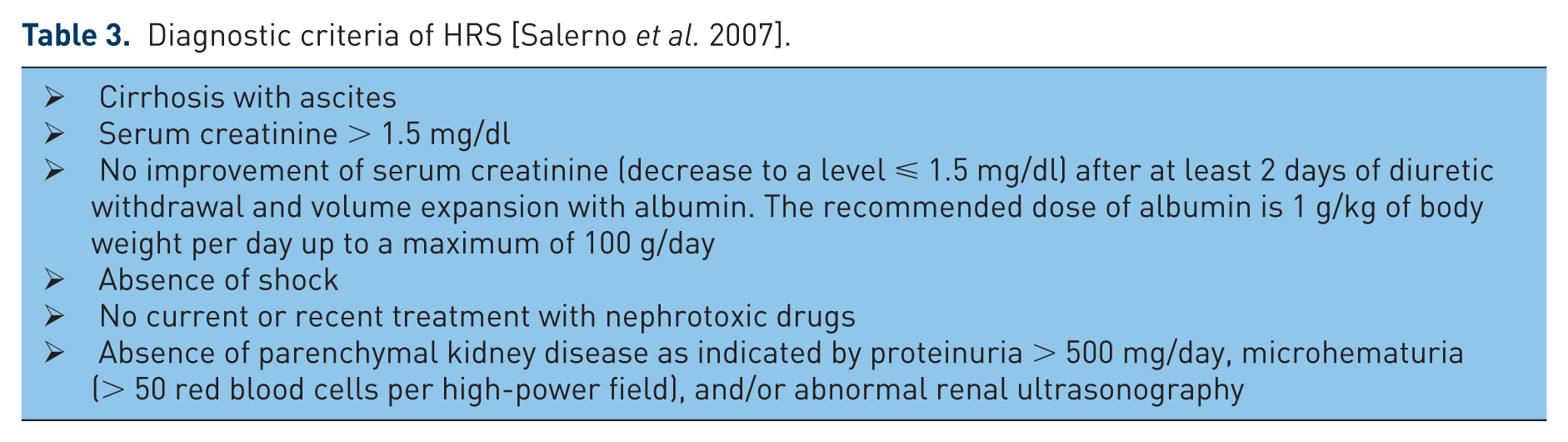
Diagnostic criteria of HRS [Salerno et al. 2007].
In selected patients, a TIPS is an alternative to serial LVP [Gines et al. 2002]. This procedure creates a shunt between the portal and suprahepatic veins, thereby reducing portal hypertension and the rate of recurrence of ascites. Use of a TIPS is associated with improved systemic hemodynamics and renal function. Many patients with refractory ascites regain the ability to excrete urinary sodium and can be maintained free of ascites without sodium restriction or diuretics. In controlled trials comparing paracentesis combined with intravenous albumin versus a TIPS in patients with cirrhosis and ascites, the TIPS was clearly worse, in terms of improvement of ascites and survival, in patients with refractory ascites than recurrent ascites [Gines et al. 2002; Lebrec et al. 1996; Rössle et al. 2000; Sanyal et al. 2003]. The most frequent complication of TIPS use is encephalopathy [Sanyal et al. 1994]; the incidence of new or worsening encephalopathy following TIPS use was found to be 20–31% [Salerno et al. 2004; Somberg et al. 1995]. In addition, a TIPS cannot be applied in some patients with advanced liver disease (Child–Pugh class C), congestive heart failure, and/or severe pulmonary hypertension owing to an increased risk of mortality, or in patients with portal thrombosis or inadequate anatomy of the portal and hepatic veins. Given that TIPS use is not associated with an improvement in survival, total paracentesis with intravenous albumin for volume replacement is considered the treatment of choice for refractory ascites in cirrhosis.
During the past 50 years, several procedures aimed at improving the treatment of refractory ascites have been developed [Arroyo, 2013]. Peritoneovenous shunting (PVS) involves a multiperforated intraperitoneal tube connected to a subcutaneously implanted unidirectional pump that permits the passage of fluid out of, but not into, the abdominal cavity, and a second subcutaneous tube that reaches the superior vena cava through the internal jugular vein [LeVeen et al. 1974]. When ascites is significant, it circulates through the prosthesis in response to a pressure gradient (positive in the peritoneum, negative in the intrathoracic circulation). However, the flow decreases or even stops if central venous pressure increases, thus preventing fluid overload. Many studies have demonstrated that PVS is associated with major improvements in circulatory and renal function and a better response to diuretics in patients with refractory ascites [Gines et al. 1991]. However, these beneficial effects did not result in a significant improvement in the long-term management of patients or in a reduction in the total time spent in hospital during follow up, because of a very high rate of shunt obstruction requiring reoperation. Moreover, these complications were not prevented by a redesign of the valve (Denver shunt) or by insertion of a thromboresistant titanium tip in the vascular end of the prosthesis [Gines et al. 1995]. Recently, an automated system was developed to remove ascites from the peritoneal cavity into the urinary bladder, from which it can be eliminated by normal urination [Bellot et al. 2013]. The pump system removed 90% of the ascitic fluid and significantly reduced the median number of LVP sessions per month in 40 patients with recurrent ascites. Larger studies are needed to confirm the safety and efficacy of this procedure.
Hepatorenal syndrome
Renal failure is a serious complication in patients with advanced cirrhosis. It occurs in about 20% of patients hospitalized with decompensated cirrhosis [Garcia-Tsao et al. 2008; Moore, 2013]. Renal dysfunction is diagnosed based on levels of creatinine and on creatinine-based equations. However, a recent study [Francoz et al. 2010] showed that creatinine-based formulae overestimate the true glomerular filtration rate, especially in patients with cirrhosis aged under 50 years and in those with ascites. In about 70% of patients, renal failure is caused by prerenal failure due to gastrointestinal hemorrhage, bacterial infection, or hypovolemia (overuse of diuretics), and in about 30%, it is caused by intrarenal causes, e.g. hepatitis B- or C-associated glomerulonephritis or toxins. In about 70% of patients with prerenal failure, renal function can be restored with fluid replacement, but the remaining 30% are unresponsive to volume expansion [Garcia-Tsao et al. 2008].
HRS is a fully reversible impairment of renal function in patients with severe hepatic failure unresponsive to volume expansion. The prevalence of HRS in patients affected by cirrhosis and ascites was found to be 18% after 1 year, increasing to 39% at 5 years [Gines and Schrier, 2009]. Before HRS is diagnosed, primary causes of renal failure, such as hypovolemia, infection, nephrotoxins, and other renal diseases, must be excluded. Nevertheless, in many patients with HRS lacking proteinuria or hematuria, some histologic changes are evident in kidney biopsies [Trawalé et al. 2010]. In almost half of patients with HRS, one or more precipitating factors were identified, including bacterial infections (57%), gastrointestinal hemorrhage (36%), and therapeutic paracentesis (7%) [Angeli et al. 2010]. Although a precipitating event is often identifiable, HRS can also develop in an apparently spontaneous way. Spontaneous HRS causes no morphologic changes in the kidney, although morphologic alterations in the course of oligoanuria may reveal a transition from intermediate to irreversible kidney damage with necrosis of the tubules. In patients with HRS scheduled for liver transplantation, it may be prudent to consider simultaneous kidney transplantation [Restuccia et al. 2004].
HRS is diagnosed by excluding other forms of renal failure. The diagnostic criteria initially proposed by the International Ascites Club in 1996 were re-examined in 2007 [Salerno et al. 2007] (see Table 4). Two types of HRS were identified.
➢ Type 1, a rapid deterioration of renal function defined as an increase in serum creatinine to a value twice the baseline and >2.5 mg/dl within 2 weeks.
➢ Type 2, a slow and gradual deterioration of renal function with serum creatinine >1.5 mg/dl, often in parallel to the worsening of cirrhosis and portal hypertension, and leading to refractory ascites.
Diagnostic criteria for kidney dysfunction in cirrhosis using the acute kidney injury definition [Wong et al. 2011].
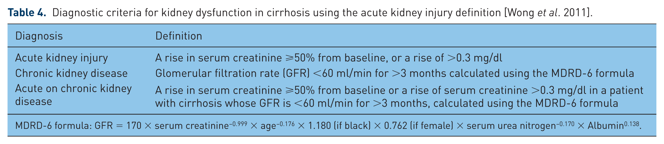
MDRD-6 formula: GFR = 170 × serum creatinine−0.999 × age−0.176 × 1.180 (if black) × 0.762 (if female) × serum urea nitrogen−0.170 × Albumin0.138.
Pathophysiology
The reduced glomerular filtration rate evident in patients with HRS is due to poor renal circulation on the one hand and a reduced filtration fraction on the other. Several factors are considered to induce the impairment of renal function (Table 5).
Pathomechanism of hepatorenal syndrome.

RAAS, renin–angiotensin–aldosterone system.
Vasodilation in the splanchnic region
This is considered the most important trigger of renal hypoperfusion. This vasodilation is associated with enhanced formation of vasodilators and vascular hyporesponsiveness to vasoconstrictors. To date, several mechanisms have been identified that may contribute to this enhanced vasodilation and impaired vasoconstriction. Portal hypertension increases the production and release of endogenous vasodilators, such as nitric oxide, carbon monoxide, and cannabinoids, into the splanchnic arterial circulation, but other factors seem to be involved as well. Even in the dormant phase of alcoholic liver disease, vasoactive substances are produced in large quantities in the liver when the production of cytokines is increased. In the presence of endotoxin stimulation, such activation is increased disproportionately. Even in patients with nonalcoholic liver disease, infiltration of endotoxins may play a role. Thus, patients with HRS prior to liver transplantation exhibited significantly higher concentrations of interleukins 2 and 6 and tumor necrosis factor than patients lacking HRS before liver transplantation [Burke et al. 1993]. Furthermore, increased endothelin concentrations have been observed, and regulatory disorders of the adenosine system have also been discussed [Gerbes et al. 1998]. Translocation of bacteria and bacterial products from the gut is likely to be a main factor underlying the development of a chronic inflammatory state [Israelsen et al. 2014]. Another mechanism seems to be defective contractile signaling in smooth muscle cells in response to vasoconstrictor stimulation [Hennenberg et al. 2008]. In addition to the liver itself, little is known about the factors responsible for these defects. The consequences are vasodilation, increased plasma volume, and increased blood flow in the splanchnic circulation, leading to a reduction in the effective arterial blood volume and a drop in arterial pressure. Vasodilation is associated with increased mesenteric blood flow, which may in fact be a useful mechanism to maintain liver function. In one study, liver function in patients who underwent transplantation for liver failure was found to be dependent on the in-vitro liver circulation [Cardoso et al. 1994].
Central underfilling
In the early stages of cirrhosis, increased cardiac output compensates for the reduction in systemic vascular resistance caused by vasodilation in the splanchnic area, and allows arterial pressure and effective blood volume to remain within normal limits. In the advanced stages of cirrhosis, splanchnic vasodilation is markedly increased, and additional increases in cardiac output can no longer compensate, leading to central underfilling. Central underfilling activates baro and volume receptors, with consequent stimulation of the sympathetic nervous system and RAAS and the nonosmotic release of vasopressin. This results in renal hypoperfusion caused by an increase in renal vascular resistance [Lenz et al. 1991], reabsorption of sodium in the tubules, and increased reabsorption of water in the distal collecting tubules.
Abolished autoregulation of kidney perfusion
Increased sympathetic nervous system activity abolishes autoregulation in the kidney [Esler and Kye, 1998]. Consequently, the renal circulation becomes pressure-dependent, even in the presence of physiologic blood pressure [Stadlbauer et al. 2008]. This is because the mean arterial blood pressure at which renin starts to be released is shifted to <110 mmHg [Kirchheim and Ehmke, 1994; Persson et al. 1990].
Hepatorenal reflex
In cirrhosis, sinusoidal pressure is elevated by increased resistance and enhanced mesenteric blood flow. Elevated pressure, secondary to swelling in the course of glutamine infusion via the superior mesenteric artery, led to a reduction of renal circulation and the glomerular filtration rate. This was due to direct hepatorenal interaction [Lang et al. 1991]. Increased intrahepatic pressure leads to enhanced activity of sympathetic efferents from the liver that, through synapses in the lumbar spinal cord, cause enhanced renal sympathetic activity (the hepatorenal reflex [Kostreva et al. 1980]). A decrease in sinusoidal blood flow also triggers the hepatorenal reflex [Ming et al. 2002]. An increase in renal sympathetic nerve activity leads to renal vasoconstriction and increased tubular sodium reabsorption. Denervation of the kidney eliminates this renal sympathetic activity, thereby reducing the impairment of renal function. This can also be achieved by hepatic vagal denervation [Levy and Wexler, 1987]. In a clinical study, Solis-Herruzo and colleagues [Solis-Herruzo et al. 1987] were able to interrupt the hepatorenal reflex with lumbar sympathetic block in patients with portal hypertension and thereby improve renal function.
Cardiac insufficiency
An increase in cardiac output is important to compensate for the reduction in systemic vascular resistance caused by vasodilation in the splanchnic area, and permits the arterial pressure and effective blood volume to remain within normal limits. There is evidence that cardiac output decreases as cirrhosis progresses. Clinical, but not overt, cardiac insufficiency, caused by underlying viral diseases, toxins such as alcohol, and/or by cirrhotic cardiomyopathy per se, seems to be one of the leading risk factors for the development of HRS. Ruiz-del-Arbrol and colleagues [Ruiz-del-Arbrol et al. 2005] showed that cardiac insufficiency can be detected at an early stage in patients developing HRS. In addition, the use of β-blockers in patients with refractory ascites has been shown to be deleterious [Serste et al. 2010]. These findings suggest a cardiorenal link in advanced cirrhosis (Krag et al. 2012b).
Relative adrenal insufficiency
This is a common subclinical condition in patients with cirrhosis, and is diagnosed as an inadequate production of cortisol in response to stimulation with synthetic adrenocorticotropic hormone. Adrenal insufficiency increases the risk of bacterial infection, septic shock, and HRS [Acevedo et al. 2013]. Cortisol plays an important role in hemodynamic homeostasis: the response of the heart and blood vessels to catecholamines and angiotensin-II is increased by cortisol. Low levels of cortisol lead to an inadequate increase in cardiac output and insufficient vasoconstriction in response to stress. Accordingly, adrenal insufficiency may be involved in the development of cardiomyopathy and HRS [Krag et al. 2012].
Treatment of renal failure
Treatment should begin as soon as renal failure is diagnosed. The criteria for diagnosis of HRS may lead to renal failure being detected too late [Nadim et al. 2012]. In addition, creatinine-based formulae may overestimate the true glomerular filtration rate, because of the low levels of muscle proteins in patients with cirrhosis and ascites [Francoz et al. 2010]. Cystatin C-based equations have proven more accurate in the evaluation of renal function and the staging of chronic renal insufficiency [De Souza et al. 2014]. Use of the new definition of acute kidney injury (AKI) developed by the Acute Kidney Injury Network as adapted for cirrhotic patients [Wong et al. 2011] (Table 6) allows renal failure to be identified much earlier [Moore, 2013]. A prospective observational cohort study of patients with cirrhosis and AKI showed that AKI is frequently progressive and associated with mortality [Belcher et al. 2013]. Therefore, the early identification and treatment of its cause are the cornerstones of therapy (Table 6). This includes withdrawal of β-blockers in patients with end-stage cirrhosis [Krag et al. 2012], discontinuation of nonsteriodal drugs and diuretics and, in patients with hypovolemia due to bleeding, prompt treatment with fluids and blood products combined with measures to stop the bleeding [Baradarian et al. 2004].
Prophylaxis against hepatorenal system.

To treat hypovolemia, the recommended dose of albumin is 1 g/kg body weight in all patients after at least 2 days of diuretic withdrawal [Salerno et al. 2007]. However, because the degree of hypovolemia varies between patients, a more precise estimation of the volume required appears logical. Indeed, using invasive instruments, Umgelter and colleagues [Umgelter et al. 2012] demonstrated that much more fluid (up to 2 g albumin/kg body weight) may be necessary to normalize renal function. The main problem in clinical practice is that invasive hemodynamic monitoring can only be performed in intensive care units. Nevertheless, if there are clinical signs that 1 g albumin/kg is inadequate, more fluid can be infused under the careful observation of noninvasive hemodynamic parameters and lung function.
Antiviral therapy may be effective in some patients with renal failure due to hepatitis C-related glomerulonephritis after consideration of the possible adverse effects of this therapy in patients with advanced cirrhosis and hepatitis B virus-associated kidney injury [Gane et al. 2014]. Bacterial infections must be identified and treated early. In patients with SBP, the addition of albumin (1.5 g/kg body weight at the time of diagnosis, followed by 1 g/kg on day 3) to antibiotic treatment reduces the incidence of renal failure [Salerno et al. 2013; Sort et al. 1999].
Ascites is a risk factor for the development of contrast-induced nephropathy. Although no reports show that cirrhosis without ascites is a risk factor, all patients with cirrhosis who undergo radiologic studies and require contrast medium should receive standard prophylactic saline hydration [Lodhia et al. 2009]. A recent study [Mahmoodi et al. 2014] involved patients with coronary heart disease and chronic renal failure with a serum creatinine level of up to 4 mg/dl who were undergoing coronary intervention, use of a bicarbonate solution (prepared by adding 154 ml of 1000 mEq/l sodium bicarbonate to 846 ml of 5% dextrose in water) conferred greater protection than hydration with normal saline against contrast-induced AKI. However, because metabolic alkalosis, mostly due to low albumin concentration, is a frequent finding in liver failure [Funk et al. 2006], further studies are required in patients with cirrhosis to confirm these results.
In end-stage cirrhosis, β-blockers may reduce survival owing to their negative impact on the cardiac compensatory reserve. The inability to increase cardiac output may compromise organ perfusion in these patients [Krag et al. 2012]. The use of β-blockers is associated with increased mortality in patients with refractory ascites at high risk of paracentesis-induced circulatory dysfunction [Serste et al. 2010]. Although data on this subject are scarce, the use of β-blockers should be avoided in patients with both cirrhosis and renal failure.
Pentoxifylline has been shown to reduce the risk of renal failure in patients with alcoholic hepatitis in the short term, but this effect is no longer evident after 6 months [Mathurin et al. 2013]. Oral antibiotics are effective in reducing the incidence of HRS in patients with advanced cirrhosis and ascites. In a randomized placebo-controlled study, primary prophylaxis with norfloxacin reduced the incidence of SBP and delayed the development of HRS in patients with cirrhosis, low-protein ascitic fluid (<15 g/l), and advanced liver failure (Child–Pugh score >9) [Fernandez et al. 2007].
Knowledge of the pathophysiologic features of HRS has led to a variety of therapeutic approaches. It would be logical to assume that renal function is improved by substances that abolish renal vasoconstriction. However, experiments performed with prostaglandin to date have produced divergent results [Wong et al. 1994]. Likewise, the administration of dopamine achieves improvements in cirrhotic patients with relatively good renal function, but not in patients with massively impaired renal function [Bennet et al. 1975; Peschl, 1987]. In patients with cirrhosis, renal failure, and tense ascites, increased abdominal pressure (IAP) may contribute to renal dysfunction. Reduction of IAP following paracentesis and albumin substitution improves renal function, probably by improving renal blood flow [Umgelter et al. 2009).
The current therapeutic approach to HRS includes the administration of vasoconstrictive agents combined with albumin (Table 7). The effect of vasopressors on renal function in this situation is due to an increase in renal perfusion, resulting from increased central blood volume and elevated blood pressure. Given that autoregulation is diminished, increased blood pressure is associated with increased renal perfusion. An inadequate blood-pressure response to vasopressor therapy is a typical finding in nonresponders [Nazar et al. 2011; Velez and Nietert, 2011].
Treatment of hepatorenal syndrome.

The available vasoconstrictors comprise vasopressin analogues and alpha-adrenergic agents such as noradrenaline and midodrine. The first studies were performed with octapressin, a vasopressin derivative, more than 40 years ago [Kew et al. 1972]. Over 30 years ago, ornipressin (8-ornithine-vasopressin: a vasopressin derivative that predominantly stimulates the vasopressin 1 receptor) was found to improve central hypovolemia through splanchnic vasoconstriction, leading to decreased activation of vasopressor hormones (norepinephrine and renin) and decreased renal vascular resistance, resulting in improved renal perfusion in patients with cirrhosis and HRS [Lenz et al. 1991]. Because ornipressin was removed from the market, subsequent studies were performed using terlipressin. Terlipressin is a triglycyl-lysine-vasopressin with a vasoconstrictive effect per se when given as a bolus and an additional long-acting vasoconstrictive effect caused by lysine-vasopressin, which is produced by triglycyl-lysine-vasopressin degradation [Ryckwaert et al. 2009]. Vasopressors such as terlipressin induce vasoconstriction in the splanchnic vasculature by acting on vasopressin 1 receptors, resulting in redistribution of blood flow to the kidneys. Vasopressin analogues have been shown to decrease serum levels of vasoconstrictor hormones such as norepinephrine and plasma renin activity, leading to a decrease in renal vascular resistance [Lenz et al. 1991]. The vasopressin analogue terlipressin has been shown not only to improve renal function by peripheral actions, but also to significantly influence cardiac filling volumes [Krag et al. 2010]. In experimental ovine endotoxemia, intermittent bolus injections of terlipressin were linked to decreased heart rate and cardiac index and increased pulmonary vascular resistance. Continuous infusion of terlipressin reversed endotoxin-induced systemic arterial hypotension and improved left-ventricular stroke-work index [Lange et al. 2007]. Therefore, continuous administration of terlipressin may be superior to bolus injection.
Norepinephrine has a positive inotropic effect, which may be important in overcoming the cardiac insufficiency induced by chronic liver disease. In patients with portal hypertension, terlipressin increased thoracic and liver blood volumes and altered portal pressure by causing vasodilation of intrahepatic vessels [Kiszja-Kanowith et al. 2004].
The effect of terlipressin can be significantly enhanced by the addition of high doses of human albumin [Ortega et al. 2002]. Although the favorable effect of albumin in this context is traditionally linked to its role in volume replacement, other mechanisms related to its nononcotic properties are likely to be involved. Albumin binds to and carries a variety of hydrophilic and hydrophobic molecules, including metals, fatty acids, metabolites, and drugs. Many potentially toxic ligands are neutralized and catabolized by binding to albumin. Furthermore, albumin is the major source of extracellular reduced sulfhydryl groups, potent scavengers of reactive oxygen species generated by oxidative stress; thus, albumin is the main circulating antioxidant in the body. Recent studies have shown that both albumin concentration and function are reduced in patients with cirrhosis [Jalan and Bernardi, 2013], and that the degree of albumin dysfunction is related to the prognosis of liver disease [Jalan and Bernardi, 2013; Oettl et al. 2013]. In addition, albumin has been shown to have a positive cardiac inotropic effect in cirrhotic rats [Bortoluzzi et al. 2013]. Substitution of albumin therefore seems to accomplish more than plasma expansion.
Many studies have been performed with terlipressin. Meta-analyses have demonstrated that the combination of terlipressin and albumin improves renal function in patients with HRS type 1 [Nazar et al. 2011; Neri et al. 2008] and reduces mortality when compared with albumin alone. The reduction in mortality was evident in patients with type I, but not type II, HRS [Gluud et al. 2010]. Therefore, terlipressin plus albumin may facilitate survival to transplantation and a better outcome after transplantation, because normalization of renal function before transplantation is associated with increased survival after transplantation [Restuccia et al. 2004]. In these studies, a fixed dose of 1 g albumin/kg body weight on the first day, with a reduction to 20–40 g on subsequent days, was used. Terlipressin was started on the first day at a dose of 0.5 mg every 4–6 hours, with an increase to a maximum of 12 mg per day until improvement of renal function occurred, defined as a serum creatinine concentration of <1.25 mg/dl. An improvement was seen within 14 days [Martin-Llahi et al. 2008; Neri et al. 2008; Sanyal et al. 2008]. Predictors of improved renal function in patients with HRS include early stage renal failure, increased blood pressure induced by the vasopressor, normal sodium concentration, and Child–Pugh score <11 [Boyer et al. 2011; Nazar et al. 2011; Velez and Nietert, 2011; Wong et al. 2004]. Given that larger amounts of albumin-containing fluids have been shown to restore kidney function more effectively [Umgelter et al. 2012], goal-oriented volume therapy, using serial echocardiography to assess changes in intravascular volume by measuring inferior vena caval diameter, right-ventricular end-diastolic volume index, left-ventricular end-diastolic area index, and global end-diastolic volume index, may be useful [Charron et al. 2006], although studies on cirrhotic patients are scarce.
Also controversial are the parameters for titrating the dose of terlipressin. Given that a close relationship exists between hemodynamics and the improvement of renal function [Nazar et al. 2011; Velez and Nietert, 2011], an increase in mean arterial pressure of ⩾10 mmHg, as used in studies with norepinephrine, may help to achieve an earlier improvement in renal function [Maddukuri et al. 2014]. Adverse effects of terlipressin (predominately ischemia) leading to the discontinuation of treatment occur in about 12% of patients [Fagundes and Gines, 2012]. Therefore, patients with coronary heart disease, peripheral vascular disease, and/or cerebrovascular disease should not be treated with terlipressin. Some of these complications may be caused by the bolus injection, because of the acute effect of the prodrug [Ryckwaert et al. 2009]. A continuous infusion may overcome this problem [Angeli et al. 2009]. Studies on endotoxic shock in animal models, in which continuous infusion of terlipressin exerted a superior effect on cardiac contractility than bolus injection [Lange et al. 2007], support the advantages of continuous infusion of terlipressin.
Alpha-adrenergic agonists, such as norepinephrine [Sharma et al. 2008; Singh et al. 2012] or midodrine, in combination with octreotide and albumin [Angeli et al. 1999; Skagen et al. 2009; Wong et al. 2004], are a reasonable alternative to terlipressin because of their low cost and wide availability. However, data on their use are limited. They are recommended in the guidelines of the American Association for the Study of Liver Diseases (AASLD) because terlipressin is not yet available in the United States [Runyon, 2012], but they are not recommended by the European Association for the Study of the Liver (EASL) [Gines et al. 2010]. If HRS reoccurs after the discontinuation of treatment, patients should be retreated with the same drug combination [Gines et al. 2010].
TIPS use was reported to be effective for HRS Type 1 in an uncontrolled study of a small group of patients [Guevara et al. 1998]. Insertion of a TIPS further improved renal function and sodium excretion in patients with HRS type 1, who responded to a combination of midodrine, octreotide, and albumin [Wong et al. 2004]. More studies are necessary to evaluate TIPS use in patients with HRS type 1. TIPS use is not recommended in the guidelines of the EASL or AASLD.
Type 2 HRS is a type of renal dysfunction that arises from the deterioration of several organ systems, and is primarily caused by liver failure and portal hypertension. It is part of a gradually developing multiorgan dysfunction, with central hypovolemia and cardiac dysfunction as the main pathogenic triggers [Lenz, 2005]. There are very few data about the use of vasopressors in combination with albumin in patients with HRS type 2. The clinical picture is dominated by therapy for refractory ascites and hyponatremia. Therefore, repeated paracentesis is the first-choice treatment; if renal function is deteriorating, the use of vasopressors should be considered [Fagundes and Gines, 2012]. Patients must be evaluated to determine whether they are candidates for liver transplantation.
Renal replacement therapy (RRT) has been used in patients with type 1 HRS waiting for liver transplantation. There are no data comparing extracorporeal therapy with vasopressors plus albumin in patients with HRS; nevertheless, RRT is not considered to be a first-line treatment, because it does not correct the underlying pathophysiologic changes. RRT should be reserved for patients planned for liver transplantation. The mortality of patients with HRS treated with hemodialysis or hemofiltration in the intensive care unit is reportedly high [Witzke et al. 2004]. In a small study [Mitzner et al. 2000], improvement of renal function followed use of an extracorporeal liver assist system (MARS®). However, in the recently published RELIEF trial, MARS therapy did not demonstrate a positive effect on patients with acute or chronic liver failure [Banares et al. 2013]. EASL guidelines state that RRT may be useful in patients who do not respond to vasoconstrictor therapy [Gines et al. 2010]. It remains unclear what modality of RRT should be the first-choice treatment for pharmacologic nonresponders awaiting transplantation.
Footnotes
Conflict of interest statement
The authors declare that they do not have anything to disclose regarding conflicts of interest with respect to this manuscript.
Funding
This research received no specific grant from any funding agency in the public, commercial, or not-for-profit sectors.