
Abstract
Injuries in football are prevalent, and while shin guards reduce these, current test standards are primarily intended to evaluate contusion risk rather than more serious outcomes such as fractures. In this study, a finite element human body model was used to assess fracture risk in the lower leg subjected to conditions representative of football impacts. Various impactor shapes, impact locations and orientations were explored to identify conditions where fractures may be more likely to occur (based on element strain) and the associated force and bending moment. The lower leg was most susceptible to fractures at the 35% tibial height. Fractures occurred most frequently from the anterolateral direction, resulting in fibula injuries. In terms of geometry, the stud impactors were the most effective at inducing fractures and fracture was highly sensitive to bone alignment. Force to fracture ranged from 1595 to 2612 N. Susceptibility to fractures was influenced by the cross-sectional area of the bone, as well as the soft tissue thickness, with increased force attenuation associated with greater tissue thicknesses. This study showed various parameters affect the fracture tolerance of the lower leg, and identified the impact energies required to induce fractures, to better inform test standards for protective equipment.
Keywords
Introduction
In football, contact with other players or the ground can lead to injuries. An injury to a single elite player is estimated to cost their club at least 500,000 euros (∼550,000 USD) a month, affecting their financial and sporting targets. 1 Leg fractures in five European football leagues have been reported to be associated with an average of 136 days of rehabilitation (18 games) prior to return to play (RTP), 2 which increased to 251 days when surgical intervention was required. 3 On average, one to two fractures occur per team, per season, 4 thus the high cost of injuries compounded with substantial time out is a concern not only from a player’s welfare perspective, but also for the club’s.
Fédération International de Football Association (FIFA) mandated the use of shin guards for association football players in 1990. These were noted to result in an overall decrease in lower leg injuries (lacerations and fractures). 5 Several shin guard performance standards exist to evaluate protection provided by this equipment, with a range of test parameters (Table 1). Across these standards, the applied impact energies never exceed 15 J. While these levels are appropriate for contusions, they are not suitable for investigating fracture resistance, which occurs at a higher energy level. As bone fractures are the most severe injuries for this anatomical region, with longer term implications, they are very important to prevent. However, they require much higher impact energies to induce, compared to lacerations or contusions. The fracture tolerance of the lower leg has been investigated previously in three-point bending, in which the proximal and distal ends were supported and mid-length was impacted.6,7 These studies, replicating automotive-pedestrian collisions, required energies exceeding 145 J 6 and 82 J 7 to generate fractures. As football injuries differ from automotive (with prior work using a single impact location, direction and bumper shape), this makes it difficult to translate their findings, owing to increased variability in parameters such as impact energy and impacting shape (stud challenge and leg on leg tackle).
Summary of methodologies used in standards and studies conducted on shin guards.

Human body models (HBM) have been developed using the finite element (FE) method to replicate the tissue mechanics and kinematic behaviour under impact. A key advantage of these models is their ability to evaluate relative injury risk under multiple conditions, in contrast to experiments, which would require numerous specimens. Therefore, HBMs can serve as a cost-effective tool to establish regions and/or impact conditions that make the leg most susceptible to fracture, which can be translated to more comprehensive shin guard test standards. One such model is the Total Human Model for Safety (THUMS)17,18 developed by Toyota Central R&D Labs Inc. and Toyota Motor Corporation.
To the authors’ knowledge, no studies have been conducted on quantifying the injury tolerance of the lower leg under football impact conditions, particularly with a focus on bone fracture. Therefore, the purpose of this study was to identify the most vulnerable conditions that may be applied to an unguarded lower leg during football impacts, investigating the effect of height, orientation, impactor shape and energy levels.
Methodology
Numerical models were run using the commercial explicit finite element solver LS-DYNA® MPP single precision version R12.2.1 (ANSYS, Canonsburg, PA, USA). A 16-core workstation was used to run the simulations, and each one required approximately 15 min to complete. No shin guards were modelled in the present study to ensure results were shin guard-independent, while allowing important metrics to be established, against which future personal protective equipment (PPE) would be evaluated. The THUMS V4.1 50th percentile male pedestrian leg was used, sectioned at the distal femur, as it had been previously validated, 19 and has been made open-source. The geometry and material modelling of the THUMS were unaltered. The soft tissues were modelled as hyperelastic, using a simplified rubber/foam material model and skin as a fabric, with data from the literature.20,21 The bones had trabecular and cortical bone modelled separately. Trabecular bones (solid elements) were given a damage material model while cortical components (shell elements) were given a piecewise linear plasticity model. Muscle activation was not included in this study. The fracture response was simulated through the built-in criterion of element erosion, whereby upon reaching a critical strain value of 2.14% 22 and 6.00% 23 for the cortical and cancellous components respectively, an element would be deleted from the model. Contact among the components within the lower limb was defined using a single surface contact.
The scenario of interest was that of a footballer’s foot planted, therefore the plantar surface was translationally constrained (Figure 1). The lower limb was sectioned at the quadriceps femoris tendon and fixed such that proximal end was not free to translate. This isolation was done to reduce computational time while still permitting natural knee motions during impact. Three impactor geometries were modelled for this study, including a stud (based upon EN 13061), a blunt cylinder (based upon EN 13061), 9 and a hemisphere (based upon NOCSAE) 8 (Figure 1). The stud was given 10 mm diameter and 16 mm height, while the blunt and spherical impactor were given diameters of 80 mm. All were defined as rigid materials and assigned densities so that their overall mass would be 5 kg. The selected mass was in agreement with the NOCSAE shin guard standard (prescribes the use of a 5 kg spherical impactor), 8 which is comparable to the approximate mass of the lower leg (foot included) for an 80 kg male. 24
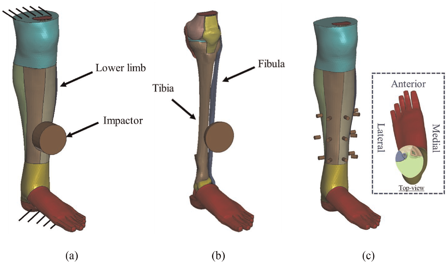
Depiction of: (a) numerical setup using a blunt impactor, (b) using a hemispherical impactor (with soft tissues hidden) at 35% tibial height in the anterior orientation and (c) stud impactors with all orientation/height combinations used in this study. Colours on the leg indicate separate parts as defined in the THUMS human body model.
A surface-to-surface contact (pinball segment based) was implemented between the impactor and the entire lower limb. A series of simulations was run where each impactor was positioned at a specific height and radial orientation. For different heights, impacts were run at 20%, 35% and 50% of the tibia length, as the majority of fractures are reported to occur between the middle and distal third of the tibia/fibula. 25 To vary the direction of impact radially, a neutral ‘anterior’ direction was defined using anatomical landmarks, and the orientations were then varied by 45°, such that impacts occurred in the anterolateral, anteromedial, lateral and medial directions (Figure 1). Each impact was constrained to travel in the required direction at a point centered width-wise on the leg.
A previous investigation identified fractures occurring when subjected to an average impact energy of 68.6 J. 6 Thus, initially impact energies of 70 J (5.29 m/s velocity) were applied and if ‘fracture’ was observed (elements in the model were deleted), the velocity was reduced in increments of 20 J, until no fractures were observed (considered the tolerance/critical limit). As few fractures occurred at 70 J, simulations were also conducted at 90 J (velocity of 6 m/s). Simulations were run up to 50 ms, to capture the peak and unloading of the event. Force data were acquired between the impactor and the lower limb and collected at 10 kHz. Bending moment was calculated using beam theory on the tibia, using the full length of the bone (391.7 mm), measured from the farthest node on each end. To calculate energy absorbed by soft tissues, the residual projectile energy as well as the energy absorbed by the skeletal components at the time of fracture were subtracted from the initial kinetic energy.
Tissue thickness of the model was measured in each orientation/height through two nodes, the first being the node on the surface of the flesh that first contacted the impactor and the second being the node on the surface of the bone closest to the impactor. Furthermore, to investigate if the cross-sectional area of the tibia and fibula at various heights contributed to fracture tolerance, the values were measured using Autodesk Inventor (Autodesk, San Francisco, CA, USA). The CAD model was developed by importing the FE mesh into Altair Hypermesh (Altair, Troy, MI, USA), where the faces of the mesh were extracted, then merged to create geometric surfaces that could be used to output a final generic CAD format.
Results
A total of 91 simulations were run, encompassing energies between 50 and 90 J. A total of eight fractures occurred at 90 J, compared to one for 70 J. Of the tests that resulted in fracture, tibia and fibula fractures were split based on the orientation of the impactor (tibia fractures for anterior and anteromedial impacts, fibula fractures for anterolateral impacts).
For the investigation of impact height, the most vulnerable condition was at 35% height (Figure 2). This location experienced a total of seven fractures, whereas the 50% and 20% heights had one fracture each. Furthermore, 35% was the only height that experienced fractures at 70 J. The average forces to generate fractures at 20%, 35% and 50% heights were 2612, 2099 and 1777 N, respectively. Bending moment to fracture was calculated using the tibial length with a range between 142.3 and 225.9 Nm.
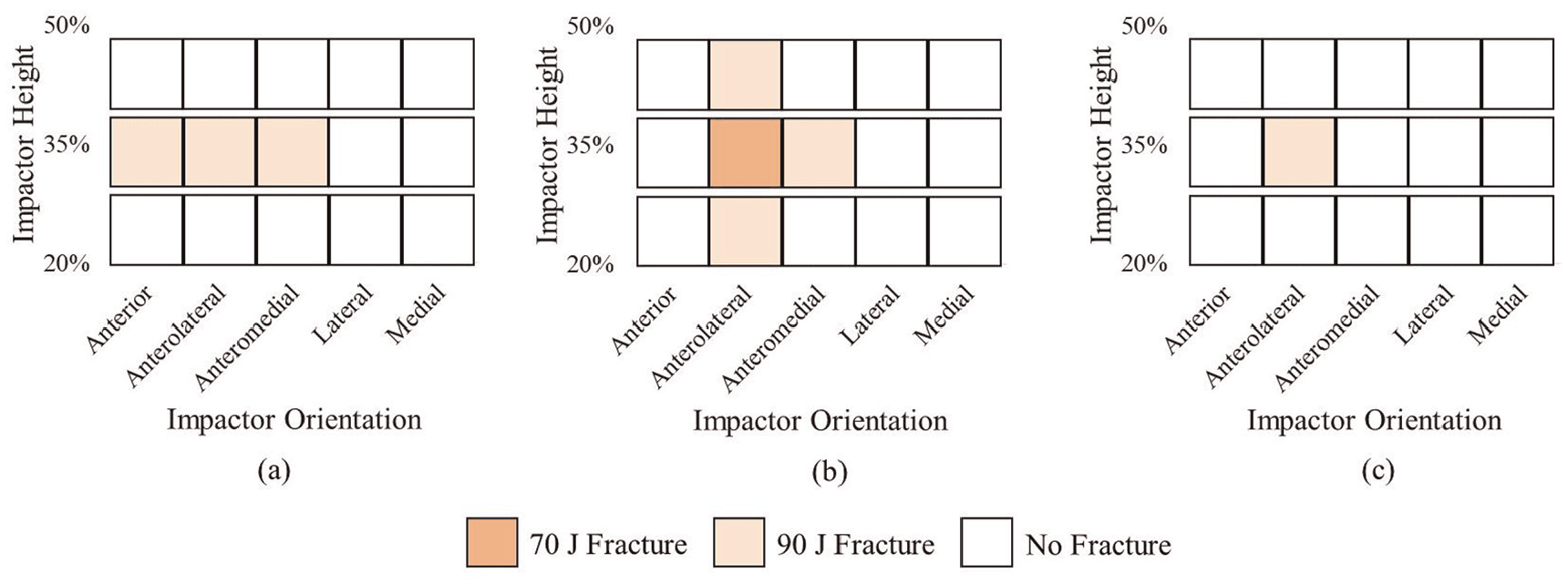
Injury matrix for impacts with the (a) hemispherical, (b) stud and (c) blunt impactors.
When comparing impactors, the stud generated the most fractures (five), followed by the hemisphere (three) and the blunt (one) (Table 2). The stud was the only impactor that generated a fracture at 70 J (at 35% height and anterolateral direction) and was most affected by positioning, due to the smaller load transmission area. The hemisphere typically generated higher forces than the stud for equivalent impacts, and generally fractured bones at higher forces as well. The blunt impactor generated higher forces than both others for most conditions (with the fewest fractures), with the average force to fracture (for all heights and orientations) for hemisphere, stud and blunt impactors being 2252, 1959 and 2538 N, respectively.
Summary of impacts resulting in fractures.
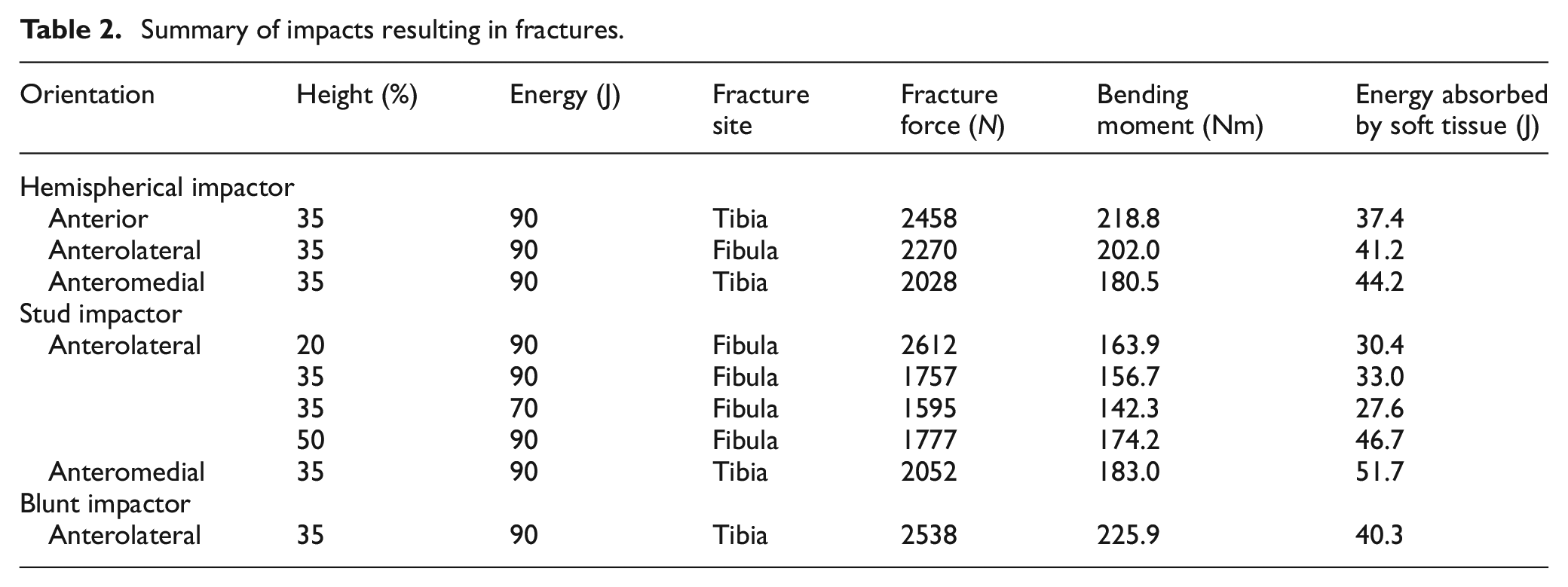
The orientation of impact strongly affected both the number of fractures and the fracture force. The most common direction for failure was the anterolateral (six fractures), followed by the anteromedial (two fractures). The anterolateral also failed at the lowest energy level (70 J). No fractures occurred for the lateral and medial impacts. The anterior direction resulted in fracture for the 35% height and 90 J condition using both hemisphere and blunt impactors. The average force to fracture (for all impactors and heights) at anteromedial, anterior and anterolateral orientations was 2040, 2458 and 2092 N, respectively.
Energy absorbed by soft tissues ranged from 27.6 to 51.7 J for all simulations that resulted in fractures (Table 2), and was affected by impactor height, shape and orientation. More energy was transferred to the soft tissues at 50% height, with an average of 51.7 J energy absorbed by soft tissues for the 50% height compared to 30.4 and 39.3 J for the 20% and 35% heights, respectively. The energy absorbed was highest for hemispherical impactors (average 40.9 J) compared to the stud (average 37.9 J) and blunt (average 40.3 J) impactors. The energy absorbed by the soft tissues was the highest in the anteromedial orientation (average 48.0 J), followed by the anterior (average 37.4 J) and anterolateral (average 36.5 J) orientation.
In general, force-displacement responses were comparable among different impactors under identical orientation, height and energy for non-injurious events (Figure 3). Peak forces for nearly all tests occurred at impactor displacements of 10-20 mm followed by a subsequent decrease in the force that then generally stabilized (Figure 3a and b). Element erosion during a fracture resulted in a sharp drop in the force response (Figure 3b and c). Von Mises stress contour plots were generated from the simulation, with peak stresses occurring at the location of impact as well as on the rear side, with failure occurring around 160 MPa (Figure 4).
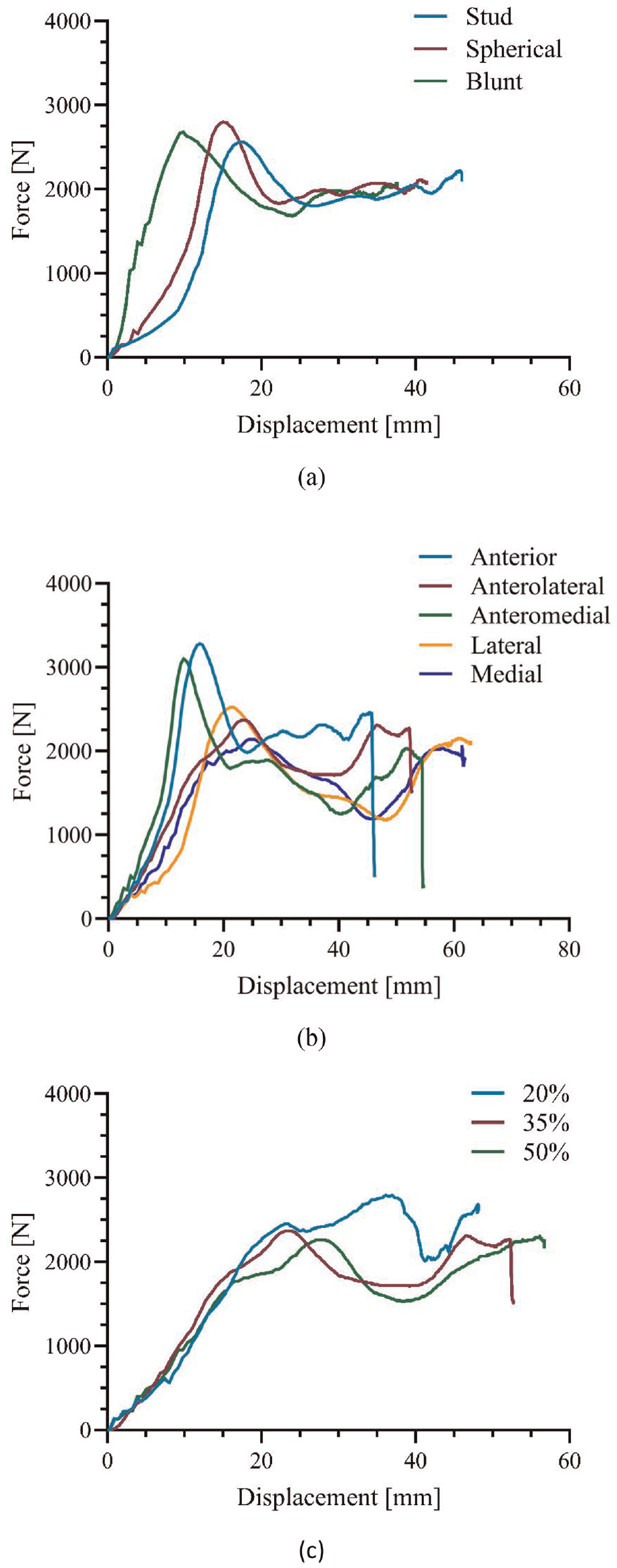
Force-displacement response showing the effect of: (a) varying impactor geometries (70 J, anterior, 35% height, no fractures), (b) varying orientations (90 J, 35% height, hemispherical impactor, several fractures) and (c) varying impactor height (90 J, anterolateral orientation, hemispherical impactor, single fracture).
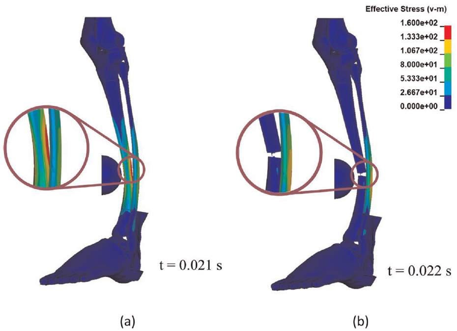
Representative stress contour plots of lower leg impacted with spherical impactor at 35% height, anteromedial orientation, highlighting: (a) peak stress just under 160 MPa under tension and (b) fracture at the same location afterwards.
Tissues were, on average, the thickest in the lateral orientation (19.4 mm) and at the 20% height (16.1 mm) (Figure 5). Maximum and minimum tissue thickness was 24.2 mm (50%, lateral) and 6 mm (50%, anterior). The 35% height was the most uniform; the range was 13.8–18.5 mm, in comparison to 6.0–24.2 mm and 9.7–22.8 mm for the 50% and 20% height, respectively. The thickness of the tissue had the greatest influence on the early portion of the response (<20 mm), in which a lower displacement to peak force was seen in ‘thinner’ regions.
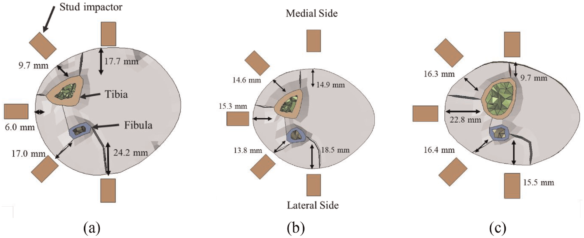
Lower leg sectioned at three heights: (a) 50% height, (b) 35% height and (c) 20% height, and the distance from the skin surface to the nearest bone; aligned with the orientation of interest.
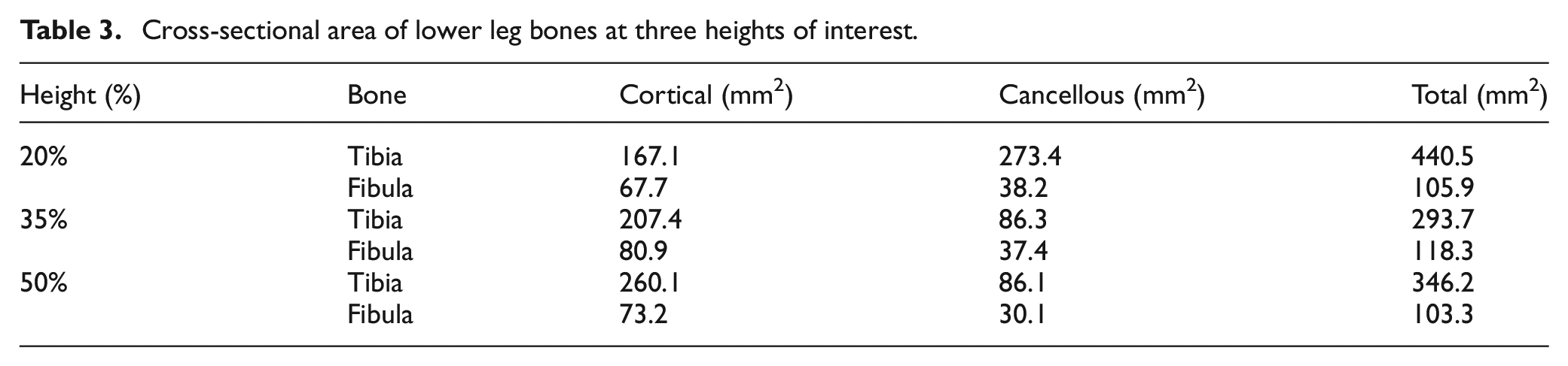
Cross-sectional area of the tibia varied substantially with height as well as the cortical/cancellous composition; however, the fibula did not vary much (Table 3). The lowest tibial overall cross-sectional value was at the 35% height, previously noted to be the most vulnerable.
Cross-sectional area of lower leg bones at three heights of interest.
Discussion
This is the first known study to investigate risk of fracture in football impacts and how they are affected by impact location and shape. The energy to fracture from previous experimental work was identified to be 68.6 J 6 and 54.8 J; 7 and while shin guards have been credited with preventing injuries,5,26 current shin guard test standards do not prescribe energies above 15 J and thus this work will provide crucial information for informing future standards to ensure shin guards are tested against the most vulnerable conditions. No shin guards were modelled in the present study, owing to pure interest in fracture behaviour of the lower limb in representative football impacts without their presence; however, these results are translatable to define preventative injury criteria for future shin guard development. Different impactor shapes can represent different loading conditions, as the stud would represent a stud on leg challenge (without a shin guard) while blunt/spherical impactors may better represent the behind-guard profile. An essential next step for this line of research should consider testing with shin guards. Overall, the fibula was the bone most prone to fractures, which required energy levels of at least 70 J to initiate fracture. Fractures were more consistent at 90 J. The findings agree with a previous study 2 on footballers in the top five European leagues, in which tibia, fibula and tibia/fibula injuries represented 20%, 36% and 17% of leg fractures. The higher proportion of infield fractures to the fibula 2 may correspond to a location of the lower leg more commonly struck during play, whereas the present study impacted 15 discrete locations to identify the regions most susceptible to injuries.
This work was conducted using the THUMS 50th percentile male model. While this model has undergone validation, 19 it was generated based on the dataset of a single 39-year-old individual. Numerical tools provide researchers the ability to investigate the mechanical response under dynamic impacts at low cost and with none of the natural variability inherent with experimental work; however, they have several simplifications and represent only the response of one anatomy. Individuals with less soft tissue thickness or more slender bones including a decrease in local cortical thickness would have a lower second moment of area and thus lower fracture tolerance. As cortical thicknesses in tibias can vary substantially (up to 27% coefficient of variation), 27 this corresponds to a large range of potential fracture forces. Furthermore, energy to fracture may differ in females, as significant differences in morphology exist between the two such as periosteal/endosteal circumference. 28 A previous study on femurs found no significant difference in cortical bone material properties between males and females, but bone porosity was identified to be influential. 29 Other variations in the general population may include differences in body mass index and bone mineral density. A combination of any of these factors will have an impact on the fracture tolerance of any subject and as such the results of the present work should be extrapolated to other geometries with caution. These differences could be captured via patient-specific FE models, including modifications to geometry or assigned material properties. Previous experimental work 6 using cadaveric specimens from both male and female donors identified energy to fracture to be higher for males, at 78.6 J compared to 58.5 J for females. As such, separate and more conservative standards may be necessary to protect females. An important future direction would be to expand the scope of work to other population groups. Muscle activation was not included in this study; however, the setup used in the present study would represent a ‘worst-case’ scenario (and thus provide greater protection to individuals), as a reduction of injuries has been previously demonstrated with the presence of muscle activation. 30 Lastly, to the authors’ best knowledge, no studies exist in the literature on the most serious and/or common football tackles, in terms of loading shape, loading location/orientation and velocity. The boundary conditions used in this study may be considered a reasonable representation of a player standing on foot and stress contour plots (Figure 4) highlighting low levels of stress at the knee joint - indicating that the system is not over constrained. The authors recognize that these conditions may not reflect all scenarios, and as such future studies could leverage real world footage data analysis to help quantify tackles and postures most likely to occur that can help develop more comprehensive guidelines.
The hemispherical impactor could be the most suitable impacting geometry for shin guard evaluation, due to its consistency in inducing fractures at different orientations (Figure 2). The stud impactor may also be an important geometry to consider, as it was capable of inducing fractures at a lower energy level. Increased injury susceptibility at lower energy levels can be attributed to the importance of concentrated loading onto the bone. To investigate if the model was sensitive to stud alignment, a sample simulation was run for the lateral orientation, at 35% height, 90 J in which the impactor was aligned to the center of the fibula. In this case fracture occurred, which did not happen when the stud was aligned to the center of the leg width. While this alignment was an important factor in determining the true fracture tolerance under stud impacts, it would not be an issue for shin guard testing where there is no ‘bone’ to which the stud would need to be aligned (as this testing generally uses a steel anvil). Lastly, the blunt impactor was ineffective for inducing fractures and thus may not be an ideal impact geometry. There were multiple instances where the loads from a blunt impactor exceeded that of a stud impactor, in identical conditions (height, orientation); however, only fractures were identified in the stud impacts. Due to the increased contact area of the blunt impactor, force overprediction was identified with respect to fracture tolerance limits. This overprediction has been previously observed, 31 where a stud-like ‘hammer’ impactor required significantly less force than a blunt ‘brick’ to initiate fractures in the head. Therefore, for each impactor geometry used in the present study, a lower limit was identified for evaluating protective equipment. Standards should tailor force requirements based on the tolerance of the lower leg and chosen impact condition, while future studies could investigate the use of alternative metrics to compliment force limits.
Anterolateral impacts were most associated with injuries (Figure 2), resulting in frequent fibula fractures, which is of concern, as this orientation has a high likelihood of occurrence during football collisions. Loading was generally concentrated onto the fibula and as the fibula possesses a lower cross-sectional area (∼30–40% compared to the tibia), this ultimately led to the increased vulnerability. The cross-sectional area of both the tibia and fibula varied as a function of height. The total area was the smallest at 35% height, noted herein as being highly susceptible to fractures. The cross-sectional area has been previously identified as a good indicator of fracture tolerance compared to other metrics. 6 Other factors weren’t explored in the present study such as bone density, due to the nature of the numerical methods (one THUMS model; one bone density value). Fracture tolerance could also be influenced by the non-uniform distribution of tissue surrounding the tibia/fibula.
The presence of soft tissue has been noted to attenuate impact force transmitted to the bone, leading to reduced fracture tolerance.6,32 In the present study, energy absorbed by the soft tissue was a function of impactor geometry, height and orientation, along with stiffness as well as bone deflection and was found to be substantial. It should be noted that the magnitude and/or percentage of soft tissue energy absorption may vary for different sized individuals and at other locations not explored in the present study.
As discussed above, several factors influence the susceptibility of injury at a given location. Cross-sectional area of the bone, tissue thickness, the loading of a specific bone among others are all influencing factors and in some cases, these factors compound, generating regions that are extremely susceptible. For example, injuries were observed across all impactors at the 35% height (lowest cross-sectional area) in an anterolateral orientation, in which the fibula fractured except for the blunt impacts (Figure 2). Future studies should focus on further developing relationships quantifying various factors and their influence on fracture tolerance.
The average force required for fracture varied with orientation; however, the range was 2040–2458 N. Previous experimental work on posterior-anterior loading of the lower leg observed fibula and tibia fractures at 2000 and 3000 N, respectively. 6 Latero-medial bending of the lower leg generated fractures as low as 2800 N (average of 3700 N), which was found to be resistant to fractures in the present study (0 fractures up to 90 J impact), indicating the presented results are generally in line with the literature. 7 In another previous study, tibias impacted at the mid-length required just under 3000 N to fracture. 13 That impactor had a similar shape to the blunt one used herein, supporting the findings that an elevated force would be required to fracture in those conditions. Overall, impactor geometry and velocity in those studies differed from that used in the present study, contributing to other differences observed. Bending moments to fracture herein were generally lower than previously reported for automotive impacts (249–302 Nm),7,33 which is attributable to the use of different impactor geometries/velocities as well as boundary conditions of the current study.
Upon examination of the force-displacement responses, it was noted that in several cases, the global peak occurred in the first 10-20 mm, which was not identified in previous experimental work. 6 This could be a consequence of the soft tissue material modelling, where the elements may have permitted larger compressions than in the experiments. Furthermore, the previous studies used different boundary conditions, in which the epiphyses (proximal and distal) were removed, both ends potted and were then simply supported 34 or given roller supports,6,7 none of which was represented in the present study. The sharp drop in force correlating to bone fracture was a characteristic identified in both the experimental work6,7 and the current study. It is important to note that while the strain-based element erosion technique can capture the bulk failure of the bone, it is highly sensitive to mesh size, would not be capable of capturing the development of microfractures and did not evaluate risk of non-fracture injuries, such as contusions. These element erosion techniques are widely used,35–37 but represent an idealization of fracture, and future implementation of novel damage models 38 may offer more insight into fracture mechanics under this type of loading. In terms of localized stress, erosion occurred at roughly 160 MPa (Figure 4), in line with tensile failure stress reported in the literature. 39
Increasing the impact energy in test standards would encourage industrial manufacturers to innovate superior protective equipment, which could further mitigate traumatic injuries that continue to be experienced by footballers. Despite the wealth of information provided in this study, challenges remain for translation into practice. Current test standards call for the use of a steel anvil to represent the athlete’s leg, which contains several undesirable properties such as a simplified geometry, the inability to represent tissue behaviour, as well as potentially unrealistic interaction of PPE with the lower leg. 40 It may be advantageous to include force attenuation requirements for stud impacts given the relatively low forces to induce fractures noted herein. Although varying heights and orientations were investigated in this study, the critical values should be used to define a conservative limit for the performance assessment of shin guards, particularly when using a device such as a rigid anvil, as it cannot mimic the complex physical characteristics of the lower leg. Furthermore, as the anvil would be substantially stiffer than the natural leg, the force values reported herein would need to be scaled to reflect this in a metallic surrogate, as is often done for anthropomorphic test devices (ATDs, or ‘crash test dummies’).41–43 For example, the current requirements for blunt impactors are not permitted to transmit more than 2000 N to the anvil, 9 which, at first glance, seem to be a good limit for testing based on the presented findings; however, further studies on correlating the forces observed here to that of a steel anvil should be undertaken. Another alternative could be the development of a deformable surrogate in lieu of the rigid anvil. 40 The inclusion of a soft tissue surrogate would be necessary for generating an accurate response, and a tissue surrogate of varying thickness would better approximate the leg’s response under different impact heights and orientations, as shown in the present study. Finally, testing of three discrete heights and five orientations remains a limitation of the current study, due to computational resources and there remains a possibility that a more vulnerable spot was overlooked.
Very little previous work has investigated the biomechanics of various challenges in football (stamping, leg on leg and stud on leg); most of the literature on lower leg injuries has focussed on automotive impacts. Future investigations should be undertaken to identify more appropriate models for approximating different loading conditions to, (1) identify relevant energy challenges (velocity and effective mass) as well as their loading shape, (2) leverage these findings for developing more representative testing methodologies and (3) define fracture tolerances for alternate population groups (e.g. small females) to create a robust, inclusive set of requirements. By improving test methodologies, as well as the biofidelity of surrogates, protective equipment will be adequately challenged and athletes kept safe.
Conclusions
This study investigated the injury tolerance of the lower leg under various football impact conditions using a finite element HBM. The stud impactor induced the most fractures, and due to the smaller contact area, lower fracture forces were reported for stud impacts in comparison to other impactor geometries. Fracture tolerance was influenced by the cross-sectional area of the bone (tibia/fibula), the thickness of the tissue, as well as the impactor geometry. HBMs are useful tools to investigate injury mechanisms and compliment cadaveric studies and thus could find further use for next generation PPE evaluation and/or other injury tolerance investigations. The authors recommend the development of a biofidelic surrogate to improve upon the shortcomings of the rigid anvil currently in use and inclusion of a force limit requirement for stud impacts and an increase in testing levels beyond ∼15 J (current maximum found in standards), to appropriately evaluate shin guards against high-energy tackles and impacts.
Footnotes
Declaration of conflicting interests
The author(s) declared no potential conflicts of interest with respect to the research, authorship, and/or publication of this article.
Funding
The author(s) received no financial support for the research, authorship, and/or publication of this article.