
Abstract
Elite cricket pace bowlers commonly sustain debilitating bone stress injuries. Lumbar bone stress injuries are more prominent in males, while lower limb bone stress injuries are more common in females. Bone stress injuries are partly attributable to bowling technique; however, scant research exists comparing bowling techniques of males and females as to better understand why males are more susceptible to lumbar bone stress injury. Three-dimensional pace bowling kinematics previously linked with lumbar bone stress injury were compared between 59 male and 19 female elite pace bowlers. Participants bowled 18 match-intensity deliveries indoors from a full run-up, whereby the mean of six deliveries was analyzed. Compared to females, males exhibited: a more extended thoraco-pelvic segment at back foot contact (p = 0.039, g = 0.46), larger shoulder counter-rotation from back foot contact to front foot flat (p = 0.021, g = 0.64), greater thoraco-pelvic lateral flexion at front foot flat (p = 0.001, g = 0.95), larger front knee flexion at ball release (p = 0.046, g = 0.57), and greater maximum front knee flexion from front foot flat to ball release (p = 0.009, g = 0.57). Australian elite male pace bowlers performed techniques linked to lumbar bone stress injury to a much greater extent than their female counterparts. A straighter front leg technique observed in elite Australian females may be why they more commonly experience lower limb bone stress injuries via possibly higher vertical loading rates. This information may assist in developing sex-specific bone stress injury risk mitigation approaches.
Introduction
The ability of a pace bowler to bowl faster is beneficial for cricket performance.1,2 Faster pace bowlers require less deliveries to take a wicket (i.e. have a lower “strike rate”) and concede fewer runs per wicket acquired (i.e. have a lower “economy”) compared to their slower counterparts. 2 Pace bowling is a physically demanding task; peak vertical and horizontal ground reaction forces upon front foot landing can range between 3.99 and 8.63 body weight (BW) and 2.55 and 6.05 BW, respectively, per delivery in elite male pace bowlers. 3 Compared with treadmill running, a delivery from a pace bowler can involve 40–50 times greater lumbar shear forces, 100 times larger peak lumbar lateral flexion moments, 25 times greater peak lumbar rotation moments, and six times larger peak lumbar flexion-extension moments.4,5 Match workload data indicates professional pace bowlers deliver between 238 and 289 balls in a 7-day span, 554 and 664 balls in a 28-day period, and 1097 and 1410 balls over 90 days. 6
Elite male pace bowlers have the highest injury prevalence (% of players unavailable through injury) of 17% compared to other specialist roles such as spin bowlers (4.2%), batters (6.5%) and wicketkeepers (3.4%). 7 Although the overall injury prevalence of elite female pace bowlers is lower than their male counterparts (6.4% vs. 10.4%, respectively), 7 female pace bowlers still have the highest injury prevalence of 7.6% followed by spin bowlers (7.5%), batters (4.9%), and wicketkeepers (2.6%). 7 Lumbar bone stress injuries are commonly experienced by males (prevalence of 2.6%, incidence of 4.3 new injuries per 100 players per season), while females more frequently experience lower limb bone stress injuries (prevalence of 0.9%, incidence of two new injuries per 100 players per season). 7 Lumbar bone stress injuries in particular can result in more than 8 months of time lost from cricket, 6 which is likely to have a negative impact on the development of a pace bowler. Due to the varied bone stress injury locations between elite male and female pace bowlers, sex-specific injury risk mitigation strategies have been advocated. 7
Although ground reaction forces do not significantly differ between elite male pace bowlers who sustain a lumbar bone stress injury versus those who do not, 8 peak lumbar flexion-extension and lateral flexion moments are discriminatory. 9 It is likely that these lumbar kinetics combined with particular lumbar kinematics increase the stress on the neural arch of the vertebrae. 10 Lumbar bone stress injury is associated with other pace bowling kinematics such as an increase in rear hip flexion angle at back foot contact, 8 an increase in lumbo-pelvic extension angle at front foot contact, 8 shoulder counter-rotation from back foot contact to front foot contact, 11 and greater contralateral thoracic lateral flexion at front foot contact and at ball release. 9 Lower limb bone stress injuries (e.g. tibial or metatarsal stress fracture) are not influenced by the magnitude of ground reaction forces as often suspected, but rather, are partly explained by greater vertical loading rates. 12 Kinematic parameters such as reduced knee flexion and faster gait speed increase vertical loading rates, 13 which may in-turn heighten the risk of sustaining a lower limb bone stress injury.
Given the importance of kinematics in understanding the etiology of bone stress injuries, it is surprising how little research has been conducted on comparing kinematics between elite male and female pace bowlers. Just one study to date has explored these differences and observed elite female pace bowlers to develop less whole-body linear momentum than males, but are able to make use of whole-body angular momentum in the delivery sequence. 14 However, that study did not investigate variables that are associated with lumbar bone stress injury in male pace bowlers such as shoulder counter-rotation and contralateral thoracic lateral flexion.9,11,15–17 Knowledge of the kinematic differences between elite male and female pace bowling deliveries may help to explain why particular bone stress injuries are more common in one sex than another. Such information can be used to develop sex-specific bone stress injury risk management approaches. Therefore, the purpose of this study was to compare pace bowling kinematics previously associated with lumbar bone stress injury between elite male and female cricketers. Observations from prior research 14 formed the hypothesis that some kinematic differences will be evident between elite male and female pace bowlers.
Method
Study design and setting
This study involved a retrospective cross-sectional design. Non-identifiable data was provided in-kind by Cricket Australia. The data was collected indoors at the BUPA National Cricket Centre (Brisbane, Australia). The indoor center housed an artificial cricket pitch and permitted a full-length run-up (Figure 1).
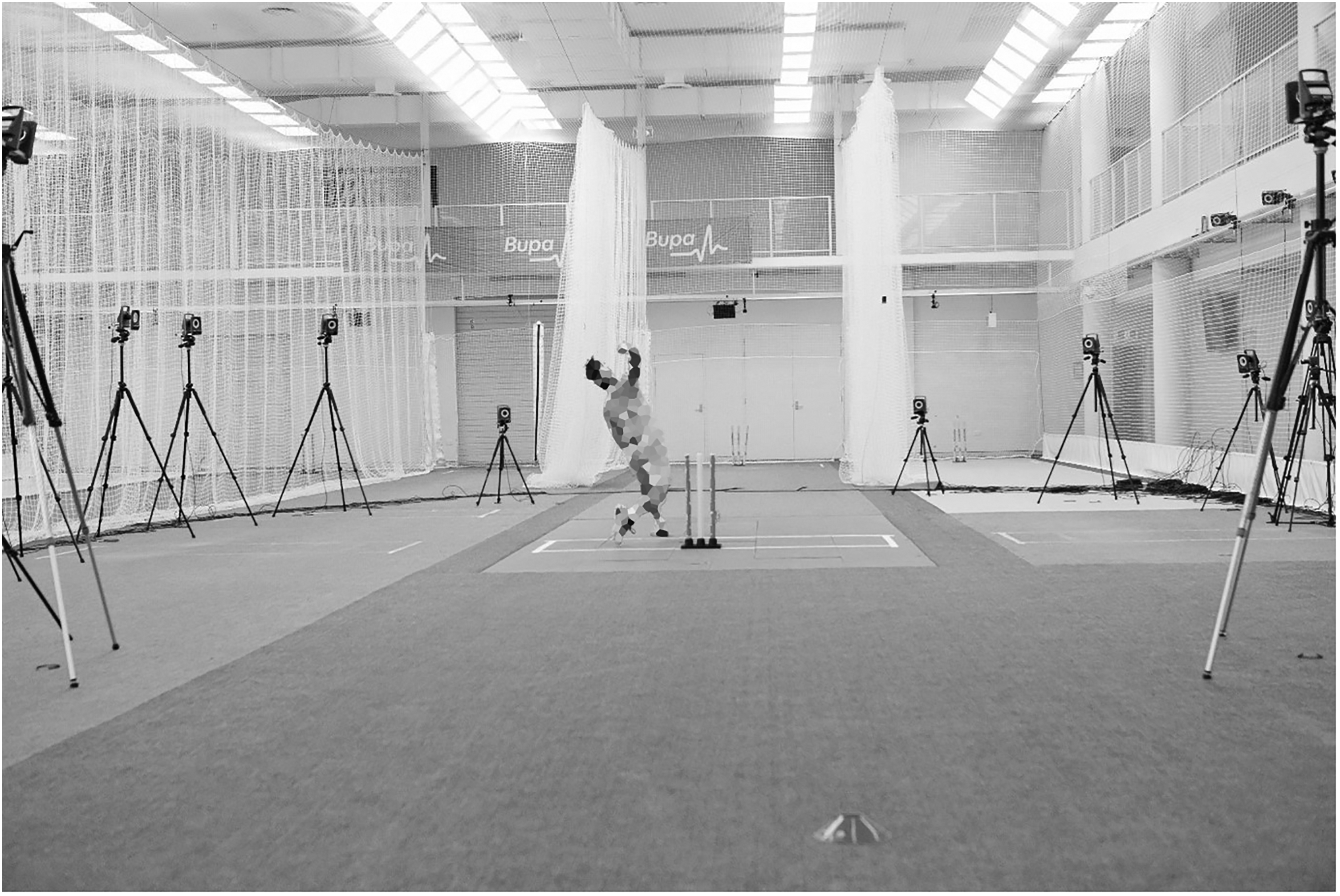
The data collection environment.
Participants
A total of 59 male and 19 female state- or national-level contracted pace bowlers (over 18 years) within Australia from 2014 to 2019 cricket seasons were included in this study. An ethics exemption was approved by Deakin University's Human Ethics Advisory Committee (project number HEAG-H 153-2020).
Procedures
Three-dimensional motion data (250 Hz) was captured via a 20-camera (T40-S) Vicon motion analysis system (Oxford Metrics Ltd, Oxford, UK) over 18 trials (Figure 1). The cameras surrounded the bowling crease in a circular manner, covering a 20 m3 volume (width = 2 m, length = 5 m, height = 2 m). This capture space was first calibrated via a calibration wand that collected 5000 frames with a tolerance of 0.2. A high-speed camera (125 Hz; Vicon Bonita 720c, Vicon Motion Systems Ltd, Oxford, UK), was placed in line with the popping crease to provide a sagittal plane view of the bowling action for purposes of identifying the moment of back foot contact and ball release.
Each participant wore sixty-three 14-mm spherical retro-reflective markers. The plug-in gait marker set, which has been used in prior cricket pace bowling research,18,19 accounted for 57 of these markers, while six additional markers were used. Of the 57 markers that formed the plug-in gait marker set, four markers were positioned on the head, five markers were situated about the thorax (C7 spinous process, T10 spinous process, right scapula, xiphoid process, and suprasternal notch), six markers were located around the pelvis and sacrum (left and right side anterior superior iliac spine, and a cluster of four markers about the sacrum), seven markers were situated on each arm (one at the acromion, upper arm, and elbow, with a marker positioned either side of the wrist, and a marker placed just proximal to the middle knuckle of the hand), and 14 markers were located on each leg (a cluster of four markers on the thigh, a marker on the medial and lateral epicondyles of the femur, a cluster of four markers on the tibia cluster, a marker on the medial and lateral malleoli, a heel and forefoot marker). Of the six additional markers, two were located on either side of the forefoot and an additional heel marker was used per foot. This customized marker set allowed full body motion to be determined using Vicon BodyBuilder (Version 3.6.1, Oxford Metrics Group, Oxford, UK) software.
One static and one dynamic calibration capture were performed. Participants stood in the anatomical position and held this for the static calibration. A set of five squats were used in the dynamic trial to define the knee joint flexion-extension axis. 20 After the dynamic calibration, joint center markers at the hip, knee, and ankle were removed as they were virtually stored in the thigh and shank clusters.
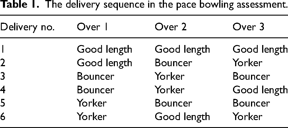
Following the static and dynamic calibration, subjects performed their own warm-up that included several warm-up balls. Once completed, participants delivered 18 balls (three overs of six deliveries each) at match intensity. These deliveries were bowled in a pre-determined order (Table 1), with six balls each at the following delivery lengths: “full” (0–4 m from batter's stumps), “good” (4–7 m from batter's stumps), and “short” (7–10 m from batter's stumps). Males bowled with a 4-piece 156 g half red/white cricket ball (Kookaburra Turf, Kookaburra, Sport, Melbourne, Australia), while females bowled with a 4-piece 142 g half red/white cricket ball (Kookaburra Turf, Kookaburra, Sport, Melbourne, Australia). A high-performance cricket coach (i.e. a coach with the Level 3 Cricket Australia Coach Accreditation) assessed bowling accuracy performance across the 18 deliveries. Time-discrete angular outputs were generated from the first six useable trials (two of each delivery length with minimal marker loss). Motion capture data were smoothed using a fourth-order low-pass Butterworth filter with a cut-off frequency of 10 Hz. 21 Pace bowling biomechanics do not appear to be influenced by the length of delivery 22 ; thereby the selection of trials at each delivery length for analysis was deemed acceptable. Note that the dataset only contained the mean data of the six trials, not trial-by-trial data for each participant.
The delivery sequence in the pace bowling assessment.
Thirteen bowling kinematic parameters were analyzed (Table 2). Reference frames were defined using three markers on each segment, allowing segment orientations and joint angles to be calculated. The thorax reference frame was defined by the C7 spinous process, T10 spinous process, xiphoid process, and suprasternal notch markers. The pelvis reference frame was defined by the bilateral anterior superior iliac spine markers and the cluster of four markers on the sacrum. Thoraco-pelvic segment orientations and joint angles could be calculated based on the thorax and pelvis local reference frames. A global coordinate system was defined with the y-axis pointing down the cricket pitch, with the x-axis pointing to the right of the cricket pitch for a right-handed bowler, and the z-axis pointing vertically upwards. The local coordinate system was defined with the y-axis pointing forwards, the x-axis pointed towards the participant's right, and the z-axis pointed upwards along the longitudinal axis of the segment. For the global orientation, the xyz rotations corresponded to tilt, drop, and twist, respectively, with orientations described relative to the anatomical position and the bowling side (anatomical position = 0°, flexion > 0°); a bowler that was in a “side-on” position was in 90° of rotation. For the joint angles, the xyz rotations corresponded to flexion–extension, abduction–adduction, and longitudinal rotation, respectively, with angles described relative to the anatomical position (anatomical position, 0°; flexion > 0°). Data for left-handed bowlers was transformed into the coordinate system of a right-handed bowler.
Pace bowling kinematic parameters and their definitions.
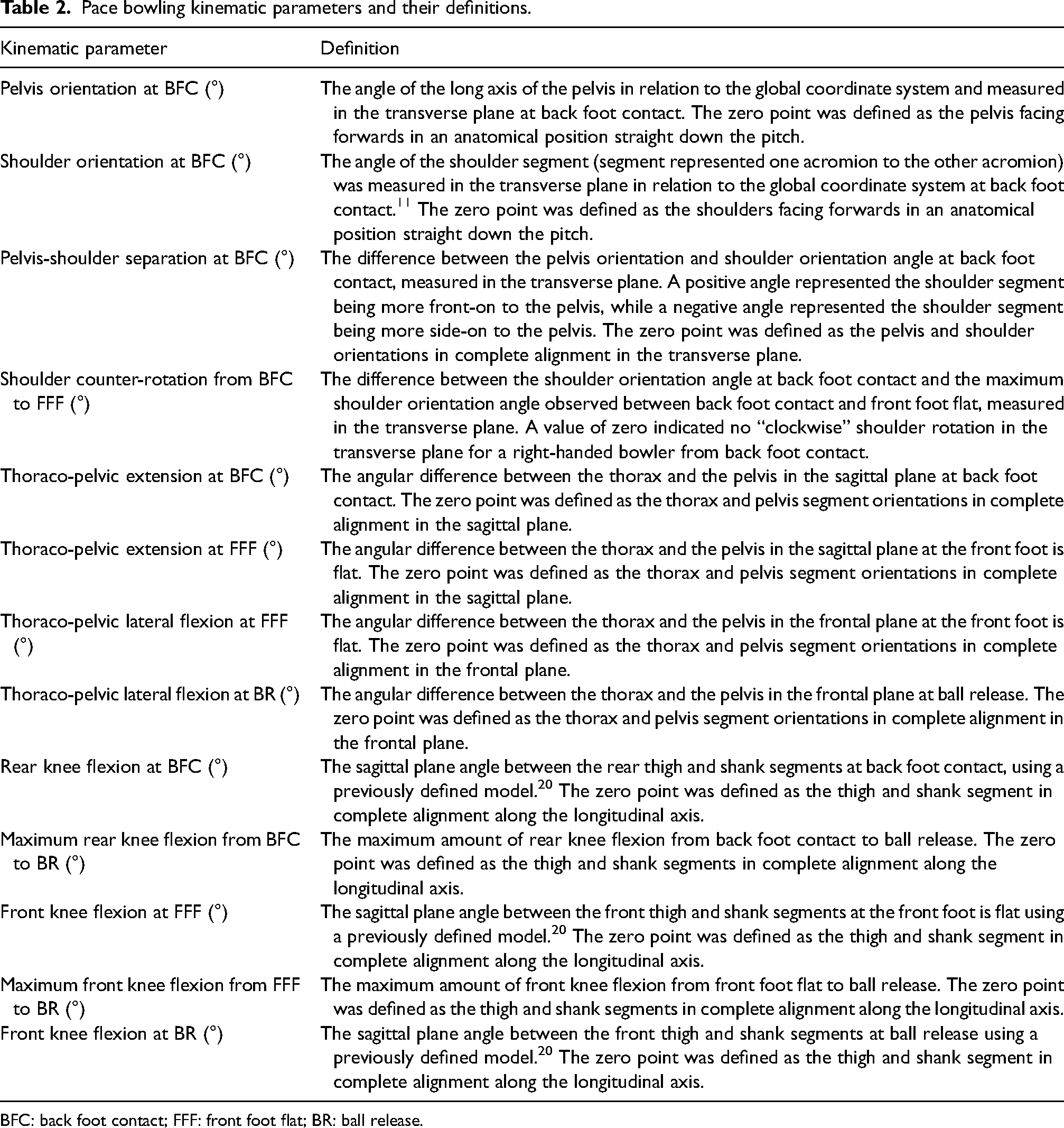
BFC: back foot contact; FFF: front foot flat; BR: ball release.
Each kinematic parameter was calculated at either back foot contact, front foot flat, or ball release. Back foot contact represented the time point when the heel markers reached their lowest point. The front foot flat was depicted as the first visible frame from the high-speed camera whereby the foot was flat. The first visible frame from the high-speed camera whereby the ball was not in contact with any part of the bowling hand was defined as the moment of ball release. Due to the differences in sampling rates between the high-speed camera (125 Hz) and Vicon cameras (250 Hz), the three-dimensional analysis involved going backwards by one frame compared to the high-speed camera to accurately determine and analyze the kinematics at front foot flat and ball release.
The mean of the six deliveries for each variable (and not the individual trial data) was included in the dataset provided by Cricket Australia. Some kinematic data were missing completely at random. Imputation for missing data was not performed. All dependent variables met the normal distribution, assessed via the Shapiro-Wilk test (i.e. p > 0.05). Independent samples t-tests were conducted to examine differences in pace bowling kinematics between males and females. Unequal variances were assumed when a statistically significant p-value (p < 0.05) was determined via the Levene's test. Hedges’ g effect sizes and 95% confidence intervals were calculated using a purpose-made Microsoft Excel (version 2016; Microsoft Corp.) spreadsheet 23 to account for the large difference in sample size between males and females and to correct the positive bias in Cohen's d effect sizes associated with smaller samples. Qualitative descriptors of standardized effect sizes (Cohen's d) were assessed using these criteria: trivial (< 0.20), small (≥ 0.20), medium (≥ 0.50), and large (≥ 0.80). Effect sizes were defined as “unclear” if the 95% confidence intervals overlapped thresholds for both small positive (> 0.2) and small negative effects (< 0.2). 24 All statistical analyses involved pairwise deletion due to a low sample size of female data. Inferential analyses were performed using IBM SPSS Statistics (version 27.0, IBM Corp, Armonk, NY). Statistical significance was accepted at p < 0.05.
Results
Elite male pace bowlers exhibited significantly greater thoraco-pelvic extension at back foot contact (mean difference ± 95% CI: 4.3 ± 4.1°, p = 0.039), shoulder counter-rotation from back foot contact to front foot flat (mean difference ± 95% CI: 8.9 ± 7.4°, p = 0.021), and thoraco-pelvic lateral flexion at front foot flat (mean difference ± 95% CI: 8.7 ± 5.1°, p = 0.001) compared to their female counterparts (Table 3, Figure 2). Elite female pace bowlers exhibited significantly less front knee flexion at ball release (mean difference ± 95% CI: −11.9 ± 11.6°, p = 0.046), and less maximum front knee flexion from front foot flat to ball release (mean difference ± 95% CI: −10.0 ± 7.4°, p = 0.009) compared to their male peers (Table 3, Figure 2).
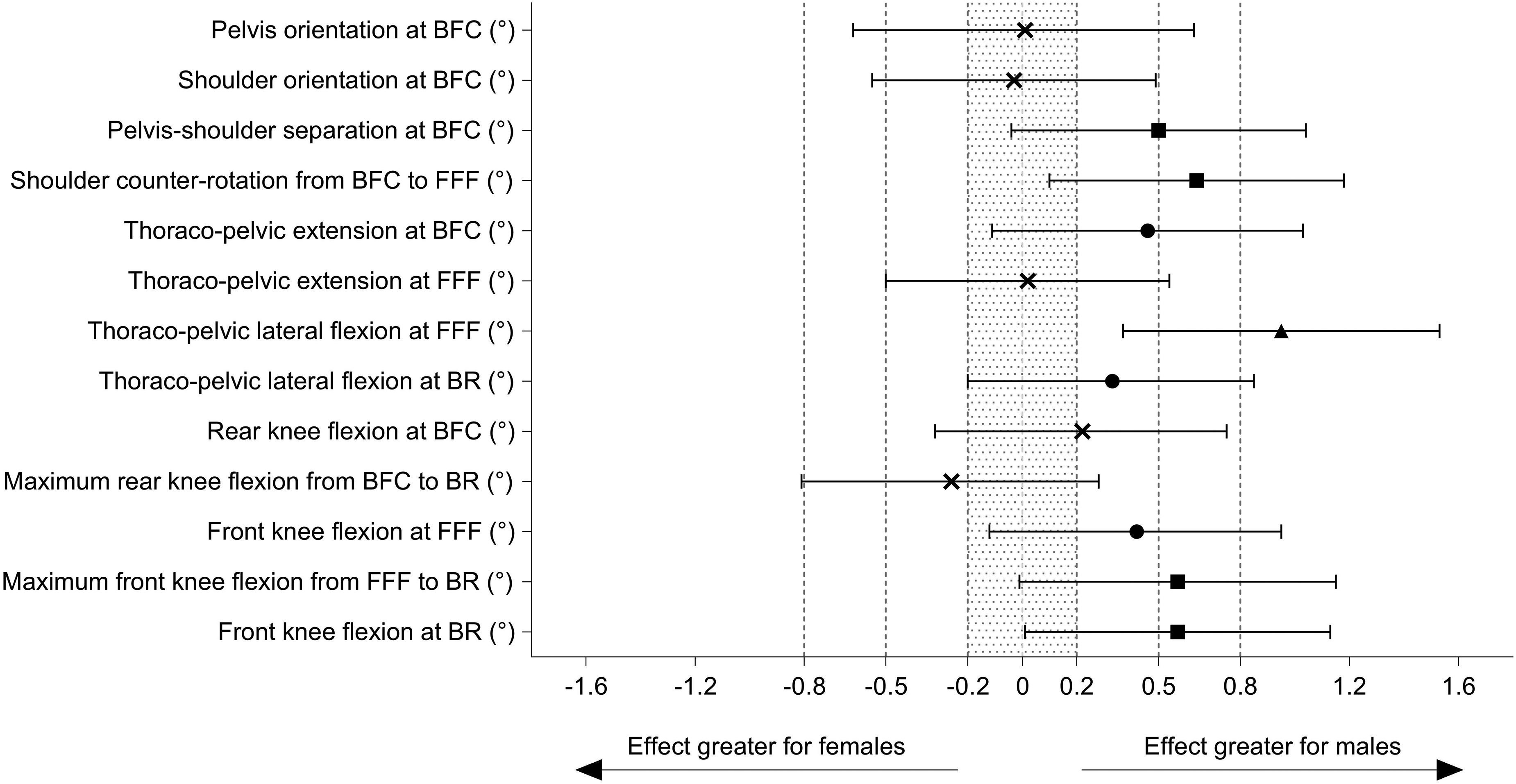
Hedges’ g effect sizes (male–female) ± 95% confidence intervals of each pace bowling kinematic parameter. The following symbols were used to assist in the qualitative interpretation of the effect size:
Descriptive data and differences in pace bowling kinematics between elite male and female pace bowlers.
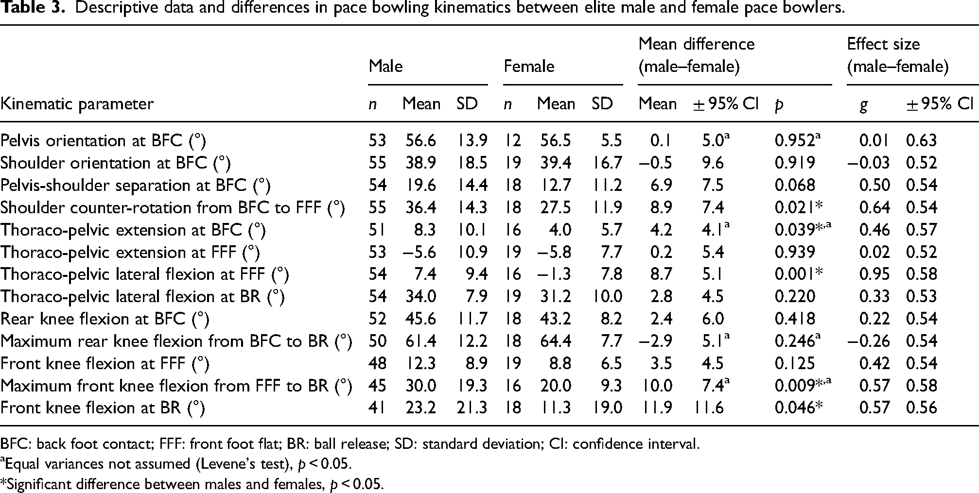
BFC: back foot contact; FFF: front foot flat; BR: ball release; SD: standard deviation; CI: confidence interval.
Equal variances not assumed (Levene's test), p < 0.05.
Significant difference between males and females, p < 0.05.
Discussion
The purpose of this study was to investigate the differences in pace bowling kinematics between elite male and female pace bowlers. In agreement with the hypothesis, this study observed several novel kinematic differences between males and females that could partly explain why males are more likely to sustain lumbar bone stress injuries and why females are more likely to sustain lower limb bone stress injuries. The findings from this investigation may have practical implications for designing sex-specific interventions to mitigate the risk of bone stress injuries.
Males counter-rotated their shoulders more than their female counterparts. Earlier research associated excessive shoulder counter-rotation and the “mixed” bowling action with lumbar bone stress injury in elite male pace bowlers.11,15–17 However, more recent conflicting evidence exists surrounding this association.8,9 Discrepancies in studies can be attributed to the limitation in projecting a three-dimensional segment orientation onto the horizontal plane. This two-dimensional kinematic parameter is a modest proxy of three-dimensional spinal kinematics. 25 Nevertheless, after applying the technique classification system with respect to the global coordinate system used by Portus et al., 11 71% of males (n = 37) were categorized to have bowled with a “mixed” bowling action compared to 47% for females (n = 8). Despite previous studies highlighting a concern with excessive counter-rotation, this data indicates excessive shoulder counter-rotation remains prevalent in elite male pace bowlers and is also potentially problematic for elite female pace bowlers too.
Males displayed greater thoraco-pelvic extension at back foot contact than females. This finding agrees with prior research. 14 Recent evidence has shown elite male pace bowlers who sustain a lumbar bone stress injury are positioned in greater lumbo-pelvic extension at front foot contact than their uninjured counterparts. 8 Bowlers who sustained a lumbar bone stress injury in that study were also observed to be in greater extension of the lower thoraco-pelvic region at back foot contact and front foot contact (medium effect size, not statistically significant), and achieved greater maximum values of extension in their lower thoraco-pelvic region from back foot contact to ball release (medium effect size, not statistically significant), compared with their uninjured counterparts. 8 Extension, lateral flexion, and rotation of the lumbar spine in conjunction with compression forces have been shown to place the pars interarticularis under the greatest amount of stress. 10 Lumbar bone stress injuries typically originate in the distal–ventral region of the pars interarticularis, and it is the ventral aspect that can experience double the stress during lumbar extension compared to the dorsal component. 26 Although speculative, the elite male pace bowlers in this study may have adopted a technique that elevated their risk of lumbar bone stress injury by adopting greater lumbo-pelvic extension upon back foot contact.
Pace bowlers perform contralateral thoraco-pelvic lateral flexion to maximize the height of ball release and subsequent bounce off the cricket pitch. 8 In this study, males were in greater contralateral thoraco-pelvic lateral flexion at the front foot flat, which may place them at greater risk of lumbar bone stress injury compared to their female counterparts. However, this result contrasts observations from prior research that compared pace bowling kinematics in elite male and female pace bowlers. 14 The discrepancy between findings may be due to differences in the measurement of front foot landing; this study measured kinematics at front foot flat which typically occurs a little later than front foot contact. Nevertheless, adolescent male pace bowlers with low back injury demonstrate excessive contralateral thoracic lateral flexion at both front foot contact and ball release, but also experience greater peak lumbar lateral flexion moments compared to their uninjured counterparts. 9 Excessive contralateral lower thoracic lateral flexion from front foot contact to ball release combined with the external ground reaction forces increases the stress on the neural arch of the lumbar vertebrae,10,27 and thereby heightens the risk of lumbar bone stress injury. Recent evidence has shown shoulder counter-rotation to be linked with contralateral thoracic lateral flexion and contralateral thoraco-lumbar lateral flexion, 25 therefore, interventions to reduce shoulder counter-rotation may also assist in limiting contralateral thoracic lateral flexion and contralateral thoraco-lumbar lateral flexion. A two-year coaching intervention study that used a constraints-led approach with the aim of reducing contralateral lower thoracic lateral flexion was shown to be ineffective 28 ; however, an eight-week exercise-based intervention program was effective in reducing contralateral trunk lateral flexion relative to the pelvic position. 29 This research highlights contralateral trunk lateral flexion (relative to the pelvis) to perhaps be more influenced by insufficient dynamic neuromuscular control of the lumbo-pelvic-hip complex.9,29
Compared to males, elite female pace bowlers were able to maintain a straighter front leg technique from front foot flat to ball release, as indicated by a lower maximum front knee flexion angle during this phase, and a lesser front knee flexion angle at ball release. Gait research conducted on healthy male subjects has shown that a more extended front knee throughout the gait cycle increases vertical loading rates compared to a more flexed knee. 13 While kinetic data was not collected as part of this study, and that human gait differs from the front leg landing mechanics of a cricket pace bowler, a straighter front leg landing technique may be positively associated with increased vertical loading rates. A systematic review and meta-analysis indicate lower limb bone stress injuries cannot be explained by the magnitude of ground reaction forces, but rather, by vertical loading rates. 12 Given elite Australian female cricketers sustain two new lower limb bone stress injuries per 100 players annually, 7 interventions to reduce the vertical loading rate upon front foot landing may be helpful to reduce the incidence of this debilitating injury. Previous research in elite male pace bowlers reveals that a more dorsiflexed ankle at the front foot upon landing to increase the time to reach vertical peak force, 30 which may have positive implications for reducing vertical loading rates.
There were several limitations to this study. As this investigation involved a secondary data analysis, conducted retrospectively, several kinematic variables linked with lumbar bone stress injury (e.g. rear hip flexion angle at back foot contact, lumbo-pelvic extension angle at front foot contact, lumbo-pelvic lateral flexion angle to the opposite side at front foot contact) or potentially associated with lower limb bone stress injury (front foot dorsiflexion angle at front foot contact) were not included in the dataset.3,8 Kinetic data was not captured, thus front-leg vertical loading rates and kinematics could not be explored. The instant of back foot contact in this study was defined as when the heel markers reached their lowest point; the true instant of back foot contact may have occurred a few frames earlier than measured in this study as pace bowlers typically land their back foot in a plantarflexed position (midfoot or forefoot) with their heel off the ground. The instants of the front foot flat and ball release were identified from high-speed camera footage operating at 125 Hz; this sampling rate is not as precise as the three-dimensional motion analysis system that operated at 250 Hz. The calculation of time-discrete kinematic data at front foot flat made it difficult to compare across other studies that investigated kinematics at front foot contact. Additional research could focus on analyzing the kinematics at when peak vertical ground reaction forces are experienced during the front leg landing of each bowler. It should be noted, however, that the analysis of time-discrete kinematic data as conducted in this study is an inferior approach compared to the analysis of continuous wave-form kinematic data; the latter can better portray “how” pace bowlers attained positions of increased injury risk. 1 Finally, the dataset provided for the secondary data analysis only comprised the mean kinematic data from the six trials per participant (with two useable trials of each delivery length). While there are some kinematic parameters that do not significantly alter with respect to the length of delivery bowled, 22 other kinematic parameters could have been sensitive to changes in delivery length, and thus the averaging of the six trials to provide representative data for each bowler would not have been appropriate due to expected lower between-trial repeatability.
Conclusion
This investigation has shown elite Australian male pace bowlers use techniques that could explain why they suffer more lumbar bone stress injuries than their female counterparts. These techniques involve greater shoulder counter-rotation, thoraco-pelvic extension at back foot contact, and contralateral thoraco-pelvic lateral flexion at front foot flat. On the other hand, elite Australian female pace bowlers bowl with a straighter front leg technique, which may partly explain why they experience more lower limb bone stress injuries, possibly through increased vertical loading rates. These findings are likely to influence sex-specific mitigation approaches to reduce the incidence of bone stress injury. Future experimental research is encouraged to adopt a multi-disciplinary approach involving biomechanics, strength and conditioning, and physiology in the mitigation of biomechanical risk factors linked to lumbar and lower limb bone stress injury in elite cricketers.
Footnotes
Acknowledgements
The authors would like to thank Cricket Australia for providing the data in-kind and for Rian Crowther's assistance regarding the protocols for the collection of three-dimensional pace bowling kinematics and ball release speed data.
Authors’ contributions
SA Feros: conceptualization, methodology, formal analysis, writing–original draft, and writing–review and editing, Visualization, supervision, and project administration. MH Gerhardy: Conceptualization, Formal analysis, Writing – original draft, Writing – review & editing, Project administration. JJ Fyfe: conceptualization, methodology, formal analysis, writing–original draft, and writing–review and editing, supervision, and project administration. DB Dwyer: conceptualization, methodology, formal analysis, writing–original draft, writing–review and editing, visualization, supervision, and project administration.
Declaration of conflicting interests
The author(s) declared no potential conflicts of interest with respect to the research, authorship, and/or publication of this article.
Funding
The author(s) received no financial support for the research, authorship, and/or publication of this article.