
Abstract
Nitroxyl, which was acknowledged as a vital constituent among the reactive nitrogen species, poses significant challenges when it comes to its swift and precise identification and measurement. Recently, a kind of BODIPY-DAP-Cu(II)-conjugate fluorescent probes has been broadly used to detect nitroxyl. However, the impact of varying linker chain lengths on the physicochemical attributes and imaging performance of dipyrromethene boron difluoride–based fluorescent probes remained uncertain. In this study, we engineered three new dipyrromethene boron difluoride–based nitroxyl fluorescent probes CuII[BD1] (3.88 Å), CuII[BD2] (5.02 Å), CuII[BD3] (6.67 Å), CuII[BD4] (10.77 Å) which were composed of dipyrromethene boron difluoride as fluorescence indicator and a tripodal-DPA-Cu(II) unit as nitroxyl recognition site, using increasing lengths of linker chain. The new probe CuII[BD3] (6.67 Å) showed fast response (within 1 min) toward nitroxyl with remarkable sensitivity, capable of detecting as low as 0.25 μM, superior specificity for nitroxyl, and relative stability under physiological pH condition. Furthermore, the probe CuII[BD3] was applied to bioimaging of nitroxyl in rat blood. In conclusion, those results revealed that the imaging characteristics of dipyrromethene boron difluoride derivatives were significantly influenced by the length of the linker chain. The fluorescence detection efficiency increases with the growth of the linker chains within 6.67 Å. Consequently, these probes hold promise as valuable chemical instruments for probing the detailed functions and mechanisms of nitroxyl within biological organisms and offered an innovative approach for the construction of dipyrromethene boron difluoride–based nitroxyl fluorescent probes.

Introduction
Nitroxyl (HNO) has been characterized as protonated species and one-electron-reduced protonated variant of nitric oxide (NO). HNO was a distinguished signaling molecule involved in numerous physiological functions, exhibiting a unique set of biological and chemical characteristics distinct from NO.1 –3 HNO can react with thiols to provide either disulfides or sulfinamides, and inhibit the enzyme’s activity.4,5 Then, HNO has recognized to serve as a potential cardiovascular function regulator that might supply a beneficial tool for cardiac ischemia-reperfusion damage 6 and cardiovascular illnesses.7,8 Currently, there is growing evidence that HNO acts an ideal biomarker for drug-induced liver injury (DILI) diagnosis 9 and an potential Parkinson’s biomarker. 10 Although great progresses regarding the biologically relevant chemistry of HNO of HNO have been achieved, certain questions continue to elude comprehensive exploration. The most captivating queries revolve around whether HNO is indeed synthesized within living organisms, the mechanisms by which it is produced, and if its generation is integral to the biological signaling processes involving redox reactions. Consequently, the creation of methods that are both sensitive and selective for the detection of HNO in biological contexts is of paramount significance.
Regrettably, the exploration of HNO’s multifaceted chemical and physiological attributes is complicated by its inherent tendency to spontaneously dimerized into N2O in aqueous environments. 11 Various conventional methodologies are available for the identification of NO and HNO, composed of headspace gas chromatography, electron paramagnetic resonance (EPR) analysis, and High-Performance Liquid Chromatography–Mass Spectrometry (HPLC-MS).12 –15 These methods often entail prolonged processing times or necessitate the disruption of cellular and tissue integrity, which limited their in vitro and in vivo applications. Fluorescence-probing serves as the most attractive approach to monitor biologically HNO owing to its remarkable sensitivity, exceptional selectivity, and real-time observation capacity.16,17 To date, a diverse array of fluorescent probes have been judiciously engineered, capitalizing on the distinctive reactivity of HNO with metal complexes18 –21 or metalloporphyrins, 22 thiols, 23 organic phosphines,24 –26 and nitroso compounds. 15 These efforts have significantly propelled the field of HNO fluorescent detection forward. Nonetheless, certain limitations of these probes remain, including slow reaction rates, 26 suboptimal sensitivity, 27 susceptibility to interference by coexisting biological molecules, 21 and the necessity for additional surfactants to enhance solubility. 28 Consequently, there remains a critical need to develop new HNO probes that feature rapid response kinetics, superior sensitivity, and exceptional selectivity, to overcome the shortcomings of current options.
Recently, the “covalent-assembly” approach is widely used to construct the fluorescent probes with a strong fluorescence “off–on” response. 29 So far, a kind of BODIPY-DPA-Cu(II)-based fluorescent probes used a dipyrromethene boron difluoride (BODIPY) moiety as fluorescence indicator, a DiPicolylAmine (DPA)-Cu(II) complex as HNO recognition site and a trizole structure as linker. 19 Those BODIPY-based probes have been widely used for the measurement of HNO levels on the basis of the reduction reaction. 30 However, the impact of varying linker chain lengths on the physicochemical attributes and imaging performance of BODIPY–based fluorescent probes remained uncertain. Therefore, in this study, we constructed four BODIPY-DPA-Cu(II)-conjugate HNO probes such as CuII[BD1], CuII[BD2], CuII[BD3], and CuII[BD4] (Chart 1) using ethyl, propyl, benzyl, and biphenyl group as linker (Scheme 1), which have gradually increasing lengths (3.88 Å, 5.02 Å, 6.67 Å, and 10.77 Å). The characterization of these constructed probes involved UV spectra, 1H NMR, HRMS (High-Resolution Mass Spectrometry), and fluorescence spectra analysis. Those probes show high sensitivities (within 1 min) toward HNO with excellent sensitivity (the lowest detection limit was 0.25 μM). In addition, our findings revealed that the linker length played a crucial role in the imaging characteristics of BODIPY-based fluorescent probes. The fluorescence detection efficiency increases with the growth of the linker chains within 6.67 Å. Moreover, those BODIPY-based probes exhibit high stability toward other species under physiological pH. Finally, the potential in vivo bioimaging application of CuII[BD3] was evaluated in the rat blood. Those notable imaging performances displayed by those BODIPY-based probes make them as useful tools to reveal HNO-related dynamic changes in live samples and supply a new methodology for the construction of novel BODIPY-based HNO fluorescent probes.

Molecular structures of CuII[BD1], CuII[BD2], CuII[BD3], and CuII[BD4].

Synthesis of CuII[BD1], CuII[BD2], CuII[BD3], and CuII[BD4].
Results and discussion
Synthesis
Synthesis of 1–3
4-bromobenzaldehyde (1.5 g, 8.1 mmol) was dissolved in a mixture dimethylformamide (DMF)/water (3:1; v/v, 120 mL). Subsequently, (4-(hydroxymethyl) phenyl) boronic acid (1.85 g, 12.15 mmol), K2CO3 (2.23 g, 16.2 mmol), and Pd (PPh3)4 (0.47 g, 0.4 mmol) were added under N2. Subsequently, the reaction mixture was elevated to 130°C and maintained under reflux conditions overnight, and the progress was monitored via Thin Layer Chromatography (TLC) until the complete consumption of the initial reactant was confirmed. Once the mixture cooled to ambient temperature, ethyl acetate (EA) was added for the purpose of extracting the crude product. Wash the organic layer with saturated NaCl, the organic layer was separated and dried over anhydrous Na2SO4, filtered. The crude compound was gained through the concentration of filtrate to dryness using rotary evaporation. Further purification of crude compound was carried out using silica gel column chromatography (EA/PE, v/v, 1:4), and the pure product
Pure product 1-2 (1.2 g, 5.65 mmol) was dissolved in dry CH2Cl2 (50 mL), thionyl chloride (1.23 mL, 16.96 mmol) was gradually introduced into the mixture via a dropping funnel, which was left under stirring at ambient temperature continuously overnight. On reaction completion, concentrated the reaction mixture to dryness using rotary evaporation to obtain the crude product. Further purification of crude compound was carried out using silica gel column chromatography (EA: PE, v/v, 1:8), and the pure product
Synthesis of B
2, 4-dimethylpyrrole (2 eq) was dissolved in dry CH2Cl2, acetyl chloride (1 eq) was gradually introduced into the mixture via a dropping funnel. The reaction mixture was agitated at ambient temperature continuously overnight. Under an ice-cold environment, Et3N (10 eq) and BF3·Et2O (10 eq) were added sequentially to the reaction mixture, which was then further agitated for extra 6 h. On reaction completion, wash the mixture with water, saturated NaHCO3, and saturated NaCl solution. The extracted organic layer was dehydrated using anhydrous Na2SO4, followed by filtration to remove the drying agent. Then, the crude product was gained through concentration of filtrate to dryness using rotary evaporation. Further purification of crude compound was carried out using silica gel column chromatography (CH2Cl2/PE) to give compound
2, 4-Dimethylpyrrole (0.9 mL, 8.66 mmol) and 1-3 (1.0 g, 4.33 mmol) were added to anhydrous CH2Cl2 (600 mL) under N2 atmosphere. As catalyst, TFA (65 μL, 0.866 mmol) was added into the mixture, which was left under stirring at ambient temperature continuously overnight. Then 2, 3-dichloro-5, 6-dicyano-1, 4-benzoquinone (DDQ, 0.98 g, 4.33 mmol) was added as oxidizing reagent. After stirring for 1 h, Et3N (6 mL, 43.3 mmol) and BF3·Et2O (5.3 mL, 43.3 mmol) were added sequentially. The reaction mixture was agitated at ambient temperature condition continuously for a duration of 6 h. On reaction completion, wash the reaction mixture with saturated NaHCO3 and saturated NaCl solution. The extracted organic layer was dehydrated using anhydrous Na2SO4, followed by filtration to remove the drying agent. Then, the crude product was gained through concentration of filtrate to dryness using rotary evaporation. Further purification of crude compound was carried out using silica gel column chromatography (PE/CH2Cl2, v/v, 2:1), the pure product
Synthesis of BD
Di (2-picolyl) amine (DPA) (1 eq), KI (1 eq) and K2CO3 (3 eq) were dissolved in dry THF, a solution of B (1.2 eq) in dry THF was gradually introduced into the mixture via a dropping funnel. Subsequently, the reaction mixture was elevated to 80°C and maintained under reflux conditions overnight, and the progress was monitored via Thin Layer Chromatography (TLC) until the complete consumption of the initial reactant was confirmed. On reaction completion, THF was eliminated using rotary evaporation, and then added CH2Cl2 to dissolve the residue. Wash the CH2Cl2 layer with water, saturated NaHCO3, and saturated NaCl solution. The extracted organic layer was dehydrated using anhydrous Na2SO4, followed by filtration to remove the drying agent. Then, the crude product was gained through the concentration of filtrate to dryness using rotary evaporation. Further purification of crude compound was carried out using silica gel column chromatography (CH2Cl2/MeOH), the compound
Synthesis of CuII[BD]
Compound
719.2668 [(M+Cu(II)+OEt−]+; 674.2328 [(M+Cu(II)-H]+
Reaction of probes with HNO
To ascertain whether these probes could serve effectively as HNO detectors, their responsiveness to HNO was thoroughly examined. Moreover, to explore the impact of varying linker chain lengths on the physical, chemical attributes, and the detection proficiency of BODIPY-derived HNO probes, a kind of BODIPY-based fluorescent probes, which used BODIPY moiety as fluorescence indicator and DPA-Cu(II) complex as HNO recognition site. CuII[BD1] (3.88 Å), CuII[BD2] (5.02 Å), CuII[BD3] (6.67 Å), and CuII[BD4] (10.77 Å) contain gradually increasing lengths of linker chain. The fluorescence intensity of CuII[BD1] (3.88 Å), CuII[BD2] (5.02 Å), and CuII[BD3] (6.67 Å) increased steadily with increasing Angeli’s salt (Na2N2O3, a HNO donor) concentration (see Figure 1) until they reached a steady state at 20 μM Na2N2O3, which aligned with 7.17-, 8.20-, 9.3-fold fluorescent enhancement individually compared to the blank [HNO] within 1 min.

Fluorescence titration of (a) CuII[BD1] (1 μM); (b) CuII[BD2] (1 μM); (c) CuII[BD3] (1 μM); (d) CuII[BD4] (1 μM) in PIPES buffer (50 mM, 0.1 M KCl, pH =7.4) in the presence of different amounts of Angeli’s salt. The fluorescence intensity was measured over an elongated duration of 30 min subsequent to adding Na2N2O3, λex: 500 nm, λem: 510 nm).
In addition, the fluorescence response of CuII[BD1] (3.88 Å), CuII[BD2] (5.02 Å),
The selectivity of probes toward HNO
Given their exceptional responsiveness to HNO, a critical next step was to assess their selectivities with different bioactive substances including reactive nitrogen species (RNS), reactive oxygen species (ROS), cations, and anions over an elongated duration of 30 min (see Figure 2). When exposed to 30-fold of NaNO2, those probes exhibited minimal fluctuation in their fluorescence emission profiles. This observation suggested that the activation observed in the presence of Angeli’s salt is attributed to the generation of HNO, rather than being influenced by the NO2− byproduct. Those probes showed a pronounced specificity toward HNO, as evidenced by their emission behaviors, which were significantly more responsive to HNO than to any other reactive species that are common constituents of the biological milieu. The biologically active compounds, including NO3−, ClO−, H2O2, HO•, O2•−, ONOO−, ROO•, and EDTA, did not result in a notable increase in the emission intensity of the CuII[BD] complex (see Figures 2 and 3). The slight increase in emission seen on exposing these probes to NO is particularly striking, highlighting the system’s potential utility in elucidating the hypothesized divergent functions of NO and HNO within biological systems. The above results indicate those copper-based probes were highly selective to HNO over other biorelated species. However, Hcy, Cys, GSH, and H2S could also restore the fluorescence signal intensity of CuII[BD3] (7.44-, 7.77-, 7.90- and 13.95-fold, see Figure S3 in the Supporting Information) because of the reduction of chelated Cu(II)-DPA. To improve the stability and selectivity of BODIPY-based HNO probes, Cu(II)-DPA group would be replaced with Cu(II)-azamacrocyclic in the following work. 31

Normalized integrated fluorescence intensity of probes (1 μM) in aqueous buffer (50 mM PIPES, 100 mM KCl, pH = 7) over an elongated duration of 30 min subsequent to adding. λex: 500 nm, λem: 510 nm.
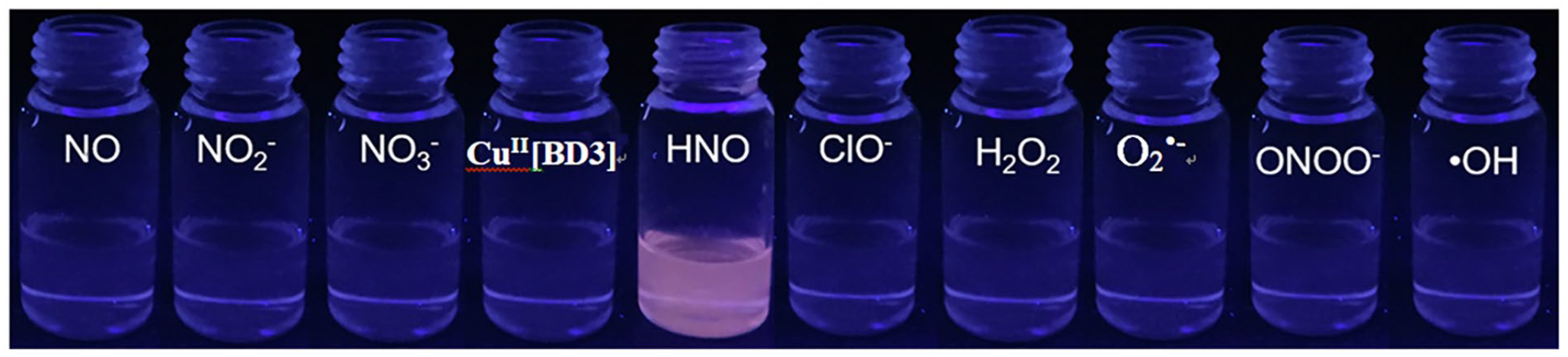
The photo of CuII[BD3] under ultraviolet lamp (365 nm) after the addition of different ROS and RNS.
Considering the common pH range of 4.5–8.0 encountered within live cellular environments, we explored the impact of pH on the emission behavior of CuII[BD3] in the presence of HNO. Acid-base fluorescence titrations revealed that the fluorescence intensity of CuII[BD3] remained constant within the pH spectrum from 6 to 9. The result suggested that this probe is well suited for functioning optimally under physiological pH condition (Figure 4).

Fluorescence responses of CuII[BD3] (10 µM) at pH 2 ~ 12. λex: 500 nm, λem: 510 nm.
To further explore in vivo application potential of those BODIPY-DPA-Cu(II)-Conjugated HNO probes, the application of CuII[BD3] in whole blood from rat was also investigated. As shown in Figure 5, CuII[BD3] exhibited minimal fluctuation in the fluorescence intensity after 6-h incubation in the blood; when exposed to 30-fold of HNO, a 8.6-fold increase in fluorescent intensity was observed. This significant enhancement demonstrated the potential in vivo application of CuII[BD3].

Fluorescence responses of CuII[BD3] (1 µM) at PBS solution, blood, and HNO.
Summary and conclusion
We have constructed a new series of BODIPY-DPA-Cu(II)-conjugate HNO probes such as CuII[BD1] (3.88 Å), CuII[BD2] (5.02 Å), CuII[BD3] (6.67 Å), and CuII[BD4] (10.77 Å) using ethyl, propyl, benzyl, and biphenyl group as linker, which contained increasing lengths of linker chains. Those probes show exceptional selectivity sensitivity toward HNO, especially probe CuII[BD3] (6.67 Å) using benzyl group as linker has the highest sensitivity (LOD = 0.25 μM). The fluorescence detection efficiency increases with the growth of the linker chain within 6.67 Å. The length of linker chain has an important influence on the detection efficiency. Apart from the increasing chain length of benzyl group, the rigidity of the benzene ring also plays an important role in enhancing the fluorescence detection efficiency. Furthermore, these BODIPY-DPA-Cu(II)-conjugate probes exhibited superior specificity for HNO, distinguishing it effectively from other biologically relevant ROS and RNS under physiological pH, which make them hold great potential for practical applications in living system. Finally, the potential in vivo bioimaging application of BODIPY-DPA-Cu(II)-conjugate HNO probe was evaluated in rat blood. Therefore, our present study provides a novel strategy for the construction of new BODIPY-based HNO fluorescent probes with enhanced characteristics which holds the potential to significantly broaden the scope of in vivo biomedical imaging practices.
Supplemental Material
sj-doc-1-chl-10.1177_17475198251313658 – Supplemental material for Synthesis and characterization of BODIPY-DPA-Cu(II)-conjugate HNO fluorescent probes: Effect of linker length on imaging properties
Supplemental material, sj-doc-1-chl-10.1177_17475198251313658 for Synthesis and characterization of BODIPY-DPA-Cu(II)-conjugate HNO fluorescent probes: Effect of linker length on imaging properties by Wenbo Liu, Yufeng Zou, Yu Zhou, Jiangwei Zheng, Xiaoguang Liu and Dengzhao Jiang in Journal of Chemical Research
Footnotes
Author contributions
W.L. contributed to conceptualization, methodology, writing, project administration, and funding acquisition; Yuf.Z. participated in investigation; Yu.Z. involved in data statistics; J.Z. performed synthesis; X.L. performed imaging test of fluorescent probe; and D.J. participated in the characterization of fluorescent probes.
Declaration of conflicting interests
The author(s) declared no potential conflicts of interest with respect to the research, authorship, and/or publication of this article.
Funding
The author(s) disclosed receipt of the following financial support for the research, authorship, and/or publication of this article: This work was supported by the Science and Technology Research Project of Jiangxi Provincial Department of Education (grant no. GJJ2201933), the Natural Science Foundation of Jiangxi Province (grant no. 20192BAB215043), and the National Natural Science Foundation of China (grant no. 21967013).
Supplemental material
Supplemental material for this article is available online.
References
Supplementary Material
Please find the following supplemental material available below.
For Open Access articles published under a Creative Commons License, all supplemental material carries the same license as the article it is associated with.
For non-Open Access articles published, all supplemental material carries a non-exclusive license, and permission requests for re-use of supplemental material or any part of supplemental material shall be sent directly to the copyright owner as specified in the copyright notice associated with the article.