Synthesis of a novel nitrogen mustard–conjugated bis-terpyridine ruthenium(II) complex as a potent anticancer agent that induces cell cycle arrest and apoptosis
Open accessResearch articleFirst published online March, 2022
Synthesis of a novel nitrogen mustard–conjugated bis-terpyridine ruthenium(II) complex as a potent anticancer agent that induces cell cycle arrest and apoptosis
A fairly small-sized aryl nitrogen mustard–conjugated terpyridine is synthesized in only two steps as a ligand to chelate with RuCl3 to afford a [Ru(tpy-CM)2]Cl2 complex. This complex exhibits prominent antiproliferative activity toward several tumor cells. Further studies conclusively show that the complex suppresses human renal clear cell carcinoma cells (786-O cells) by inducing G1 phase cell cycle arrest and apoptosis. This work provides a synthetic and therapeutic model for nitrogen mustard-containing metal complexes.
In the search for novel non-platinum-based antitumor agents with a wide range of activity and fewer side effects than those of platinum drugs and their analogues, ruthenium (Ru) complexes appear to be the most popular alternatives.1,2 Over the last few decades, interest in ruthenium polypyridyl complexes as promising in vitro antitumor agents has grown. These Ru polypyridyl complexes are commonly analogues such as [Ru(tpy)Cl3] (tpy = 2,2′:6′,2″-terpyridine), [Ru(tpy)2]2+, [Ru(azpy)2Cl2]2+, (azpy = 2-phenylazopyridine), [Ru(bpy)2Cl2]2+, (bpy = 2,2'-bipyridine), and [Ru(bpy)3]2+.3–7 These Ru complexes exhibit their antiproliferative activity mostly through inhibition of DNA replication,8 protein or enzyme activity,9,10 and result in cell arrest or apoptosis.11 Terpyridine ruthenium complexes are well known as effective non-covalent DNA binders.12 They recognize DNA bases within DNA grooves via motifs and effectively slot into the grooves of the DNA double-helix by non-covalent binding, which blocks the DNA replication and translation.13 Such complexes show varying degrees of cytotoxicity against a wide variety of tumor cell lines, and the cytotoxicity generally changes on varying the ligand structure. Therefore, to improve the antitumor activity, ligand modification is of significant importance.
Nitrogen mustards, a family of DNA alkylating agents discovered in 1942, opened a new avenue in cancer chemotherapy.14 This meritorious category of alkylating agent shows antitumor activity by typically binding to DNA, blocking DNA replication and ultimately leading to cell death.15 To date, research on nitrogen mustards has enabled the development of more effective nitrogen mustard analogues.16,17 Studies on ways to enhance the cytotoxic efficacy of nitrogen mustard analogues is currently a trending topic in research.
In this study, a novel and fairly small-sized aryl nitrogen mustard–conjugated terpyridine ligand was designed and synthesized as an active ligand by only two steps. Two equivalents of the ligand were then used to chelate with RuCl3 and FeCl3 to afford the corresponding nitrogen mustard–conjugated bis-terpyridine complexes [Ru(tpy-CM)2]Cl2 (4a) and [Fe(tpy-CM)2]Cl2 (4b), respectively, as candidates for further research on antitumor activity. The in vitro antiproliferative activity of complexes 4a and 4b against several tumor cells (MDA-MB-231, 786-O, A549, and HepG2) was investigated. In addition, the inhibition effects on tumor cell migration, invasion, crawl and colony, as well as cell cycle arrest and induction of apoptosis by [Ru(tpy-CM)2]Cl2 (4a) were studied. It is expected that this study will provide important ideas and strategies for the research and development of novel nitrogen mustard drugs and ruthenium complexes.
Results and discussion
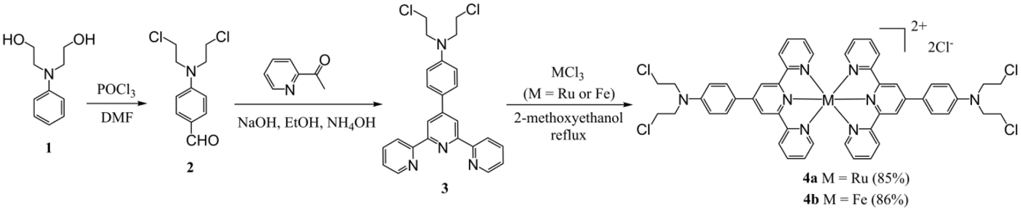
The synthetic pathway toward the target compounds 4a and 4b is illustrated in Scheme 1. The complexes were prepared as follows. First, dichloride 2 was synthesized by the Vilsmeier–Haack reaction18 of 2,2'-(phenylazanediyl)bis(ethan-1-ol) (1), excess POCl3, and DMF. Dichloride 2 was then converted into compound 3 in EtOH at room temperature by via a Knoevenagel condensation reaction,19 in which compound 2, 1-(pyridin-2-yl)ethan-1-one, and NH4OH were involved as the reactants and NaOH as the base. It is noteworthy that compound 3 is a small-sized ligand incorporating an aryl nitrogen mustard and terpyridine. Complexes 4a and 4b were obtained in high yield by chelation reactions between compound 3 and FeCl3 or RuCl3 under reflux in 2-methoxyethanol.
Synthesis of nitrogen mustard–conjugated bis-terpyridine complexes 4a and 4b.
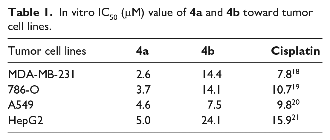
The cytotoxic activities of complexes 4a and 4b were evaluated against MDA-MB-231, 786-O, A549, and HepG2 cell lines using the CCK8 assay. The results are shown in Figure 1(a) and the IC50 values are listed in Table 1. Although complexes 4a and 4b both exerted obvious cell cytotoxicity against the above tumor cells, 4a exhibited more potent activity than 4b. For example, complex 4a showed relatively strong antiproliferative activities against the MDA-MB-231, 786-O, A549, and HepG2 cell lines with IC50 values of 2.6, 3.7, 4.6, and 5.0 μM, respectively, whereas the IC50 values for 4b were 14.4, 14.1, 7.5, and 24.1 μM. Meanwhile, cisplatin from the literatures20–23 was used as a competitor, the IC50 values of which against MDA-MB-231, 786-O, A549, and HepG2 cell lines were 7.8, 10.7, 9.8, and 15.9 μM, respectively, the results are presented in Table 1. Hence, the nitrogen mustard–conjugated terpyridine Ru complex exerted better antiproliferative activities than the corresponding Fe complex 4b and cisplatin. The reasons for their different activities are first attributed to the different lone-pair electron density of N atom in N,N-bis(2-chloroethyl)aniline unit. Specifically, the 1H NMR results show that the chemical shift of α-methylene of N,N-bis(2-chloroethyl)aniline unit in the Ru complex is 3.96 (δ) (Figure S6) but 4.01 (δ) in the Fe complex (Figure S9), indicating the higher electron density of the Natom of N, N-bis(2-chloroethyl)aniline unit in the Ru complex than in the Fe complex. Thus, the N,N-bis(2-chloroethyl)aniline unit in the Ru complex is more likely to form the cyclic aminium ions. Naturally, the N-nucleophilicity in Ru complex is stronger than in the Fe complex, which subsequently leads to a better antiproliferative activity of the Ru complex than that of Fe complex. The second reason may be due to the preferential non-covalent binding property between the terpyridine ruthenium complexes with DNA.12,13 Based on the above results, compound 4a was selected for further study using the 786-O cell line. We next ran a colony formation assay (Figure 1(b)), and the results revealed that the proliferation ability of 786-O cells was markedly reduced after treatment with 4a. For instance, the number of colonies was less than one-fifth of control (Figure 1(c)) after treating cells with 4a (16 μM).
In vitro IC50 (μM) value of 4a and 4b toward tumor cell lines.
The antiproliferative activity of complexes 4a and 4b. (a) The cytotoxicity of complexes 4a and 4b toward the MDA-MB-231, 786-O, A549, and HepG2 cell lines. Cells were treated with the indicated concentrations of 4a or 4b for 72 h and cell viability was determined by the CCK8 assay. (b) Colony formation assay; 786-O cells were treated with 8 and 16 μM of 4a, respectively, and incubated for 14 days. (c) Statistical bar chart of the colony numbers. The data are presented as the mean value ± SD of three replicates per group.
To investigate the inhibitory effects of 4a on the migration and invasion 786-O cells, transwell migration and invasion assays were carried out. As shown in Figure 2(a), the migration and invasion numbers of 786-O cells were dramatically diminished in a dose-dependent manner after treatment with 4a compared with the control group. This can be confirmed by the statistical bar chart of the numbers of migration (Figure 2(c)) and invasion (Figure 2(d)) cells. Moreover, a wound-healing assay (Figure 2(b)) revealed that the wound-healing capacity of 786-O cells was also reduced. For instance, without treating with 4a, up to 70% of wound width was healed; however, when treating with 8 and 16 μM of 4a, only 31% and 20% wound width was healed, respectively (Figure 2(e)). These results collectively indicate that complex 4a can restrain the migration, invasion, and wound-healing capacity of 786-O cells.
Complex 4a inhibits the migration, invasion, and wound-healing ability of 786-O cells. (a) Transwell migration and invasion assays. Cells were treated with 8 and 16 μM of 4a, respectively, and incubated for 24 h. (b). Wound-healing assay; 786-O cells were treated with 8 and 16 μM of 4a, respectively, and incubated for 24 h, then photographs were taken with an optical microscope. Statistical bar charts of the number of migration (c) and invasion (d) cells. (e) Statistical bar chart of wound healing. The data are presented as the mean value ± SD of three replicates per group.
To study the influence of complex 4a on 786-O cell cycle progression, the cellular DNA content was measured by flow cytometry. The percentages of G1, S, and G2/M phases were calculated, and the results are presented in Figure 3(a). The G1 phase rise ascends from 48.3% to 67.4% when treated with 8 μM of 4a compared with the control, and this upward tendency rises to 75.7% as the concentration of 4a increases to 16 μM. Furthermore, the western blot analysis of cycle arrest-related proteins such as cyclin A1, cyclin E, and CDK124 was investigated and the results are shown in Figure 3(b). The results show that these proteins are all markedly downregulated by 4a, which leads to the conclusion that complex 4a gave rise to cycle arrest at the G1 phase.
Cell arrest and apoptosis on 786-O cells induced by complex 4a. (a) Cell cycle analysis of 786-O cells. Cells were treated with 8 and 16 μM of 4a, respectively, stained with propidium iodide (PI) and analyzed with a flow cytometer. (b) Western blot analyses of proteins on 786-O cells after treatment with 4a for 48 h. (c) Apoptosis analysis using an Annexin V-FITC/PI Apoptosis Detection Kit. (d) Statistical bar chart of the apoptosis rate.
An Annexin V-FITC/PI Apoptosis Detection Kit was used to evaluate the apoptosis-inducing efficacy of complex 4a on 786-O cells (Figure 3(c)). The upper right section represents the late apoptotic cells, and the lower right section represents the early or middle apoptosis. Compared with the control, the late apoptosis upper right section percentage increased from 5.47% to 8.33% and 19.02%, respectively, as the concentration of 4a increased up to 8 μM and then 16 μM. The total apoptosis percentage increased from 11.92% to 18.84% and 28.15%, respectively (Figure 3(d)). Moreover, as Figure 3(b) shows, the pro-apoptotic proteins Bim (a Bcl-2 interacting mediator of cell death)25 was significantly upregulated by 4a. These results demonstrate that ruthenium (II) complex 4a induced 786-O cell death by apoptosis.
Conclusion
Nitrogen mustard–based DNA alkylating agents as effective anticancer agents against many forms of cancer have been studied for decades. However, only a few studies have reported nitrogen mustard conjugates with metal complexes. Often, long and intricate reaction steps and sophisticated separation methods are frequently inevitable. In this study, we have designed and synthesized a fairly small-sized aryl nitrogen mustard–conjugated terpyridine as a biologically active ligand via only two steps. The ligand was then smoothly chelated with RuCl3 and FeCl3 to afford the nitrogen mustard-conjugated bis-terpyridine Ru and Fe complexes 4a and 4b. Four tumor cells—MDA-MB-231, 786-O, A549, and HepG2—were selected to screen the cytotoxicity of complexes 4a and 4b via the CCK-8 assay. The results revealed that both 4a and 4b exhibited potent cytotoxicity. However, with IC50 values of 2.6–5.0 μM against the above tumor cells, Ru complex 4a exerted stronger cytotoxicity than 4b (IC50 values ranging from 7.5 to 24.1 μM). Hence, although chelating the same active group, the nitrogen mustard–conjugated terpyridine ligand, the Ru complex presented superior cytotoxicity than the corresponding Fe complex against the four tumor cells. The reasons for the different activities are attributed to a stronger N-nucleophilicity In the Ru complex than in the Fe complex, and the preferential non-covalent binding property between the terpyridine ruthenium complexes with DNA. Further studies on Ru complex 4a demonstrated that it significantly restrained the wound-healing ability of 786-O cells, markedly suppressed the migration and invasion capacity as well as notably inhibiting the colony formation of 786-O cells in a dose-dependent manner. The synthetic Ru complex is pharmacologically composed of the bis-terpyridine ruthenium(II) complex moiety and the aryl nitrogen mustard moiety. They synergistically exerted their anticancer activities as a whole. These results attest to the outstanding antiproliferative activity of 4a.
Experimental
General information
All solvents and reagents were commercially available and used without further purification. The spectra including 1H NMR, 13C NMR, and high-resolution mass spectrometry (HRMS) of all compounds are presented in Supplemental Figures S1–S11. 1H NMR and 13C NMR spectra were recorded on a Bruker spectrometer (600 and 150 MHz) in DMSO-d6. Mass spectra were obtained using an AB SCIEX X500R QTOF instrument. All cell lines were cultured in a humidified atmosphere of 5% CO2 at 37 °C. The cancer cell lines were maintained in Dulbecco’s modified Eagle’s medium (DMEM).
Synthesis
4-[Bis(2-chloroethyl)amino]benzaldehyde (2)
DMF (1.49 g, 20.4 mmol) was added to a dry round-bottom flask and POCl3 (1.39 g, 9.1 mmol) was slowly added while stirring at ice/water bath temperature. The reaction was stirred at the same temperature for a further 30 min. A solution of N,N-(2-hydroxyethyl)-aniline (0.50 g, 2.8 mmol) in DMF (2 mL) was added and the mixture stirred at 100 °C for 3 h. After cooling to room temperature, the solution was poured into ice water (200 mL), neutralized with NaOH (1 mol/L) solution. The precipitated solid was collected by filtration, washed twice with ethanol/water mixture (v/v = 1:1). Recrystallization from ethanol/dichloromethane (v/v = 1:1) mixture afforded the product as a light yellow solid. Yield 85%, m.p. 81~83 °C. 1H NMR (600 MHz, DMSO-d6): δ = 9.72 (s, 1H, CHO), 7.72 (d, J = 8.86 Hz, 2H, ArH), 6.90 (d, J = 8.80 Hz, 2H, ArH), 3.85 (t, J = 5.75 Hz, 4H, CH2CH2Cl), 3.79 (t, J = 5.61 Hz, 4H, CH2CH2Cl). HRMS (ESI): m/z [M + H]+ calcd. for C11H14Cl2NO: 246.0452; found: 246.0450.
4-(Bis(2-chloroethyl)amino)benzaldehyde (2) (1.23 g, 10 mmol), 2-acetylpyridine (1.21 g, 20 mmol), and NaOH (0.80 g, 20 mmol) were added to ethanol (80 mL). After stirring for 30 min at room temperature, 25% ammonia water (15 mL) was added and the mixture stirred for 12 h. The solvent was concentrated to about a third of its original volume and then the mixture was filtered. The filter cake was washed twice with water/ethanol (v/v = 1:1), dried, and recrystallized from CH3OH/CH2Cl2 (v/v = 1:1) to give the product as a light yellow solid. Yield: 42%, m.p. 134.1~135.1 °C. 1H NMR (600 MHz, DMSO-d6): δ = 8.76 (d, J = 4.26 Hz, 2H, H1), 8.66 (overlap, 4H, H5, H4), 8.02 (ddd, J = 8.09, 7.39, 1.42 Hz, 2H, H3), 7.81 (d, J = 8.71 Hz, 2H, H6), 7.52 (dd, J = 7.39, 4.26 Hz, 2H, H2), 6.95 (d, J = 8.71 Hz, 2H, H7), 3.83~3.80 (overlap, 8H, H8, H9). 13C NMR (150 MHz, DMSO-d6): δ = 155.92, 155.68, 149.76, 149.52, 148.03, 137.84, 128.44, 125.58, 124.84, 121.33, 116.99, 112.85, 52.35, 41.49. HRMS (ESI): m/z [M + H]+ calcd. for C25H23Cl2N4: 449.1300; found: 449.1305.
[Ru(tpy-CM)2]Cl2 (4a)
Compound 3 (1 mmol) and RuCl3·3H2O (0.5 mmol) were suspended in 2-methoxyethanol (30 mL). The mixture was allowed to reflux at 124 °C for 4 h. The solvent was removed and the remaining solid was washed with diethyl ether (2 × 10 mL) and ultrapure water (10 mL) and then dried to give [Ru(tpy-CM)2]Cl2 (4a). Yield 85%. 1H NMR (600 MHz, DMSO-d6): δ = 9.42 (s, 2H, H5), 9.15 (d, J = 8.25 Hz, 2H, H4), 8.40 (d, J = 9.10 Hz, 2H, H1), 8.06 (ddd, J = 8.25, 7.75, 1.60 Hz, 2H, H3), 7.52 (d, J = 8.91 Hz, 2H, H6), 7.28 (ddd, J = 9.10, 7.75, 1.12 Hz, 2H, H2), 7.10 (d, J = 8.91 Hz, 2H, H7), 3.96 (t, J = 6.60 Hz, 4H, H8), 3.89 (t, J = 6.60 Hz, 4H, H9). 13C NMR (150 MHz, DMSO-d6): δ = 158.78, 155.30, 152.51, 148.89, 147.38, 138.34, 129.57, 128.08, 125.22, 124.20, 119.92, 112.77, 52.22, and 41.80. HRMS (ESI): m/z [M − 2Cl + 2H]2+ calcd. for C50H48Cl4N8Ru: 500.0822; found: 500.0820.
[Fe(tpy-CM)2]Cl2 (4b)
Compound 3 (1 mmol) and FeCl3 (0.5 mmol) were suspended in 2-methoxyethanol (30 mL). The mixture was allowed to reflux at 124 °C for 4 h. The solvent was removed and the remaining solid was washed with diethyl ether (2 × 10 mL) and ultrapure water (10 mL) and then dried to give [Fe(Tpy-CM)2]Cl2 (4b). Yield 86%. 1H NMR (600 MHz, DMSO-d6): δ = 9.63 (s, 2H, H5), 9.13 (d, J = 7.76 Hz, 2H, H4), 8.53 (d, J = 8.26 Hz, 2H, H1), 8.04 (dd, J = 7.76, 15.54 Hz, 2H, H3), 7.27 (d, J = 8.42 Hz, 2H, H6), 7.22 (dd, J = 8.26, 15.54 Hz, 2H, H2), 7.17 (d, J = 8.42 Hz, 2H, H7), 3.96 (t, J = 6.71 Hz, 4H, H8), 3.89 (t, J = 6.71 Hz, 4H, H9). 13C NMR (150 MHz, DMSO-d6): δ = 160.01, 158.65, 153.15, 149.43, 149.23, 139.03, 129.75, 127.89, 124.44, 123.92, 119.92, 112.83, 52.20, and 41.81. HRMS (ESI): m/z [M − 2Cl + 2H]2+ calcd. for C50H48Cl4N8Fe: 477.0975; found: 477.0970.
Cytotoxicity assay
Cell viability was measured using the CCK8 assay kit.26 Cells were seeded in 96-well plates (2 × 10⁴ cells/well) with 100 μL of medium per well. After 24 h, different concentration of drugs was added to the plate (diluted with a medium containing 10% fatal bovine serun) and incubated at 37 °C for 72 h. The medium was removed and the CCK8 reagent was added to the plate (100 μL/well), which was then incubated at 37 °C for 2 h. The plate was read using an enzyme plate analyzer at OD450.
Cell cycle analysis
786-O cells were seeded in a 6-cm dish (1 × 10⁶ cells/dish), and after 24 h, they were exposed to [Ru(tpy-CM)2]Cl2 (4a) (final concentration: 0, 8, and 16 μM). After incubation for 48 h, the cells were harvested and fixed in 70% ethanol at 4 °C for 12 h. They were then stained with PI (300 μL) and incubated for 30 min while avoiding light.27 The cell cycle was tested by flow cytometry (Beckman Coulter, Inc.250 S. Kraemer Boulevard Brea, CA, USA).
Annexin V-FITC/PI double-staining assay
Cell apoptosis was detected using an Annexin V (FITC)/PI Apoptosis Detection Kit.28 Briefly, 786-O cells were treated with the indicated concentrations of [Ru(tpy-CM)2]Cl2 (4a), then washed three times with PBS. After dissociation, the cells were resuspended in PBS and the PBS was removed by centrifugation. Binding buffer (50 μL), Annexin V-FITC (2.5 μL), and PI (2.5 μL) were then added and the mixture incubated away from light for 15 min. Binding buffer (150 μL) was added and the contexts were well mixed. The supernatant was removed, binding buffer (200 μL) was added, and the mixture was analyzed by flow cytometry.
Wound-healing assay
Cells were seeded in six-well plate (3 × 10⁵ cells/dish). The monolayer cells were scratched with a 200-μL pipette tip to create the wound, and then they were washed with PBS to remove floating cells and debris.29 Serum-free medium was added followed by the addition of complex 4a. The wound-healing area was photographed and calculated at the indicated time point.
Transwell migration and invasion assay
786-O cells were seeded in six-well plate. After adherence, different concentrations of [Ru(tpy-CM)2]Cl2 (4a) were added. After incubation for 48 h, abandon the culture medium and the plate was washed with PBS. The cells were resuspended with serum-free medium and then cells (5 × 10⁴) were seeded into the upper chamber (8-μm pore size, Millipore, Billerica, MA, USA) and supplemented with serum-free medium to 300 μL for the invasion assay. The upper chambers were coated with 50 µL of 0.5 mg/mL Matrigel. The lower chamber was treated with 1 mL of medium containing 10% FBS and cultured in a 24-well plate. After incubation for 24 h, the transwell chamber was extracted and fixed with methanol for 15 min and then stained with 0.1% crystal violet. The cells that did not pass through the chamber were wiped with a cotton swab.30 The cells were photographed and counted under an inverted microscope.
Colony formation assay
786-O cells in logarithmic growth phase were planted in six-well plates. After the cells had adhered to the wall, different concentrations of 4a were added. After incubation for 48 h, the medium was discarded and the cells were digested with trypsin. The cells were seeded in six-well plates (800 cells/well), and then added to the medium to incubate for 10–14 days. The medium was absorbed and discarded, and the excess medium was washed with PBS and fixed with methanol for 30 min. The excess methanol was washed off with PBS, and then the cells were stained with 1 mM of crystal violet dye for 10 min. The excess dye was washed off with PBS and the cells photographed.31
Western blot analysis
The expression levels of proteins were determined by the western blot analysis.32 The protein concentrations were determined with a BCA kit (Sigma-Aldrich, St. Louis, MO, USA) and the protein bands were detected on X-ray film. β-Actin was used to ensure comparable amounts of proteins were in each lane.
Supplemental Material
sj-docx-1-chl-10.1177_17475198221085482 – Supplemental material for Synthesis of a novel nitrogen mustard–conjugated bis-terpyridine ruthenium(II) complex as a potent anticancer agent that induces cell cycle arrest and apoptosis
Supplemental material, sj-docx-1-chl-10.1177_17475198221085482 for Synthesis of a novel nitrogen mustard–conjugated bis-terpyridine ruthenium(II) complex as a potent anticancer agent that induces cell cycle arrest and apoptosis by Yuanwei Liang, Weiting Huang, Siqi Wang, Weiming Su, Qianyi Situ and Luxin He in Journal of Chemical Research
Footnotes
Declaration of conflicting interests
The author(s) declared no potential conflicts of interest with respect to the research, authorship and/or publication of this article.
Funding
The author(s) disclosed receipt of the following financial support for the research, authorship, and/or publication of this article: This research was funded by the Guangdong Basic and Applied Basic Research Foundation (2019A1515110313), the Science and Technology Plan Project of Zhanjiang City (2019A01012 and 2021A05045), the Program for Scientific Research Start-up Funds of Guangdong Ocean University (R19057), and the College Students Innovation and Entrepreneurship Training Program of Guangdong Ocean University (CXXL2020291).
ORCID iD
Yuanwei Liang
Supplemental material
Supplemental material for this article is available online.
References
1.
SpencePFieldenJWallerZAE. J Am Chem Soc2020; 142: 13856.
2.
GillMRGarcia-LaraJFosterSJ, et al. Nat Chem2009; 1: 662.
3.
WangXXiaoRAiJ, et al. J Chem Res2017; 47: 576.
4.
WangJZhaoHChenM, et al. J Am Chem Soc2020; 142: 21691.
5.
HotzeACGVan Der GeerEPLCaspersSE, et al. Inorg Chem2004; 43: 4935.
6.
Lenis-RojasOAFernandesARRoma-RodriguesC, et al. Dalton Trans2016; 45: 19127.
7.
CerfontaineSTroian-GautierLDuezQ, et al. Inorg Chem2021; 60: 366.
8.
HavrylyukDHeidaryDKNeaseL, et al. Eur J Inorg Chem2017; 1687.
9.
RenCBobstCEKaltashovIA. Anal Chem2019; 91: 7189.
10.
ZamoraADenningCAHeidaryDK, et al. Dalton Trans2017; 46: 2165.
11.
McQuaidKAbellHGurungSP, et al. Angew Chem Int Ed2019; 58: 9881.
12.
JiangCWChaoHLiH, et al. J Inorg Biochem2003; 93: 247.
13.
GillMRThomasJA. Chem Soc Rev2012; 41: 3179.
14.
MoreGSThomasABChitlangeSS, et al. Anticancer Agents Med Chem2019; 19: 1080.
15.
Diethelm-VarelaBAiYLiangD, et al. Curr Top Med Chem2019; 19: 691.
16.
ChenWFanHBalakrishnanK, et al. J Med Chem2018; 61: 9132.
17.
Sinha RoyKGoudDRChandraB, et al. Anal Chem2018; 90: 8295.
18.
LiuBSuDWeiZ, et al. Chem Lett2017; 46: 249.
19.
ChenHFanLHuT, et al. Inorg Chem2021; 60: 5005.
20.
YinLLWenXMLaiQH, et al. Oncol Lett2018; 15: 6469.
21.
WangZLiuSDingK, et al. Tumour Biol2016; 37: 15133.
22.
MonroeJDHodzicDMillayMH, et al. Molecules2019; 24: 3889.
23.
PascaleFBedouetLBaylatryM, et al. Anticancer Res2015; 35: 6497.
24.
BoyleEMDeshpandeSTytarenkoR, et al. Nat Commun2021; 12: 293.
25.
WangZLiuWWangC, et al. Cancer Lett2020; 471: 116.
26.
JiaXCongBZhangJ, et al. Eur J Immunol2014; 442: 489.
27.
LiangYZhouYDengS, et al. Chemmedchem2016; 11: 2339.
28.
FisherALSroleDNPalaskasNJ, et al. J Biol Chem2021; 297: 101156.
29.
HuangWJiaoJLiuJ, et al. Cell Death Discov2020; 6: 84.
30.
LiangYZengDYouY, et al. ACS Med Chem Lett2020; 11: 1421.
GheghianiLWangLZhangY, et al. Cancer Res2021; 81: 1293.
Supplementary Material
Please find the following supplemental material available below.
For Open Access articles published under a Creative Commons License, all supplemental material carries the same license as the article it is associated with.
For non-Open Access articles published, all supplemental material carries a non-exclusive license, and permission requests for re-use of supplemental material or any part of supplemental material shall be sent directly to the copyright owner as specified in the copyright notice associated with the article.