Two novel ZnII complexes based on a tripodal carboxamide-containing ligand, and (where LL-2 = N′,N″,N′″-tris(pyrid-2-ylmethyl)-1,3,5-benzenetricarboxamide) are synthesized and structurally characterized. The first complex is a zero-dimensional structure which was further connected into a three-dimensional structure via hydrogen bond interactions, while the second complex features a one-dimensional tube-like structure, where the structural diversities were mainly caused by the different anions. The solid-state luminescent properties of the complexes were investigated and powder X-ray diffraction and thermal gravimetric analysis were performed.
Two novel ZnII complexes based on a tripodal carboxamide-containing ligand, (1) and (2) (where LL-2 = N′,N″,N′″-tris(pyrid-2-ylmethyl)-1,3,5-benzenetricarboxamide) are synthesized and structurally characterized. The crystal structures and luminescent properties of the complexes were investigated and powder X-ray diffraction and thermal gravimetric analysis were performed.
Introduction
Metal-organic complexes (MOCs), which contain both inorganic metal ions and organic ligands at the same time, usually feature a variety of structures1,2 and interesting distinct properties that pure inorganic or organic compounds do not have. This results in MOCs having potential applications in many fields, including optics,3 magnetic materials,4 porous materials.5
A literature survey shows that many factors can influence the formation and structures of MOCs, leading to complexity and unpredictability in the final structures.6 Therefore, it is important in crystal engineering and coordination chemistry to study the influence of various factors on coordination polymer structures during the self-assembly of complexes.7 Meanwhile, d10 metal ions have been widely used in the study of coordination compounds and many interesting structures have been obtained, such as one-dimensional spiral chains, molecular zippers, two-dimensional grids, penetrating and non-penetrating three-dimensional structures.8 Furthermore, most of these complexes have fluorescent properties.9,10
In our previous work, we synthesized a series of d10 metal complexes based on the tripodal carboxamide-containing ligand N′,N″,N′″-tris(pyrid-4-ylmethyl)-1,3,5-benzenetricarboxamide and investigated their structural diversity and physical properties.11 In order to extend the research on this system, in this paper, we present two MOCs, namely, (1) and (2). Studies on these complexes showed that that the anionic species have an effect on the structure of the complexes, and hydrogen bonds play an important role in the frameworks of the stable coordination compounds. The solid-state fluorescence of the complexes at room temperature is also presented.
Results and discussion
The crystal structure of complex 1
Single crystal X-ray analysis revealed that 1 crystallized in the triclinic system, space group Pī; selected bonds and angles are listed in Table 1. As depicted in Figure 1(a), there are two zinc ions with different coordination modes in complex 1: Zn1 is six-coordinated, where four coordination positions are occupied by two oxygen atoms, two pyridine nitrogen atoms from two different LL-2 ligands, and the other two coordination atoms are water molecules. The bond lengths of Zn1–O and Zn1–O range from 2.031(4) to 2.150(4) Å, while the corresponding bond angles vary from 88.25(13)° to 180.0°. Therefore, the coordination configuration of Zn1 is a distorted octahedron, and the coordination mode is N2O4. On the contrary, Zn2 is also six-coordinated with different coordination environments, consisting of oxygen atoms O71 and O72 from a nitrate, two coordinated water molecules O2 and O4, an amide-oxygen O1, and a pyridine nitrogen atom N2 from the LL-2 ligand. The corresponding bond lengths and angles range from 2.039(4) to 2.445(4) Å and 54.62(12)° to 172.68(16)°, respectively.
Selected bond distances (Å) and angels (°) for complexes 1 and 2.a
Complex 1
Zn1–O6
2.031(4)
Zn1–N4
2.150(4)
Zn1–O3
2.191(3)
Zn2–O2
2.006(4)
Zn2–N2
2.039(4)
Zn2–O4
2.105(4)
Zn2–O1
2.105(3)
Zn2–O71
2.113(3)
Zn2–O72
2.445(4)
O6#1–Zn1–O6
180.0
O6#1–Zn1–N4#1
90.30(16)
O6–Zn1–N4#1
89.70(16)
O6–Zn1–N4
90.30(16)
N4#1–Zn1–N4
180.0
O6#1–Zn1–O3
90.25(15)
O6–Zn1–O3
89.75(15)
N4#1–Zn1–O3
88.25(13)
N4–Zn1–O3
91.75(13)
O3–Zn1–O3#1
180.0
O2–Zn2–N2
102.2(2)
O2–Zn2–O4
86.09(19)
N2–Zn2–O4
93.44(17)
O2–Zn2–O1
88.95(15)
N2–Zn2–O1
92.87(14)
O4–Zn2–O1
172.68(16)
O2–Zn2–O71
106.03(19)
N2–Zn2–O71
151.62(16)
O4–Zn2–O71
91.31(17)
O1–Zn2–O71
84.88(13)
O2–Zn2–O72
157.68(17)
N2–Zn2–O72
98.15(15)
O4–Zn2–O72
83.63(18)
O1–Zn2–O72
99.13(13)
O71–Zn2–O72
54.62(12)
Complex 2
Zn1–O101
2.210(16)
Zn1–O3
2.068(8)
Zn1–N11
2.098(12)
Zn2–O103
1.978(8)
Zn2–N31
1.993(13)
Zn2–N51#2
2.012(13)
Zn2–O1
2.112(7)
Zn2–O5#2
2.155(8)
O3–Zn1–O3#1
164.7(4)
O3–Zn1–N11
95.5(4)
O3–Zn1–N11#1
92.8(4)
O3–Zn1–N11
95.5(4)
O3–Zn1–O101
86.1(5)
O3–Zn1–O101#1
82.4(5)
N11–Zn1–O101
82.1(6)
N11–Zn1–O101#1
164.5(6)
N11–Zn1–N11#1
113.3(7)
O101#1–Zn1–O101
82.4(9)
O103–Zn2–N31
116.7(5)
O103–Zn2–N51#2
113.2(5)
N31–Zn2–N51#2
130.0(5)
O103–Zn2–O1
85.1(3)
N31–Zn2–O1
92.6(4)
N51#2–Zn2–O1
93.3(4)
O103–Zn2–O5#2
86.0(3)
N31–Zn2–O5#2
91.4(4)
N51#2–Zn2–O5#2
90.2(4)
O1–Zn2–O5#2
171.1(3)
Symmetry transformations used to generate equivalent atoms: For 1: (#1) –x + 2, –y, –z; For 2: (#1) –x + 1, y, –z + 1/2; (#2) x, –y + 1, z + 1/2.
(a) The coordination environment around the ZnII centers in complex 1, where the ellipsoids are drawn at 30% probability. Uncoordinated anions and H atoms are omitted for clarity. (b) (Top) The one-dimensional infinite chain formed by O–H···N hydrogen bonds in 1; the anions are omitted for clarity; (bottom) schematic drawing of the one-dimensional chain in which the three-connecting LL-2 ligands are represented by three spokes radiating from a solid point and the ZnII by solid balls. The hydrogen bonds are indicated by dashed lines. (c) Schematic drawing of the two-dimensional network of complex 1 viewed along the c axis, in which the four-connecting LL-2 ligands are represented by four spokes radiating from a solid point and the ZnII by solid balls. The hydrogen bonds are indicated by dashed lines.
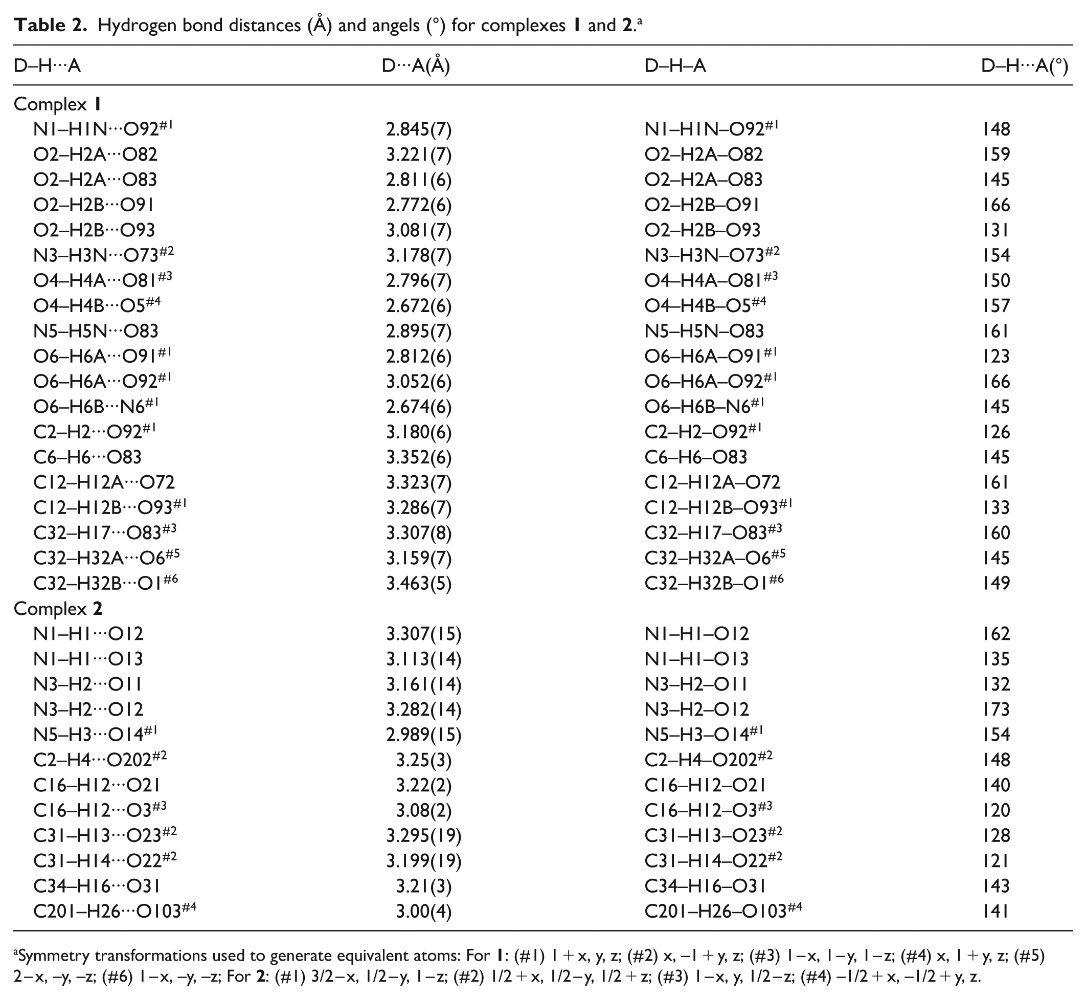
In complex 1, the LL-2 ligand adopts a cis, trans, trans-conformation, and one arm does not participate in coordination with the metal centers, forming a zero-dimensional tri-nuclear molecular compound (Figure 1(a)). The dihedral angles of the three pyridine rings and the central benzene ring of the ligands are 62.9°, 84.8°, and 78.0°, respectively. An O6–H6B···N6 hydrogen bond is formed between the uncoordinated pyridine nitrogen atom and a water (O6) molecule which coordinates with Zn1 on the adjacent unit (Table 2), resulting in a one-dimensional chain structure along the a direction (Figure 1(b)).
Hydrogen bond distances (Å) and angels (°) for complexes 1 and 2.a
D–H···A
D···A(Å)
D–H–A
D–H···A(°)
Complex 1
N1–H1N···O92#1
2.845(7)
N1–H1N–O92#1
148
O2–H2A···O82
3.221(7)
O2–H2A–O82
159
O2–H2A···O83
2.811(6)
O2–H2A–O83
145
O2–H2B···O91
2.772(6)
O2–H2B–O91
166
O2–H2B···O93
3.081(7)
O2–H2B–O93
131
N3–H3N···O73#2
3.178(7)
N3–H3N–O73#2
154
O4–H4A···O81#3
2.796(7)
O4–H4A–O81#3
150
O4–H4B···O5#4
2.672(6)
O4–H4B–O5#4
157
N5–H5N···O83
2.895(7)
N5–H5N–O83
161
O6–H6A···O91#1
2.812(6)
O6–H6A–O91#1
123
O6–H6A···O92#1
3.052(6)
O6–H6A–O92#1
166
O6–H6B···N6#1
2.674(6)
O6–H6B–N6#1
145
C2–H2···O92#1
3.180(6)
C2–H2–O92#1
126
C6–H6···O83
3.352(6)
C6–H6–O83
145
C12–H12A···O72
3.323(7)
C12–H12A–O72
161
C12–H12B···O93#1
3.286(7)
C12–H12B–O93#1
133
C32–H17···O83#3
3.307(8)
C32–H17–O83#3
160
C32–H32A···O6#5
3.159(7)
C32–H32A–O6#5
145
C32–H32B···O1#6
3.463(5)
C32–H32B–O1#6
149
Complex 2
N1–H1···O12
3.307(15)
N1–H1–O12
162
N1–H1···O13
3.113(14)
N1–H1–O13
135
N3–H2···O11
3.161(14)
N3–H2–O11
132
N3–H2···O12
3.282(14)
N3–H2–O12
173
N5–H3···O14#1
2.989(15)
N5–H3–O14#1
154
C2–H4···O202#2
3.25(3)
C2–H4–O202#2
148
C16–H12···O21
3.22(2)
C16–H12–O21
140
C16–H12···O3#3
3.08(2)
C16–H12–O3#3
120
C31–H13···O23#2
3.295(19)
C31–H13–O23#2
128
C31–H14···O22#2
3.199(19)
C31–H14–O22#2
121
C34–H16···O31
3.21(3)
C34–H16–O31
143
C201–H26···O103#4
3.00(4)
C201–H26–O103#4
141
Symmetry transformations used to generate equivalent atoms: For 1: (#1) 1 + x, y, z; (#2) x, –1 + y, z; (#3) 1 – x, 1 – y, 1 – z; (#4) x, 1 + y, z; (#5) 2 – x, –y, –z; (#6) 1 – x, –y, –z; For 2: (#1) 3/2 – x, 1/2 – y, 1 – z; (#2) 1/2 + x, 1/2 – y, 1/2 + z; (#3) 1 – x, y, 1/2 – z; (#4) –1/2 + x, –1/2 + y, z.
In addition, the amide-oxygen atoms of the uncoordinated arms in the LL-2 ligand also participate in the formation of hydrogen bonds. Also, a hydrogen bond is formed between O4–H4B···O5 with coordination of a water molecule O4 on Zn2 (Table 2). Through these hydrogen bonds, the above one-dimensional chain structure is further connected on the ab plane into a two-dimensional grid structure (Figure 1(c)). Uncoordinated is present between the layers and forms N–H···O, O–H···O and C–H···O hydrogen bonds with the skeleton of the complex (Table 2), which further stabilizes the structure of the complex. In complex 1, although one of the pyridine arms does not directly participate in coordination with the metal ions, it does form hydrogen bonds with the corresponding water molecules instead, and plays an important role in the whole stabilization of the structure.12
The crystal structure of complex 2
Complex 2 crystallized in the C2/c space group, monoclinic crystal system. The coordination environment of 2 is shown in Figure 2(a). Again, there are two ZnII ions in the complex with different coordination environments: Zn1 is six-coordinated, among which four coordination atoms are two amide-oxygen atoms and two pyridine nitrogen atoms of two different ligands, and the remaining two coordination positions are occupied by water molecules. The bond lengths of Zn1–O and Zn1–N are 2.210(16), 2.068(8), and 2.098(12) Å, while the O–Zn1–O, O–Zn1–N, and N–Zn1–N bond angles ranged from 82.1(6)° to 164.7(4)°. So, the coordination configuration of Zn1 is a distorted octahedron of O4N2 donor set. Zn2 is five-coordinated, with O3N2 donor set consisting of one water molecule, two amide-oxygen atoms of two different ligands, and two pyridine nitrogen atoms. The corresponding bonds and angles around Zn2 range from 1.978(8) to 2.155(8) Å and 85.1(3)° to 171.1(3)°, respectively.
(a) The coordination environment around the ZnII centers in compound 2, where the ellipsoids are drawn at 30% probability. The H atoms, anions, and uncoordinated solvent molecules are omitted for clarity. (b) Side (left) and top (right) view of a 32-membered macro ring of complex 2. (c) (Left) one-dimensional tube-like structure of 2 and (right) schematic drawing of the tube-like structure of 2, in which the LL-2 ligands are simplified as straight lines from the centroids of the central benzene rings to the coordinated zinc atoms, water molecules are omitted for clarity.
In complex 2, the LL-2 ligand is also in cis, trans, trans-conformation. In addition, four ZnII ions and four ligands (each ligand connects two metals by using its two arms) form a 32-membered ring (Figure 2(b)), where the Zn–Zn distances vary from 7.564(4) to 12.348(6) Å, and the distances between C5–C2 and C6–C6A are 11.001(18) and 19.099(18) Å. The above cyclic units of 2 are stacked along the direction of c and are connected into a one-dimensional pipe-like structure by the coordination of the third arm of the ligand with the ZnII centers (Figure 2(c)). The cavity size of the pipeline is smaller than the previously reported cavity of similar one-dimensional tubular complexes (Zn(TITMB)(OAc))(OH)·8.5H2O.13 Furthermore, the one-dimensional pipelines were arranged in the ab plane. The uncoordinated perchlorate and solvent molecules occupy cavities in or between the pipelines and form abundant hydrogen bonds with the main framework of the complex stabilizing the overall structure of complex 2. The structural differences in the complexes 1 and 2 may be caused by the different anions.
Thermogravimetric analysis and powder X-ray diffraction
Thermal gravimetric analysis (TGA) was also carried out to examine the thermal stabilities of complexes 1 and 2 (Supplemental Figures S1 and S2). For 1, a weight loss of 6.80% (calcd 6.60%) is observed from ca. 130°C to 160°C, which may be attributed to the loss of coordinated water molecules. The next weight loss follows closely, corresponding to the decomposition of the anhydrous complex. For complex 2, as shown in Supplemental Figure S2, a weight loss of about 7.30% is observed from room temperature until ca. 190°C, which can be attributed to the loss of solvent molecules. Distinction between the loss of the coordinated and uncoordinated solvent molecules was not clearly observed from the TGA data. Due to the high volatility of ethanol molecules, the first weight loss is lower than the calculated value. This non-distinctive behavior is also reported in literature.14 The second weight loss of complex 2 begins at ca. 320°C where the decomposition of the residue starts.
In order to confirm that the phase purity of these bulk materials can be represented by the crystal structures, powder X-ray diffraction (PXRD) experiments were carried out for complexes 1 and 2 (Supplemental Figures S3 and S4). For complex 1, the experimental patterns of the as-synthesized samples is consistent with the simulated example, indicating the phase purity of the sample.15 For 2, despite a few unindexed diffraction lines and some lightly broadened peaks, the experimental patterns of the synthesized sample can be considered comparable to the corresponding simulated example, also indicating the phase purity of the sample. Combined with the results of TGA curves, we think that these unindexed diffraction lines and some lightly broadened peaks in 2 may be assigned to the slight change of the framework after the partial loss of highly volatile solvent molecules at room temperature.16 The TGA and PXRD results make the title complexes potential candidates for practical applications.
Fluorescence properties
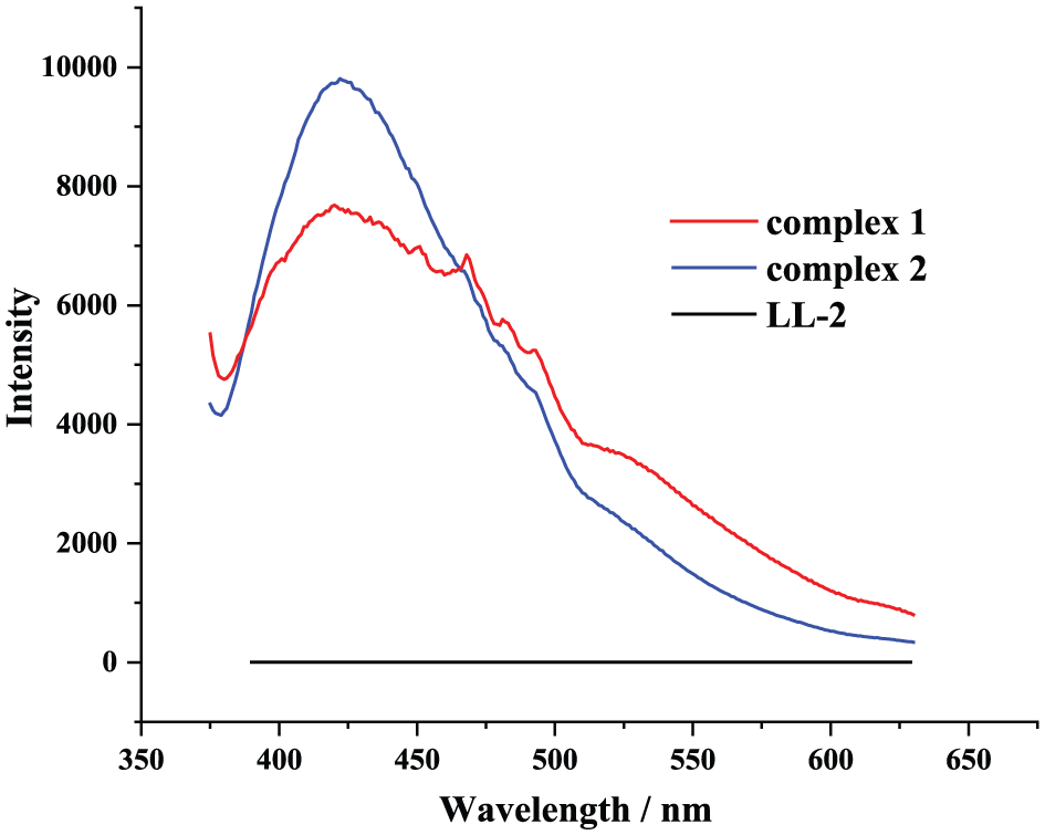
For MOCs containing zinc metal centers, the luminescent properties have attracted more interest due to their good applications in various areas, including chemical sensors, photochemistry, and electroluminescent displays.17 The solid fluorescence spectra of ligand LL-2 and complexes 1 and 2 at room temperature are depicted in Figure 3. Intense broad emission bands at 420 nm for 1 and 425 nm for 2 are observed at an excitation wavelength of 360 nm. Meanwhile, the free LL-2 ligand exhibited no emission under the same excitation conditions (Supplemental Figure S5). No matter the organic ligands have fluorescence or not, similar fluorescence emission peaks around 420 nm have been reported for d10 metal complexes, and which were assigned to the charge transfer from ligand to metal. Thus, the fluorescence emission of the complexes can be similarly attributed to the ligand-to-metal charge transfer (LMCT) of the complexes.18,19 These results show that complexes 1 and 2 may be candidates for potential photoactive materials.
The fluorescence emission spectra of free LL-2, 1 and 2 in the solid state.
Conclusion
Through reactions of tripodal carboxamide-containing ligand LL-2 with Zn(NO3)2·6H2O and Zn(ClO4)2·6H2O, respectively, two novel coordination compounds 1 and 2 have been synthesized and characterized. In these complexes, the ligands adopt different coordination modes resulting in different crystal structures, which may be influenced by the different anions in the self-assembly process. The fluorescence properties of complexes 1 and 2 have also been studied, showing these complexes have potential applications as photoactive materials.
Experimental
All commercially available chemicals except THF (tetrahydrofuran) are of reagent grade and were used as received without further purification. THF was purified according to the standard methods. C, H, and N analyses were performed on a Perkin-Elmer 240C elemental analyzer at the analysis center of Nanjing University. PXRD patterns were recorded on a RigakuD/max-RA rotating anode X-ray diffractometer with graphite monochromated Cu-Kα (λ = 1.542 Å) radiation at room temperature. TGA was performed on a Netzsch STA-409PC instrument under an N2 flow with a heating rate of 20°C/min. The luminescence spectra for the powdered solid samples were measured at room temperature on a Hitachi F-4500 fluorescence spectrophotometer with a xenon arc lamp as the light source.
Synthesis of LL-2 ligand
The ligand LL-2 was prepared according to the literature.11 To a dry CH2Cl2 solution (50 mL) of 2-aminomethyl pyridine and triethlyamine was slowly added 1,3,5-benzenetricarbonyl trichloride in dry THF (25 mL) at –4°C. The solution was then stirred for one day at room temperature and the obtained precipitate was filtered. Recrystallization of LL-2 was carried out in ethanol/THF solution.
Synthesis of (1)
A solution of Zn(NO3)2·6H2O (14.9 mg, 0.05 mmol), and LL-2 (24.0 mg, 0.05 mmol) in EtOH (5 mL) was placed in a Teflon-lined stainless steel vessel. The mixture was sealed and heated at 80°C for 72 h, and then the reaction system was cooled to room temperature. The resulting clear yellow solution was filtered. After slowly diffusing n-hexane into the filtrate, yellowish bulky crystal of complex 1 was obtained after about 3 weeks. Yield: 45%. Elemental analysis (C27H30N9O15Zn1.5), calcd (%): C, 39.61; H, 3.70; N, 15.40; found (%): C, 39.88; H, 3.84; N, 15.21.
Synthesis of (2)
The synthetic procedure for complex 2 was similar to that of 1, except that Zn(ClO4)2·6H2O (18.5 mg, 0.05 mmol) was used. Yield (based on Zn): 62%. Elemental analysis (C62H85Cl5N12O37Zn3), calcd (%): C, 37.92; H, 4.37; N, 8.56; found (%): C, 38.22; H, 4.28; N, 8.69.
Single crystal structure determination
The X-ray diffraction data for 1 were collected on a Bruker Smart Apex II CCD equipped with a Mo-Kα radiation source (λ = 0.71073 Å). The data were integrated by using the SAINT program,20 which was also used for the intensity corrections for the Lorentz and polarization effects. An empirical absorption correction was applied using the SADABS program.21 The structures were solved by direct methods with the SHELXS-97 program and refined with SHELXL-97 by full-matrix least-squares techniques on F2. All non-hydrogen atoms were refined anisotropically and all hydrogen atoms were refined isotropically. The hydrogen atoms except for those of water molecules were generated geometrically. All calculations were performed on a personal computer (Lenovo Thinkpad X1) with the SHELXL-97 crystallographic software package.22,23
The diffraction data of complexes 2 were collected on a Rigaku RAXIS-RAPID single-crystal diffractometer with graphite monochromated Mo-Kα ray (λ = 0.71075 Å) as the X-ray diffraction source. The crystal structure of complexes 2 was analyzed by direct methods (SHELX-97 program).22 The coordinates and anisotropic temperature factors of all non-hydrogen atoms in the complexes are modified by the full-matrix least-square method, and hydrogen atoms on non-water molecules are theoretically hydrogenated. All calculations were carried out on an SGI workstation using the teXsan crystallographic software package of the Molecular Structure Corporation.24 In complex 2, the water molecule O101 and the alcohol molecule C203, C204 are disordered into two positions, with occupancy rates of 0.66 (2), 0.34 (2); 0.69 (4), 0.31 (4); and 0.69 (4), 0.31(4), respectively. The details of the crystal parameters, data collection, and refinement for the compounds are summarized in Table 3. Selected bond lengths and angles with their estimated standard deviations are listed in Table 1. The Cambridge Crystallographic Data Centre (CCDC) numbers are 1955730 (1) and 1955732 (2).
Crystal data and structure refinement parameters for complexes 1 and 2.a
supporting_information – Supplemental material for Synthesis, structure, and fluorescence properties of two metal-organic complexes based on a tripodal carboxamide-containing ligand
Supplemental material, supporting_information for Synthesis, structure, and fluorescence properties of two metal-organic complexes based on a tripodal carboxamide-containing ligand by Yan Wang, Xiner Xu, Yong Zhao, Hai Yu, Yanyan Zhang, Wei Xu, Zhiqiang Liu and Rongyi Huang in Journal of Chemical Research
Footnotes
Declaration of conflicting interests
The author(s) declared no potential conflicts of interest with respect to the research, authorship, and/or publication of this article.
Funding
The author(s) disclosed receipt of the following financial support for the research, authorship, and/or publication of this article: We are very thankful for the financial support from the Provincial Natural Science Foundation of Anhui (No. 1408085QB32, No. 1908085QB47), the Key Project of Anhui Provincial Fund for Distinguished Young Scholars in Colleges and Universities (No. 2013SQRL059ZD), the Natural Science Foundation of Anhui Education Committee (No. KJ2013B126, No. KJ2018A0371), and the Program for Innovative Research Team in Anqing Normal University.
ORCID iD
Yan Wang
Supplemental material
Supplemental material for this article is available online.
References
1.
ZhangXMLiuFGaoW, et al. Cryst Eng Comm2018; 20: 1985–1996.
2.
LiuZQChenKZhaoY, et al. Cryst Growth Des2018; 18: 1136–1146.
3.
LiJFXuXDZhouL, et al. J Chem Res2018; 42: 424–427.
4.
ShangKXJing-Sun, HuDC, et al. Cryst Growth Des2018; 18: 2112–2120.
5.
LyuJZhangXOtakeKI, et al. Chem Sci2019; 10: 1186–1192.
6.
XuCBiCZhuZ, et al. Cryst Eng Comm2019; 21: 2333–2344.
Please find the following supplemental material available below.
For Open Access articles published under a Creative Commons License, all supplemental material carries the same license as the article it is associated with.
For non-Open Access articles published, all supplemental material carries a non-exclusive license, and permission requests for re-use of supplemental material or any part of supplemental material shall be sent directly to the copyright owner as specified in the copyright notice associated with the article.