
Abstract
In this article, a new “turn-off” fluorescent sensor N-n-butyl-4-{2-[(ethylimino)methyl]phenol}-1,8-naphthalimide (
Chemosensor

Introduction
It is well known that CuII ions are representative transition metal elements, which are widely distributed in biological tissues. In addition, copper is the second most important trace element in the human body.1–4 To sustain normal health, it is highly essential to intake a limited amount of dietary copper. However, excess concentrations of CuII ions can easily burden the organs in the body, resulting in metabolism becoming disordered, which in turn may be associated with many neuronal cytoplasmic neurodegenerative diseases like Wilson’s disease, dyslexia, and Alzheimer’s disease.5–19 The results of toxicity data and scientific studies by the World Health Organization (WHO) and United States Environmental Protection Agency (US EPA) determined the acceptable limits of CuII ions in drinking water to be 31.5 and 20 μM, respectively.11,20 Since excessive CuII ions may pose a threat to life and sanitation, it is important to develop efficient and convenient methods to detect CuII ion concentrations.
Due to high sensitivity, low cost, simplicity, and high efficiency, fluorescence detection has broad applications, and is commonly applied in many fields including chemistry, electronics, materials science, analytical biochemistry, cell biology, medical diagnostics, and environmental control.4–8,21 The 1,8-naphthalimide derivatives are currently commonly used as precursors or fluorescent sensors because of their high photostability and high quantum yields, strong absorption bands in the visible region, and large Stokes shifts, 22 which give this method advantages over other traditional metal ion detection and analysis methods. 23 As a result of this, derivatives of 1,8-naphthalimide have been successfully utilized in the production of fluorescent whitening agents, liquid crystal displays, fluorescent dyes, laser-active media, electroluminescent materials, photoconductive materials, and fluorescent switches and sensors.24–39Until now, many excellent fluorescent sensors based on naphthalimide and its derivatives for detection of CuII have been reported,40–55 but most were not sensitive enough to determine low concentration levels of CuII and only function in organic solvents, with only a very few examples working in aqueous media.56–59 Many copper compounds that coordinate with naphthalimide and its derivatives have been reported and their structures have been determined, but few have been used to explain the relationship between structure and fluorescence quenching mechanisms.60–70 Due to the lack of crystal structures of copper(II) complexes with sensors, it is difficult to speculate on the fluorescence quenching mechanism. It is still important and challenging to develop 1,8-naphthalimide-based sensors which can detect CuII rapidly in aqueous media and provide specific binding modes.
Herein, we have designed and synthesized a new “turn-off” fluorescent sensor toward CuII ions with 1,8-naphthalimide as the chromophore and a Schiff base as the recognition group, which exhibits a large fluorescence quenching upon binding to CuII. The sensor showed good sensitivity and selectivity for the detection of copper(II) ions over other transition metal ions.
Results and discussion
The route to the synthesis of N-n-butyl-4-{2-[(ethylimino)methyl]phenol}-1,8-naphthalimide (

Synthesis of sensor
It is essential to investigate the optimization of pH on the efficiency of the sensor
Selectivity and interference study
The performance ability of an ideal chemosensor is greatly influenced by its selectivity parameter. Therefore, the binding properties of
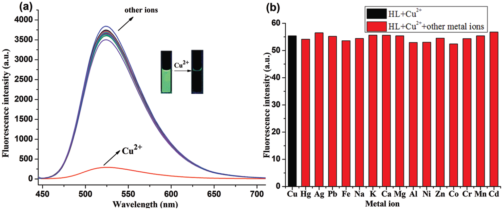
(a) Fluorescence spectra of
In order to further verify the sensing potency of sensor
Fluorescence titrations
To further investigate the characteristics of the sensor

(a) Fluorescence spectra of

(a) Determination of the association constant of
Figure 3(b) shows good linearity between the emission at 524 nm and the concentrations of CuII ions in the range from 0.5 to 5.0 μM, which indicates that
The value of the detection limit was further calculated based on the following equation:
77
DL = 3σ/k. Herein, σ is the standard deviation of the blank solution and k is the slope of the intensity versus sample concentration. DL was calculated to be 3.30 × 10⁻⁸M, which is far lower than the WHO and US EPA regulated limits of 31.5 and 20 μM, respectively. This result validates that sensor
Mechanism of recognition
Although we have obtained a stoichiometric ratio of approximately 1:2 between the CuII ions and

Job’s plot for the determination of the stoichiometry of the [L–Cu2+] system in acetonitrile–HEPES (1:1, v/v, HEPES buffer, 10 μM, pH 7.4).
To further demonstrate the stoichiometry between
Combining the obtained fluorescence titration, Job’s plot, and MALDI-TOF mass spectrometry, we have proposed a sensing mechanism of the sensor

Proposed mechanism for detection of Cu(II) by
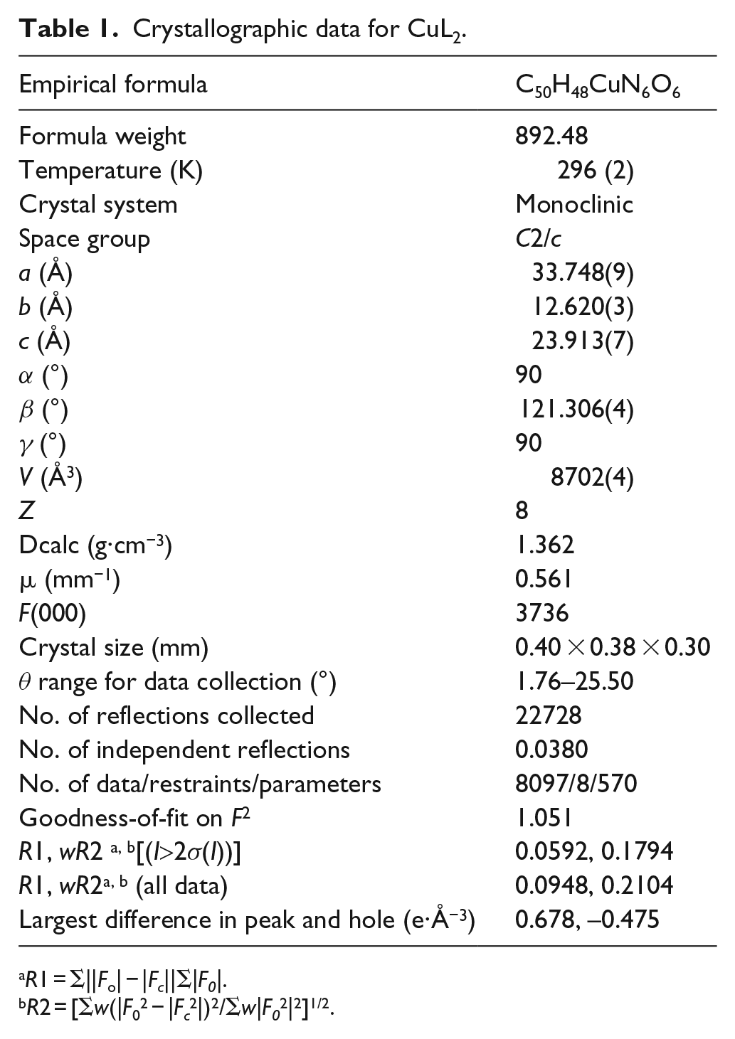
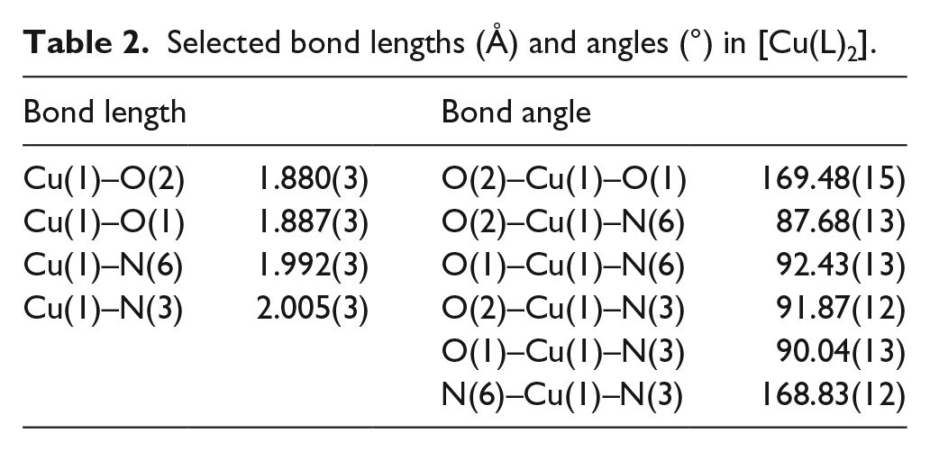
Crystal structure of the CuL2 complex
To investigate the binding mode between

Molecular structure (ORTEP) of [Cu(L)2] in the crystal with displacement ellipsoids at the 15% probability level; hydrogen atoms are omitted for clarity.
Crystallographic data for CuL2.
R1 = Σ||Fo| − |Fc||Σ|F0|.
R2 = [Σw(|F02 − |Fc2|)2/Σw|F02|2]1/2.
Selected bond lengths (Å) and angles (°) in [Cu(L)2].
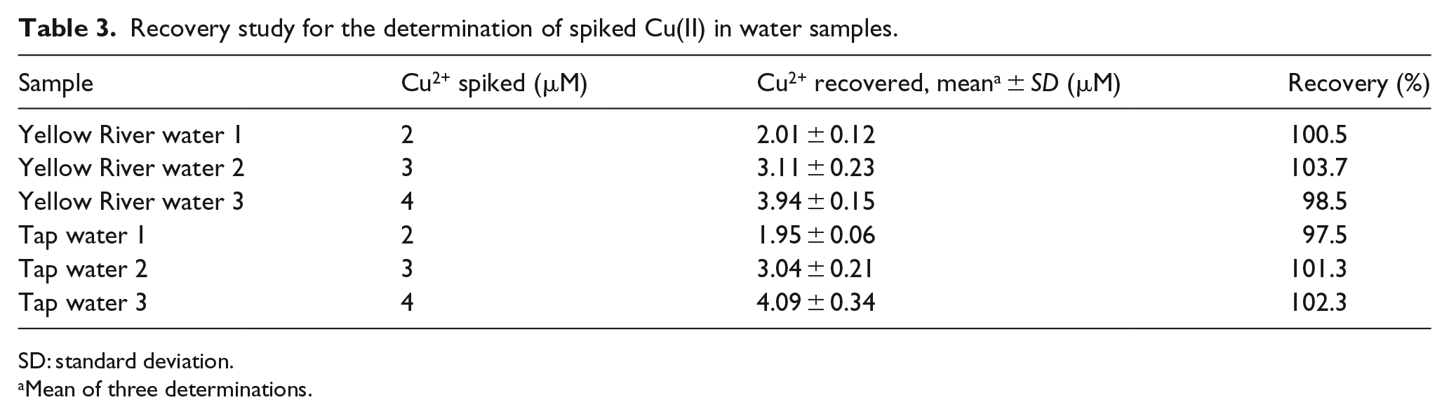
Application of HL for CuII ion analysis in water samples
The effectiveness of the designed sensor
Recovery study for the determination of spiked Cu(II) in water samples.
SD: standard deviation.
Mean of three determinations.
Conclusion
In summary, a new fluorescent sensor has been synthesized for the highly selective and sensitive detection of CuII ions. The experimental results indicate that
Experimental
Materials and Methods
All chemicals were obtained from commercial sources, and except if specified, they are of analytical reagent grade and were used without any further purification. Adjustment of the pH was carried out by addition of dilute hydrochloric acid (HCl) and sodium hydroxide (NaOH). The HEPES buffer (pH = 7.4) was prepared by using double-distilled water. Solutions of Na+, K+, Ca2+, Mg2+, Al3+, Pb2+, Fe3+, Ni2+, Zn2+, Hg2+, Ag+, Co2+, Cr3+, Mn2+, Cd2+, and Cu2+ were prepared from their nitrate salts. Double distilled water was used for the preparation of metal ion (1 mM) nitrate stock solutions.
The C, H, and N content analyses were carried out by using a Carlo Erba 1106 elemental analyzer. An XD-4 digital micro-melting point apparatus was used for determining the melting points of compounds. Thin-layer chromatography (TLC) was carried out on silica gel 60F254 plates (Merck KGaA). The 1H NMR and 13C NMR spectra were recorded on Mercury plus 400 MHz NMR spectrometer (Palo Alto, CA, USA) with tetramethylsilane (TMS) as the internal standard and dimethyl sulfoxide-d6 (DMSO-d6) as the solvent. The infrared (IR) spectra (4000–400 cm−1) were recorded on a Nicolet FT-VERTEX 70 spectrometer as KBr pellets. The UV-Vis spectra were obtained using a Lab-Tech Bluestar Plus spectrophotometer. Fluorescence spectra were measured with a Lengguang Tech. F97 Pro spectrofluorometer. ESI-MS were obtained on a Bruker microTOF-Q system.
Synthesis of N-n-butyl-4-{2-[(ethylimino)methyl]phenol}-1,8-naphthalimide (HL)
The intermediate compounds
Preparation of a single crystal of complex Cu(L)2
To a stirred solution of
X-ray crystallography
The intensity data of single crystals mounted on glass fibers were collected on a Bruker Smart CCD diffractometer with graphite-monochromated MoKα radiation (k = 0.71073 Å) at 296 K. Data reduction and cell refinement were carried out by using the SMART and SAINT programs. 89 Data was refined by using SHELXTL software through full-matrix least squares program. 90 The CCDC 1810957 contains the supplementary crystallographic data for this article. These data can be obtained free of charge from The Cambridge Crystallographic Data Centre via www.ccdc.cam.ac.uk/data_request/cif.
Supplemental Material
Supplementary_data – Supplemental material for A new 1,8-naphthalimide-based fluorescent “turn-off” sensor for detecting Cu2+ and sensing mechanisms
Supplemental material, Supplementary_data for A new 1,8-naphthalimide-based fluorescent “turn-off” sensor for detecting Cu2+ and sensing mechanisms by Yao Qu, Yancong Wu, Cong Wang, Kun Zhao and Huilu Wu in Journal of Chemical Research
Footnotes
Declaration of conflicting interests
The author(s) declared no potential conflicts of interest with respect to the research, authorship, and/or publication of this article.
Funding
The author(s) disclosed receipt of the following financial support for the research, authorship, and/or publication of this article: This work was supported by the Natural Science Foundation of Gansu Province (grant no. 17JR5RA090), Foundation of A Hundred Youth Talents Training Program of Lanzhou Jiaotong University (grant no. 152022), and the National Natural Science Foundation of China (grant no. 21367017).
Supplemental material
Supplemental material for this article is available online.
References
Supplementary Material
Please find the following supplemental material available below.
For Open Access articles published under a Creative Commons License, all supplemental material carries the same license as the article it is associated with.
For non-Open Access articles published, all supplemental material carries a non-exclusive license, and permission requests for re-use of supplemental material or any part of supplemental material shall be sent directly to the copyright owner as specified in the copyright notice associated with the article.