A new hydrazone-based colorimetric Cu2+ chemosensor is synthesized. Its structure was confirmed by single-crystal X-ray diffraction. Its binding properties towards various metal ions are examined through absorption spectroscopy. In aqueous THF solution, the chemosensor exhibits selective recognition towards Cu2+ over other metal ions with a colour change from colourless to pink. The complex formed between the chemosensor and Cu2+ ions forms a 2:1 stoichiometry with an association constant of 2.46 × 10⁸M−2. The analytical detection limit for Cu2+ by the naked eye is as low as 10.0 μM.
In recent years, the development of highly selective and sensitive chemosensors for transition metal ions has attracted significant interest due to their vital role in various biological and environmental processes.1–3 Among these metal ions, copper ions play a critical role in various biological processes4,5 and are also significant environmental pollutant in modern society. The over-accumulation of copper ions can cause a series of severe diseases such as Alzheimer’s, Wilson’s and Menke’s diseases.6,7 Therefore, it is very important to detect rapidly Cu2+ in environmental and biological systems.
Up to now, many fluorescent probes8–16 have been reported. However, most of them usually require expensive instruments, involve complicated syntheses or are insoluble in aqueous solutions. For practical applications, it is necessary to develop Cu2+ sensors that are easily prepared and that can be easy detected rapidly without the help of instruments. In this respect, colorimetric chemosensors would appeared to be the most attractive and could be widely used owing to the low cost and lack of equipment required. Moreover, a colour change can easily be observed by the naked eye.17 However, so far, reported colorimetric Cu2+ probes are still relatively rare,18–23 and it is still a challenge to develop colorimetric chemosensing molecules to detect Cu2+ in aqueous solution.
In this study, we reported a new hydrazone compound (1), 2-(benzo[d]thiazol-2(3H)-ylidene)hydrazono-1,2-diphenylethanone, which can be used as a highly selective colorimetric naked-eye sensor for Cu2+ in aqueous solution.
Result and discussion
The synthesis of chemosensor 1 is shown in Scheme 1; first, hydrazinobenzothiazole (2) is synthesized from benzothiazol-2-ylamine; second, benzil (4) is synthesized from benzaldehyde through two-step reaction involved benzoin condensation and further oxidation reaction; finally, the reaction of 2-hydrazinobenzothiazole (2) and benzil (4) under reflux in ethanol gives chemosensor 1, the structure of which is identified by NMR, ESI-MS (Figure S1–S3 in the Supporting Information) and X-ray diffraction analysis. The molecular view of 1 is shown in Figure 1. The crystal packing is stabilized by the water molecules via intermolecular N–H1 … .O2, O2–H (2B) … .N2 and O2–H (2A) … .O1 hydrogen bonds (Figure 2). The water molecules serve as both hydrogen-bond donors and acceptors.
Synthesis of the sensor 1.
The molecular structure of 1 with displacement ellipsoids drawn at the 30% probability level.
A packing diagram for 1 viewed along the c-axis. Dashed lines show arrays of hydrogen bonds.
The interaction of 1 (10 μM) with various metal ions in 9:1 (v/v) THF–HEPES buffer solution (pH = 7.0) was investigated by UV-Vis absorption spectrometry. As shown in Figure 3, free 1 exhibits a broad band at 358 nm. The addition of Cu2+ to 1 led to a red shift of its absorption maxima and formed a new absorption band at 524 nm. However, sensor 1 showed almost no change in its absorption peak on addition of other metal ions. Moreover, as shown in the insert in Figure 3, the Cu2+ sensing and the concomitant absorption change were clearly visible to the naked eye, which indicated that 1 can act as a colorimetric chemosensor for the naked-eye detection of Cu2+ in an aqueous solution.
UV-Vis spectral changes of compound 1 (10 μM) in 9:1 (v/v) THF–HEPES buffer solution (10 mM, pH = 7.0) upon addition of various metal ions (20 μM). Insert: Visual colour change of 1 (10 μM) upon addition of 2.0 equiv. of Cu2+.
The UV-Vis titration of 1 with Cu2+ was carried out. As shown in Figure 4, upon the addition of Cu2+ (0–5 equiv.) to a solution of 1, the absorbance peaks at 358 nm gradually decreased and meanwhile new absorption peaks at 524 nm appeared with a clear isosbestic point at 400 nm. This red shift from 358 to 524 nm may result from the Cu2+ coordination-enhanced LMCT (ligand-to-metal charge-transfer) effect.24
UV-Vis absorption spectra of 1 (10 μM) in 9:1 (v/v) THF–HEPES buffer solution (10 mM, pH = 7.0) upon the addition of Cu2+ (0–50 μM) at 25 °C. Each spectrum is obtained 5 min after Cu2+ addition.
A Job plot25 analysis was used to determine the stoichiometry of 1−Cu2+. Figure 5 shows a plot of absorbance at 524 nm against the mole fractions of Cu2+ at a constant concentration of [1] + [Cu2+]. As shown in Figure 5, when the molar fraction [Cu2+]/([1] + [Cu2+]) was about 0.33, the concentration of the 1–Cu2+ complex reached a maximum value, which indicated a 2:1 stoichiometric complexation of 1 with Cu2+ ions. The association constant (Kass) between compound 1 and Cu2+ was calculated to be 2.46 × 10⁸M-2 by non-linear fitting of the titration curve of a 2:1 binding model (Figure 6).
Job plot of a 2:1 complex of 1 and Cu2+. [1] + [Cu2+] = 10 mM. The absorbance is monitored at 524 nm.
Plot of C0/(A524-εmC0) against 1/C0Cg at 524 nm. See text for a definition of the parameters.
The analytical detection limit of Cu2+, defined as the lowest concentration of Cu2+ at which the colour change of 1 (10 μM) in 9:1 (v/v) THF–HEPES buffer solution can be detected by the naked eye,18 is as low as 10.0 μM (Figure 7), which is lower than the limit of copper in drinking water (~20 μM).
Visual colour change of 1 (10 μM) in 9:1 (v/v) THF–HEPES buffer solution (10 mM, pH = 7.0) upon addition of 10–50 μM of Cu2+ ions.
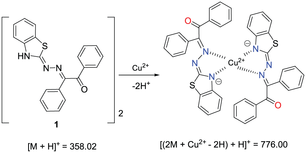
The mode of interaction between 1 and Cu2+ ions was investigated through 1H NMR spectroscopy and mass spectrometry upon the addition of Cu2+. The 1H NMR spectra of sensor 1 revealed that the imine proton (Ha) signal at 9.33 ppm almost completely disappeared upon the addition of 5 equiv. of Cu2+ (Figure 8), which indicates that Cu2+ induces deprotonation of the active NH of the imine group. The appearance of a peak at m/z 776.00 (calcd for 776.12, [(2 receptor 1 + Cu2+−2H+) + H+]) in the electrospray ionization mass spectrum (Figure S4) further confirmed the formation of a 2:1 complex between 1 and Cu2+ ions.
1H NMR (300 MHz) spectra of 1 in the presence of Cu2+ in CDCN.
Based on the above results of the Job plot analysis, the mass spectrum, and the 1H NMR study, a possible coordination mode of receptor 1 with Cu2+ is proposed in Scheme 2.
Proposed coordination mode of sensor 1 with Cu2+.
Conclusion
In conclusion, a new hydrazone-based colorimetric Cu2+ chemosensor 1 was synthesized and its structure was confirmed by X-ray diffraction analysis. The recognition of Cu2+ brought about colour changes from colorless to pink. The analytical detection limit for Cu2+ by the naked eye is as low as 10.0 μM, which suggests chemosensor 1 has great potential in colorimetric Cu2+ detection.
Experimental
Infrared (IR) spectra were recorded on a Perkin-Elmer PE-983 IR spectrometer as KBr pellets. NMR spectra were recorded on a Varian Mercury 300 spectrometer. Electro-spray ionization mass spectra were measured on a Finnigan LCQ Advantage Max spectrometer. Absorption spectra were measured on a Hitachi U-3010 spectrometer. The crystal structure of 1 was determined on a Bruker SMART APEX CCD system. HEPES buffer solution (pH = 7.0) was prepared using 4-(2-hydroxyethyl)-1-piperazine ethanesulfonic acid (HEPES) and aqueous sodium hydroxide. 2-Hydrazinobenzothiazole (2)26 and benzil (4)27 were prepared according to reported procedures.
Synthesis of 2-(benzo[d]thiazol-2(3H)-ylidene)hydrazono-1,2-diphenylethanone (1)
A solution of 2-hydrazinobenzothiazole (2) (1.0 mmol) and benzil (4) (1.0 mmol) in ethanol (40 mL) was heated under reflux for 3 h. After cooling, the solvent was evaporated and the desired product 1 (0.25 g) was obtained in 70% yield via flash chromatography with petroleum as the eluent; m.p.: 94–95 °C. IR (νmax, KBr, cm-1): 3450, 1666, 1606, 1575, 1539, 1471, 1441, 1271, 1223, 1130, 1075, 731, 519; 1H NMR (DMSO-d6): δ 12.10 (s, 1H, NH), 7.85–7.88 (m, 2H, Ar-H), 7.55–7.71 (m, 6H, Ar-H), 7.43–7.49 (m, 3H, Ar-H), 7.20–7.25 (m, 1H, Ar-H), 6.99–7.08 (m, 2H, Ar-H),13C NMR (DMSO-d6), δ 191.2, 155.1, 147.6, 144.0, 138.5, 138.0, 131.8, 130.5, 129.8, 129.6, 129.5, 128.9, 127.8, 117.9, 108.4. ESI-MS: m/z 358.02 [M+ H]+; M+ calculated 357.09.
X-ray diffraction study of compound 1
A yellow crystal of the compound 1, obtained by slow evaporation from 95% ethanol solution, was mounted on a glass fibre in a random orientation at 295(2) K. The determination of the unit cell and the data collection were performed with MoKɑ radiation (λ = 0.71073 Å) on a Bruker Smart Apex-CCD diffactometer with a ‘ɷ’-(j) scan mode. The structure was solved by direct methods with SHELXS-97 and expanded by the Fourier technique. The non-hydrogen atoms were refined anisotropically, and the hydrogen atoms were placed at calculated positions. Crystal data for C21H15N3OS. H2O: crystal size: 0.2 × 0.1 × 0.1 mm, M = 375.44, Monoclinic, space group P2(1)/n. a = 9.253(2) Å, b = 7.983(2) Å, c = 25.931(7) Å, V = 1912.3(8) Å3, Z = 4, T = 298(2) K, θmax = 30.00o, 18,963 reflections measured, 5544 unique (Rint = 0.0559). Final R indices (I >2σ(I)): R1 = 0.0524, wR2 = 0.1273 and GOF = 1.029. CCDC 924965 contains the supplementary crystallographic data for this article. These data can be obtained free of charge from the Cambridge Crystallographic Data Centre via www.ccdc.cam.ac.uk/data_request/cif.
Binding studies
A stock solution of compound 1 was prepared by dissolution in 9:1 (v/v) THF–HEPES buffer solution (10 mM, pH = 7.4) (1.0 × 10⁻⁵ M). The solutions of metal ions were prepared from Al2(SO4)3, Pb(NO3)2 and the chlorides of Na+, K+, Co2+, Cr3+, Fe3+, Hg2+, Cd2+, Mg2+, Cu2+, Zn2+, Ni2+ and Mn2+, respectively, and were dissolved in water (3.0 × 10⁻³ M). Fluorescence titrations were performed on a 3-mL solution of compound 1 in a quartz cell of 1-cm optical path length, by adding portionwise different stock solutions of cations into the quartz cell using a microsyringe.
Supplemental Material
Supplemental_material – Supplemental material for A new hydrazone-based colorimetric chemosensor for naked-eye detection of copper ion in aqueous medium
Supplemental material, Supplemental_material for A new hydrazone-based colorimetric chemosensor for naked-eye detection of copper ion in aqueous medium by Shao-Min Shi, Qiao Li and Sheng-Li Hu in Journal of Chemical Research
Footnotes
Declaration of conflicting interests
The author(s) declared no potential conflicts of interest with respect to the research, authorship, and/or publication of this article.
Funding
The author(s) disclosed receipt of the following financial support for the research, authorship, and/or publication of this article: This study was supported by the Hubei Province Education Ministry Foundation of China (No. B2016157) and the Science Foundation for Creative Research Group of Hubei Normal University (HBNU) (No. T201501).
ORCID iD
Sheng-Li Hu
Supplemental material
Supplemental material for this article is available online.
References
1.
de SilvaAPGunaratneHQNGunnlaugssonTet al. Chem Rev1997; 97: 1515.
Please find the following supplemental material available below.
For Open Access articles published under a Creative Commons License, all supplemental material carries the same license as the article it is associated with.
For non-Open Access articles published, all supplemental material carries a non-exclusive license, and permission requests for re-use of supplemental material or any part of supplemental material shall be sent directly to the copyright owner as specified in the copyright notice associated with the article.