
Abstract
Pulmonary fibrosis is a chronic progressive disorder in which excessive deposition of extracellular matrix leads to irreversible scarring to interstitial lung tissue. In this study, we search to evaluate the therapeutic effect of flaxseed oil (FO) in experimental bleomycin (BLM)-induced pulmonary fibrosis. During our study, 30 male Wistar rats (weight range, 180–220 g) were divided into three groups: the control group (W) received no treatment; the second group (C) received BLM; and the third group (T) received BLM and FO for 21 days. Metabolites present in the bronchoalveolar lavage fluid (BALF) marking the changes obtained following treatment with FO were determined, histological changes in the lungs were evaluated, fatty acids present in lungs and erythrocytes of rats groups were determined by gas chromatography, and oxidative stress and antioxidant enzyme activity in the lung tissue were also recorded. Our results displayed that rat body weight decreased while fibrosis score and inflammatory index in lung tissue were significantly increased after bleomycin instillation. Administration of bleomycin followed by FO treatment reduced bleomycin-induced weight loss, increased proline, glucose, and glycerid rates in BALF and which are characterized by their anti-inflammatory effect and confirming the histological results proved by a decrease in inflammatory index and fibrosis score. This oil also significantly reduced thiobarbitunic acid reactive substance levels in the lungs of rats and increased levels of SOD and CAT and increased fatty acids levels promoting anti-inflammatory reactions especially in erythrocytes (linoleic, arachidonic, docosapentaenoic, and dihomo-γ-linoleic acids). In conclusion, these findings indicate that FO treatment significantly attenuated the increased pulmonary damage induced by bleomycin.
Introduction
Linum usitatissimum L. is an herb belonging to the Linaceae family, popularly known as flaxseed or linseed. The seeds of flax are used as a source material for the production of oil and meal, which is rich in fiber, protein, and fat. Its oil is arguably one of the richest in polyunsaturated fatty acids and essential fatty acids (EFAs), with 56.6% linolenic acid (w-3 [C18: 3cis9,12,15]) and 13.2% linoleic acid (w-6 [C18: 2cis 9,12]), in addition to 17.8% monounsatured fatty acids (MUFAs) oleic acid (w-9 [C18: 1cis 9]). 1
The fixed oil of Linum usitatissimum has several health benefits and disease preventive properties, which include hypocholesterolemic, anticancerous, antiviral effects, bactericidical activity, reduction of inflammation, hypoglycemic effects, and laxative effects. Also, it has an effect on coronary heart disease and neurological and hormonal disorders. Pulmonary fibrosis (PF) is a progressive and lethal lung disease, characterized by the accumulation of extracellular matrix in interalveolar septum to result in destruction of the normal lung architecture. The common treatment of PF uses corticosteroids in combination with immunosuppressant, anti-inflammatory, antifibrosis, antioxidant, and anticoagulative drugs but the results of therapy are not satisfying. Despite a number of advances in basic and clinical research, currently PF is still a progressive and lethal disease and no effective medical therapies are available to show the decline of pulmonary functions or reduce the mortality. 2
Within this context, the present study was designed to investigate the curative effect of flaxseed oil (FO) on bleomycin-induced PF in wistar rats.
Materials and methods
Animals and treatment
Thirty male Wistar rats (Pasteur Institute, Tunis, Tunisia) aged 3 months and weighing between 180 and 220 g were housed in our animal facility under closely controlled environmental conditions (12 h light–dark cycle; room temperature 22°C) and fed ad libitum (food and water). They were treated in accordance with the European Convention (2010) for the protection and use of vertebrate animals and all possible efforts were made to reduce suffering and minimize the number of rats used. Animals were divided into three equal groups (n = 10: 5 rats/cage): W, control group; C, lung fibrosis group (BLM); and T, treated lung fibrosis group.
After weighing, rats were anesthetized by intraperitoneal injection of pentobarbital sodium solution (100 mg/kg). The W group received 0.9% saline (intratracheal, 2 mL/kg body weight); the C group received an intratracheal instillation of bleomycin solution (4.8 mg/kg body weight), 3 days later they received by gavage 0.9% saline (2 mL/kg body weight) once daily for 21 days; the T group received BLM solution intratracheally (4.8 mg/kg body weight), 3 days later they were treated by FO gavage (2 mL/kg body weight) once a day for 21 days (Figure 1).

Schematic representation of animal handling and sample collection.
Extraction of flaxseed oil
The flaxseed was cleaned carefully, dried for 12 h at 105°C in an oven, and then rushed with HCCl3-MeOH (2:1, V/V) in a mortar. A total of 10 g of the crushed flaxseed powder was mixed with n-hexane and put into the water bath with a controlled temperature. 3 After each extraction, extract was filtered through the Whatman filter paper under vacuum and the solution was collected and concentrated with a rotary evaporator at 40°C to acquire the FO. The acquired FO was further dried in a vacuum dryer to remove the residual n-hexane.
Gas chromatographic (GC) analysis revealed that FO was with the following fatty acid composition: 5.91% C16:0, 4.31% C18:0, 22.55% C18:1, 12.83% C18:2, and 53.55% C18:3: it is rich in Omega 3 and Omega 6.
Organ sampling and bronchoalveolar lavage fluid
At the end of the experimental period, animals were anesthetized with the same procedure described during induction of fibrosis; animals were euthanized by injection of a lethal dose of sodium pentobarbital (200 mg/kg bw Nembutal ® Ceva Animal Health) and several samples were taken. Sections of the diaphragm and anterior thorax allowed us to extract the heart-lung block. The left lung was drawn and weighted. Same samples were homogenized (1:2, w/v) in 50 mM Tris buffer (pH = 7.4) containing 150 m M NaCl using an Ultra-Tunax. Homogenates were centrifuged at 5000× g for 25 min at 4°C and aliquots of supernatant were kept at −30°C until analysis.
Before the extraction of the heart-lung block, the trachea was exposed and cannulated. Intratracheal injection of saline (4×5 mL) was performed via this catheter and the liquid was sucked back by gentle suction injected between two fractions (bronchoalveolar lavage fluid [BALF]). It was then centrifuged (Jouan, MR23i) at 250 g for 10 min to exclude cells and waste that may be present in the sample. 4 A volume of 7 mL of BALF was introduced into a flask placed in liquid nitrogen to freeze the sample. After forming a thin layer of ice on the surface of the balloon, the whole is freeze-dried. Then the lyophilisate is suspended in 800 μL of D2O. A total of 50 μL of a solution of TSP (0.7 mm final concentration) were then added to the total. Lyophilization is used to make metabolites in BALF (too dilute) observed by the NMR technique.
Determination of antioxidant enzymes activities and lipid peroxidation level and protein quantification
Catalase (CAT) activity and Superoxide-Dismutase (SOD) activity were performed using kits. Levels of lipid peroxidation in lung were estimated by measuring the formation of thiobarbitunic acid reactive substances (TBARS).
Protein content was assayed as described by Lowry et al., using bovine serum albumin as standard. The Lowry Assay “Protein by Folin Reaction” has been the most widely used method to estimate the amount of proteins (already in solution or easily-soluble in dilute alkali) in biological samples. First, proteins are pre-treated with copper ion in alkali solution, and then aromatic amino acids in the treated sample reduce the phosphomolybdic acid and phosphotungstic acid presents in the Folin Reagent. The end product of this reaction has a blue color. The amount of proteins in the sample can be estimated via reading the absorbance (at 750 nm) of the end product of the Folin reaction against a standard curve of a selected standard protein solution (in our case; Bovine Serum Albumin [BSA] solution). 5
Histological and immunohistochemical analysis
For histological studies, the lungs were perfused through their main bronchus with fixative solution (10% neutral-buffered formalin), immersed in the fixative for 24 h, and the blocks were taken thereafter. Tissue blocks were placed in formalin dehydrated in a graded series of ethanol, embedded in paraffin, cut into 4 mm thick serial sections, and stained with hematoxylin & eosin (H&E) to identify the inflammatory cells of Masson’s trichrome for collagen deposition. Histological grading of lesions was performed using a blinded semi-quantitative scoring system for extent and severity of inflammation and fibrosis in lung parenchyma. The severity of inflammation was estimated using the semi-quantitative grading system which considers the following categories: Grade 0, absence of inflammation; Grade 1, minimal inflammation; Grade 2, minimal to moderate inflammation; Grade 3, moderate inflammation with thickening of alveolar walls; Grade 4, moderate to severe inflammation; and Grade 5, severe inflammation with presence of follicles which replace the parenchyma. The severity of interstitial fibrosis was also determined using the semi-quantitative grading system described by Ashcroft et al. and modified by Ralf-Harto Hübner et al. 6 The entire lung section was observed at a ×100 magnification and a score ranging from 0 (normal lung) to 8 (total fibrosis) was assigned. The adopted categories of grading pulmonary fibrosis were as follows: Grade 0, normal lung; Grade 1, minimal fibrous thickening of alveolar or bronchial walls; Grades 2 and 3, moderate thickening of walls without obvious damage to lung architecture; Grades 4 and 5, increased fibrosis with definite damage to lung architecture and formation of fibrous bands or small fibrous mass; Grades 6 and 7, severe distortion of structure and large fibrous areas, “honeycomb lung” was placed in this category; and Grade 8, total fibrotic obliteration of the field. The mean score of all fields was taken as the fibrosis score of that lung section.
The immunohistochemical studies were performed on one representative block from each case. Sections of 3–4 μm were deparaffinezed with xylem and ethanol. Endogenous peroxides activity was blocked with 3% hydrogen peroxide for 10 min. Microwave epitope retrieval was used. Immunohistochemical analysis was performed using TGFβ antibody (R&D System Laboratories, France). The density of TGFβ in lung tissue was scored on a scale in the range of 0–3: 0, absent; 1, low; 2, medium; and 3, important. Micrographics were obtained by Nikon Cooplix 4500 camera.
GC analysis
Fatty acid compositions of lung and red cells were determined using GC after derivatization to fatty acid methyl esters (FAME). The preparation of FAME was performed via saponification in 0.5 M NaOH–MeOH solution and methylation with 14% BF3–MeOH (Sigma, USA), according to the 5509 ISO method FAME separation and identification were carried out on the gas chromatograph (6890 N, Agilent Technologies, USA) equipped with a flame ionization detector and capillary column HP-Innowax (30 m × 0.32 mm × 0.25 m).
The amount of each sample injected was 1.0 L. Nitrogen, at a constant flow of 1.0 mL/min, was used as the carrier gas and a spilt/spiltless injector was used with a split ratio of 50:1. The injector temperature was 230°C and the detector temperature was 280°C. The column temperature was programmed according to the following: initial temperature was 150°C for 1 min and then increased 15°C/min to 210°C and maintained for 5 min before being readjusted upward again 5°C/ min to 250°C and then maintained until the end of the analysis that takes 25 min.
Fatty acid methyl esters were identified by comparison with the standard fatty acid methyl esters (Sigma, USA). Fatty acid methyl esters were quantified as percentages of the total methyl ester peak areas.
1H-NMR spectroscopic measurement of BALF
A total of 550 μL of each BALF sample were placed in a 5-mm NMR tube to be analyzed by proton NMR spectroscopy (1H-NMR). Measurements performed on BALF were performed on AVANCE 500 spectrometer (Bruker) at a magnetic field of 11.75 T corresponding to a Larmor frequency of 500 MHz for proton. 7 After obtaining the lock signal (deuterium) and optimization of shims to correct inhomogeneities in the magnetic field, rotation (spinning) of the sample was applied to homogenize the signal at the time of acquisition. A first pulse sequence was used to determine the exact resonance frequency of the protons of the water molecule, which will in the second pulse sequence selectively saturate the resonance and to minimize the water signal. For samples of BALF, 128 very dilute scans were necessary. Once the recorded electrical signals, a Fourier transform was applied to switch the time domain to the frequency domain.
Statistical analysis
SPSS 17.0 was used in data analysis. Data were analyzed with one-way analysis of variance (ANOVA) followed by a post hoc test (LSD alpha) for multiple comparisons. Alveolitis, fibrosis scores of lung tissue, and TGFβ density were evaluated using the Mann–Whitney U-test and fatty acids comparison were evaluated using independent sample t-test. The data were expressed as mean ± standard deviation (SD). P values <0.05 were considered statistically significant.
Results
Mortality and morbidity
No rats died during the 3-week dietary treatment after bleomycin induction. The morbidity of rats was noted after bleomycin induction and decreased gradually until the return to the normal state during the first week.
Body weight and food and water intake during treatment
Data in Figure 1a showed a significant decrease (P = 0.04) in the body weight of the C group compared to the W group. After FO treatment, the body weight gain of the T group did not increase significantly (P >0.05) compared to the C group.
The consumed food quantity, after BLM induction, decreased significantly in the C group compared to the T (P = 0.04) and W (P = 0.03) groups (Figure 2b). Consumed water volume showed no significant difference (P >0.05) between all groups (Figure 2c).
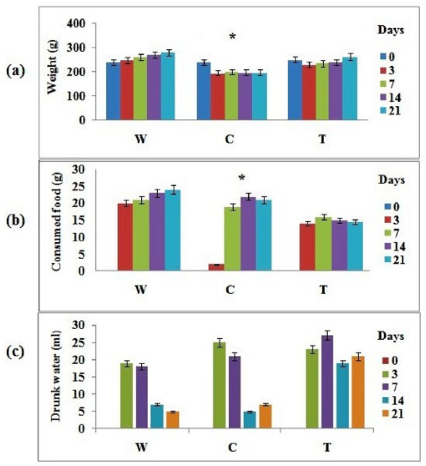
(a) Body weight gain or loss (g) shown by experimental group before and after bleomycin treatment.
Effect of FO on lipid peroxidation levels and enzymatic activities in lung
TBARS level in lung tissue was significantly increased (P = 0.04) in the C group compared to the W group. FO improved lipid peroxidation by decreasing TBARS level significantly (P = 0.04) compared to C group.
SOD and catalase activities were significantly reduced in lung tissue after BLM intoxication (C group) compared to the W group (P = 0.03). FO given in the T group caused a significant increase in SOD and catalase activities compared to the C group (P = 0.032) (Table 1).
Variation of TBARS levels and SOD and catalase activities; values are expressed as means ± SD; n = 10.

Significant compared to the W group.
Significant compared to the T group.
Histopathological observation
The histological study showed that the C and T groups are characterized by an inflammatory lymphocytic reshuffle with a disrupted alveolar architecture by fibrous alterations as well as the presence of small nodules made by fibrous remodeling.
Immunohistochemically, all groups showed a positive immunostaining of TGFβ, for rare lymphocytes in C group, intense in some lymphocytes in T group (Figure 4).
FO effect on fibrosis score, inflammatory index, and lung TGFβ density
FO treatment after fibrosis induction showed no effect to report on fibrosis score and inflammatory index (Table 2).
Fibrosis score, inflammatory index, and TGFβ density in different lung areas; values are expressed as means ± SD; n = 10.

Significant compared to the W group.
Significant compared to the W and T groups.
Significant compared to the W and C groups.
The TGFβ immunostaining evaluation (Figure 4 and Table 2) in different lung regions (alveoli, macrophages, peribronchial epithelium, fibrocytes, and inflammatory infiltrate), showed a net decrease of this cytokine distribution in lungs alveoli, inflammatory infiltrate, and fibrocytes of rats in T group compared to W and C groups.
FO effect on fatty acids composition
From the results obtained by GC, we compared the peaks which reflect the concentrations of different fatty acids in lung. This analysis showed that the induction of fibrosis has altered substantially the percentage of mono-unsaturated fatty acids (MUFAs) and polyunsaturated fatty acids (PUFAs) by increasing the levels of some acids (dihomo-γ-linoleic acid, docosadienoic acid) and reducing the rate of others (linoleic acid) in the C group compared to the W group. However, in red cell profiles, fatty acid composition of fibrotic rats showed a difference compared to the almost totally normal control rats while noting an increase in rates of saturated fatty acids and MUFAs (caprylic acid, palmitic acid, and oleic acid) against a decrease of PUFAs rate (linoleic acid, eicosadeinoic acid, γ-linoleic acid, arachidonic acid, and docosadienoic acid) in fibrotic rats unlike the control rats (Table 3).
Comparison of peaks heights (cm) corresponding to fatty acids lungs and red blood cells in different samples obtained by GC; values are expressed as means ± SD; n = 10.

Significant compared to the W group.
Significant compared to the C group.
In lung tissue, analysis revealed that FO treatment decreased rates of SFAs and MUFAs coupled with increased rates of PUFAs compared to the control group. Also, analysis by GC of red cells in rat’s blood in different groups showed a decrease in the rate of SFAs and an increase of PUFAs in rats of the treated group compared to control group (Table 2).
FO effect on BALF metabolites
Spectra corresponding to the samples of BALF of different groups which were obtained following a H+NMR analysis, are then studied by principal component analysis to the high variability between spectra. The amounts of metabolites present in BALF vary depending on the group. The results obtained after processing the spectra Mestrenova and Simcap software showed that the C group was characterized by the important presence of β-hydroxybutyrate, taurine, lactate, dimethylamine, isobyturate, and choline compared to the W group (Figure 3). Spectra H+NMR corresponding to the samples of the T group was marked by perturbation in endogenous metabolites: proline, fructose, glucose, glyceride, and malate.
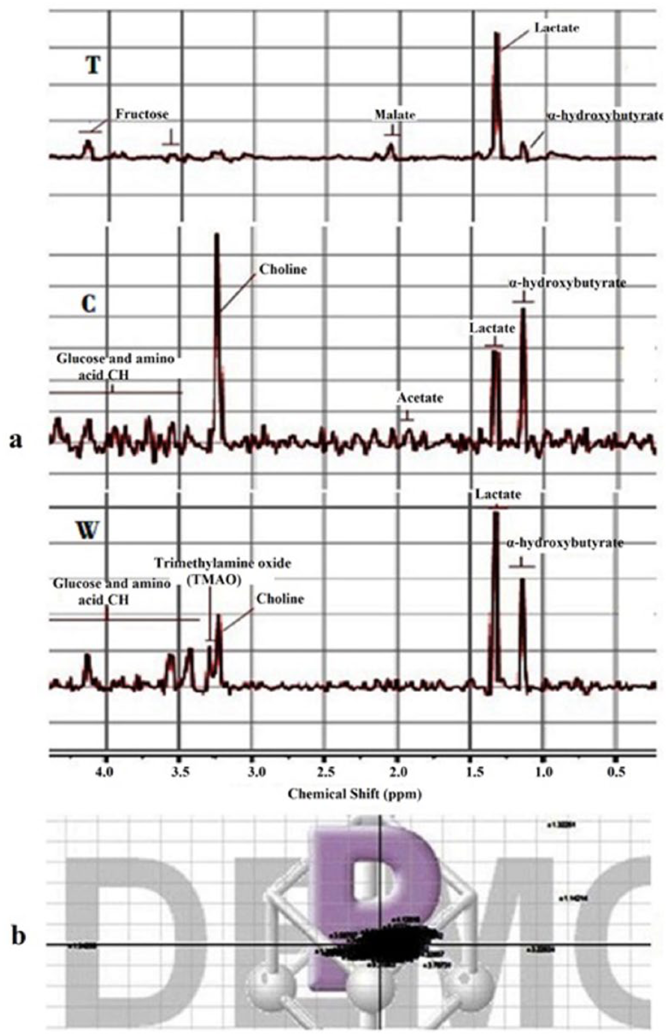
(a) 600 MHz presaturated 1H NMR spectra (δ9.0–0.5) of BALF samples following the effect of FO (2 mg/kg BW). (W) normal control group, (C) BLM group, (T) treated group. (b) Principal components analysis score derived from 1H NMR spectra of BALF samples. The scores plot showed that most of points corresponding to the spectra of BALF metabolites are grouped into one region.
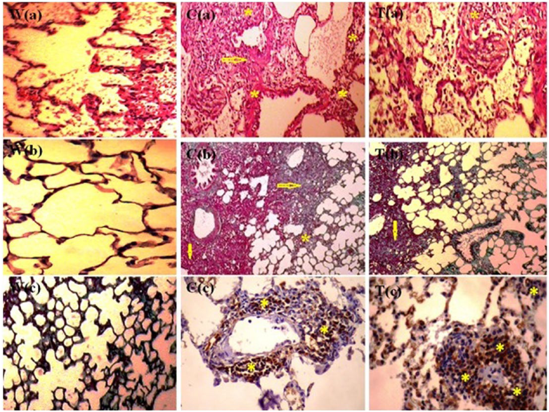
(a) Histological analysis (H&E) of lung tissue from a healthy rat (W), a rat receiving BLM (C), and a rat receiving BLM + FO (T). Sections note intense inflammation (*), disruption of alveolar architecture with presence of lymphoid follicles (arrow), and significant interstitial thickening in the control group (C). A marked regression of inflammation was showed in treated group. One representative example is shown for each group. Original magnification ×400. (b) Masson trichrome: section of lung tissue from healthy rat (W), rat received BLM (C), and rat received BLM + FO (T). Sections note inter alveolar edema (*), inflammatory infiltrate of septa (arrow). Original magnification ×400. (c) Immunohistochemical analysis: section of a healthy rat (W), a rat receiving BLM (C), and a rat receiving BLM + FO (T). The immunostaining was concentrated on lymphocytes and macrophages (yellow asterisk “*”). One representative example is shown for each group. Original magnification ×400.
Discussion
PF is a chronic progressive disorder in which excessive deposition of extracellular matrix leads to irreversible scarring to interstitial lung tissue. The etiology remains unknown, but growing evidence suggests that disequilibrium in oxidant/antioxidant balance contributes significantly. 8
In our study, following the intratracheal administration of BLM, the architecture of the extracellular matrix is totally disorganized compared to the normal lung compliance and it showed a significant inflammatory response as already demonstrated in the literature. Moreover, the choice of flaxseed oil (Linum usitatissimum) is based primarily on its anti-inflammatory effect played by the high fatty acid content. In fact, the FO fatty acid profile is characterized by a high α-linolenic acid richness that may play a major role in the inhibition of inflammatory lung disease by a decrease in LDL. In addition, fatty acid composition of this oil showed a low rate of saturated fatty acids but essentially palmitic acid which may be another factor inhibiting LDL and blood platelet aggregation. FO can reduce blood cholesterol levels and helps to avoid the risk of cancer while acting as an antioxidant. So, its composition, rich in omega 3 and omega 6, has conferred an inhibitory effect of development of malaria. 9
SOD is the only enzymatic system decomposing superoxide radicals to H2O2 and is hypothesized to play a significant role against oxidant stress, especially in the lung. Data from the present study revealed that BLM significantly decreased SOD (10.85 ± 3.21 U/mg protein) and catalase (14.79 ± 1.05 mol/H2O2/min/mg protein) activities in lung tissues compared to the FO treated group (consecutively 19.04 ± 2.30 U/mg protein and 31.98 ± 2.01 mol/H2O2/min/mg protein). Our finding was in line with the results of Jangale, which suggest that BLM exposure caused the significant decrease in antioxidant enzyme activity and therefore an increase of lipid peroxidation. 10 Kim et al., in 2006, suggested that MDA concentration in the lung was significantly higher in the fibrosis group (0.27 ± 0.13 nmol/mg protein) than in the control group (0.13 ± 0.07 nmol/mg protein) in a model of paraquat-induced lung injury. 11 FO administration strongly attenuated those changes. These results indicated that FO effects on lung fibrosis were associated with oxygen free radicals.
The use of BLM alone led to significantly higher mean lung levels of TBARS (10.85 ± 1.02 nmol/mg/protein). These data confirm those of previous published results indicating that the BLM-induced inflammatory process involves increases in lung lipid peroxidation. FO administered to the treated group led to values close to WG (3.70 ± 0.43 nmol/mg/protein). 12 Because oxidant-induced damage gives rise to a high level of inflammation, our results suggested that FO could also exhibit an anti-inflammatory activity, which should be relate to antipulmonary fibrosis effect of this oil.
In our study, after 3 days of BLM induction, there was a disruption of the histological lung parenchymal structure. In fact, we were showed a severe histopathological changes (score of fibrosis and inflammatory index) accompanied with a decrease in body weight. In order to reduce this damage, treatment with FO can be effective. In histology, this oil has raised high efficiency while reducing the inflammatory index in T group compared to C group. In addition, an immunohistochemical study showed a significant reduce of the density of distribution of TGFβ in alveoli (1.4 ± 1.07), inflammatory infiltrate (1.6 ± 1.17), and fibrocytes (0.0 ± 0.00) in the lung-treated rats compared to the fibrosis group (consecutively 4 ± 1.05; 2.4 ± 1.07; 0.4 ± 0.51). This finding was confirmed by Yacoubi et al. in 2011. 13
TGFβ is a cytokine which play a role in the initiation of the immune response and tissue repair. A prolonged production of TGFβ is involved in the development of fibrosis. It can stimulate or inhibit the proliferation depending on cellular context, controlling the turnover of extracellular matrix: the rate of this cytokine increases in lung fibrosis. By stimulating the biosynthesis of fibronectin and collagen, TGFβ exerted a powerful role in fibroblast proliferation. It also promoted the growth of fibroblasts and regulates their differentiation into myofibroblasts. The increased density of TGFβ has also been reported in experimental fibrosis induced by bleomycin. There exists a positive correlation between the tissue levels of TGF β and the severity of fibrosis induced by BLM. Also, the role of TGFβ in the excessive synthesis of collagen in lung fibrosis is demonstrated. 14 So, intense production of this cytokine and its attachment to its specific receptor type II favored the induction of the inflammatory response and subsequent installation of fibrosis.
These inhibitory effects of FO on the fibrosis score and the TGFβ distribution may be mainly due to high levels of omega 3 and 6 in both lung and blood of studied rats. Thus, compared to normal, BLM leads to almost complete modification on fatty acid levels in red blood and the lung cells. In fact, fibrotic rats showed a PUFAs rate reduction, which are an inflammation promoters and they were rewarded by a SFAs and MUFAs increase.
Indeed, treatment with linseed oil, which is rich in omega 3 and omega 6 and known for its therapeutic benefits, seems more effective after induction of fibrosis by increasing the rate of PUFAs and decreasing the SFAs and MUFAs mainly in red blood cells. This level of fatty acid modification may be the cause of an important anti-inflammatory effect. 15
Given the wealth of FO in polyunsaturated fatty acids, the effect of treatment with the latter can be explained by its role in modifying the lipid composition of cells and the biosynthesis of eicosanoids. In fact, fatty acid metabolism occurred in different organs such as the liver, kidney, brain, and heart, which proved the high rate of arachidonic acid (12.56 ± 6.20 vs. 7.96 ± 6.95 in the W group) docosapentaenoic acid (0.66 ± 0.42 vs. 1.12 ± 0.39) and DGLA (0.45 ± 0.17 vs. 0.20 ± 0.10 in the C group and 0.40 ± 0.13 in the W group) in the lungs and especially in the red blood cells. Our results were confirmed by Gadaria-Rathod et al. which suggested in 2013 that in the ω3 group, the median change from baseline was +1.46% for eicosapentaenoic acid (EPA) (P = 0.03) +1.49% for docosahexaenoic acid (DHA) (P = 0.08) and −1.91% for arachidonic acids (AA) (P = 0.02). 16 The essential polyunsaturated fatty acids (linoleic “9.67 ± 2.33 vs. 6.91 ± 1.81 in C group and 11.09 ± 1.34 W group” and alpha linoleic) undergo different reactions of desaturation and elongation. These polyunsaturated fatty acids were converted into prostaglandins and leukotrienes which gave them their anti-inflammatory and antiproliferative effects. Prostaglandin derived from arachidonic acid, dihomo-γ-linolenic acid, and eicosapentaenoic acid under the effect of cyclooxygenase thus induces a significant presence of these fatty acids in the blood during the inflammatory and fibrosis reactions as favoring vasoconstriction, platelet aggregation, and dilatation. Leukotrienes are derived from arachidonic acid and eicosapentaenoic as a result of lipoxygenases thus promoting anti-inflammatory response. 17 The conversion of PUFAs in prostaglandin and leukotrienes are associated with different physiological and pathophysiological reactions, given FO’s effect on the change in the density of TGFβ which causes a decrease in the inflammatory index and fibrosis score in the treated groups compared to fibrosis group. In this line, Romieu and Trenga showed in 2001 that TGFβ decreases the inflammatory index and the fibrosis score in the group receiving a rich omega 3 diet compared to the normal group. 18
The BALF analyses agree well with those of histology, immunohistochemistry, and GC. In fact, the fibrotic group (CG) showed an increase in taurine, proline, and lactate which marked a significant inflammation after fibrosis induction. Subsequently, difficulties in oxygenation were revealed by intermediates of citric acid cycle and stimulation of collagen secretion by proline which causes inflammation. Taurine accumulated in the cytoplasm neutrophils and other leukocytes present in the respiratory airway prove inflammation and reaction against trying to perform its role of detoxification in the presence of functional lung trouble. For cons, the FO-treated groups are marked by an excessive presence of glucose, choline glyceride, and malate which participate in the underlying synthesis of phospholipids characterizing the development of fibrosis. The FO effect on pulmonary fibrosis can be summarized in the increased secretion of liver choline which may confer an anti-inflammatory role in order to reduce inflammation and trying to ensure the integrity of alveolar membranes. 19
The results showed that FO acted before the initiation of inflammation, so a diet rich in this oil to very specific levels helped to avoid toxicity of the latter in high doses. FO acted, after the induction of the disease, by the reduction of the inflammatory process. Finally, it affects the pro-inflammatory cytokines activity, leading to the installation of fibrosis by differentiation of fibroblasts into myofibroblasts and produced intense collagen, which are the main fibrotic factors. We deduced, then, a positive correlation between the rate of polyunsaturated fatty acids with an anti-inflammatory effect and the severity of lung inflammation and subsequent fibrosis score and TGFβ density in lung parenchyma. Obtained results are very encouraging; for cons, the toxicity effects on other organs should be checked and exact determination of active components in the oil and their therapeutic involvement should be accomplished.
FO enriched in PUFA (ALA) and phytoestrogenic lignans provide one such dietary source of biologically active components that has been shown to be co-preventive 20 and co-therapeutic 10 in a wide variety of ailments. The two main fatty acids, linoleic and alpha linolenic acid, are like vitamins: we cannot make them. They are essential and must be in our daily diet, and it would seem that a balance between the two is important. Thus, the clinical treatment by this oil on patients with PF seems to be advised. This is a simple and natural way that any toxic effect of this oil has been reported and thereafter randomized controlled trials should be performed to determine and specify the best way of using this oil to treat lung diseases.
Conclusion
Our study revealed that the PF treatment by FO (Linum usitatissimum) acted well after the installation of fibrosis, while lowering the fibrosis score but also during the inflammatory phase by the immunostaining decrease of TGFβ in lung fibrocytes. These effects may be due to the wealth of this oil in omega 3 and omega 6 which are held in high value in the lung but mainly in the red cells and thus promoting an anti-inflammatory response. FO also attenuates pulmonary oxidative stress. These results supported the ability of FO to attenuate bleomycin-induced lung fibrosis and provide evidence that FO may serve as a target for potential protective treatment of the lung.
Footnotes
Acknowledgements
The authors are grateful to all people who helped to conduct this study. The authors also thank Nadia Hichem for participating in all the corrections assigned to this article.
Declaration of conflicting interests
The author(s) declared no potential conflicts of interest with respect to the research, authorship, and/or publication of this article. The plant material was identified by Smaoui Abderrazek, Professor in the Faculty of Science of Bizerte, Tunisia.
Funding
This study was funded by the Tunisian Ministry of Higher Education. The authors are grateful to all persons who helped to conduct this study. Also, we thank Nadia HICHEM for participating in all the corrections assigned to this article.