
Abstract
Studies have demonstrated that fibrinogen-like protein 2 (Fgl2) has been involved in immune and inflammatory responses which might contribute to the pathogenesis of immune-mediated diseases. Experimental autoimmune myocarditis (EAM) provided a model that mimics the pathophysiology of human giant cell myocarditis. We observed that the effect of Fgl2 overexpression on the heart function of autoimmune myocarditis rats using echocardiography and detected the level of inflammatory cytokines using enzyme-linked immunosorbent assay. Fgl2 overexpression decreased the heart function of EAM model rat, increased the inflammatory cell infiltrations, and increased the level of brain natriuretic peptide (BNP). Tumor necrosis factor-α (TNF-α), interleukin-6 (IL-6), and interleukin-17 (IL-17). These results indicate that Fgl2 is a potent target for the treatment of EAM. In conclusion, Fgl2 is a potent target to treat inflammatory dysfunctions of the heart.
Keywords
Introduction
Studies have demonstrated that Fgl2 has been involved in immune and inflammatory responses which might contribute to the pathogenesis of immune-mediated diseases. Our study found that fibrinogen-like protein 2 (Fgl2) gene silencing activates angiopoietin/Tie system and induces myocardial microvascular endothelial cell proliferation and cell migration. We also found that serum Fgl2 levels were elevated in patients with acute coronary syndrome (ACS). These results are preliminary, but very meaningful.1,2 Researchers discovered that excessive induction of Fgl2 under certain medical conditions (e.g. pathogen invasion) could trigger complement activation, inflammatory response, cellular apoptosis, and immune dysfunctions.
Experimental autoimmune myocarditis (EAM) can be induced in rats by immunizing them with cardiac myosin together with complete Freund’s adjuvant, providing a model that mimics the pathophysiology of human giant cell myocarditis. It is known that interleukin (IL)-6, 3 tumor necrosis factor (TNF)-a and IL-17 play an important role and are highly expressed in hearts with myocarditis,4,5 thus it is important to improve heart function that decrease inflammatory cytokines in EAM model rat, and this animal model can be a good tool to innovate new therapeutic approaches for the treatment of myocarditis in humans. EAM in rats is a T cell-mediated autoimmune disease. Activated T cells secrete various chemokines and cytokines which recruit and activate other inflammatory cells, such as macrophages, neutrophils, and mast cells. An excess amount of cytokines induced by inflammatory stimuli contributes to the progression of myocardial damage in patients with myocarditis. We did not know the role of Fgl2 in the process of EAM. So, in the present study, we showed the first time that overexpression of Fgl2 increased the level of inflammatory cytokines and deteriorate the heart function of EAM.
Experimental design
This study is an experimental study and has been approved by the first affiliated Hospital of Nanchang University Research and Ethical Committee. All studies were carried out using 7-week-old male Lewis rats weighing about 190–210 g. Seven-week-old male Lewis rats were injected into the footpads with antigen-adjuvant emulsion according to the procedure described previously. In brief, porcine cardiac myosin was dissolved in phosphate-buffered saline at 5 mg/mL and emulsified with an equal volume of complete Freund’s adjuvant with 11 mg/mL Mycobacterium tuberculosis H37RA (Difco Lab., Detroit, MI, USA). EAM in rats was induced by immunization with 0.1 mL emulsion once by subcutaneous injection into their rear footpads (0.1 mL into each footpad). The morbidity of EAM was 100% in rats immunized by this procedure. Rats immunized with myosin became ill and immobile in the second week. After myosin injection, rats were divided randomly into EAM group the same amount of PBS was injected through tail vein on day 1 immunized with myosin) (n = 8), EAM-GFP group (5×10⁷ IU/mL GFP-lentiviral vector was injected through tail vein on day 1 immunized with myosin) (n = 8), EAM-Fgl2 group (5×10⁷ IU/mL Fgl2-lentiviral vector was injected through tail vein on day 1 immunized with myosin) (n = 8). Fgl2-lentiviral vector and GFP- lentiviral vector were provided by Jikai company. Age-matched Lewis rats without immunization were used as normal controls (n = 8). Rats were maintained with free access to water and chow throughout the period of study, and animals were treated in accordance with the ‘Guidelines for Animal Experimentation’ of our institute.
Echocardiography
A trans-thoracic echocardiographic analysis was performed on day 60 using a linear array transducer in vivo imaging system (GEvivid7) with a 12-MHz imaging transducer. Briefly, rats were anesthetized with an intraperitoneal injection of pentobarbital sodium (20–35 mg/kg). An M-mode echocardiography was performed at the papillary muscle level under spontaneous ventilation. The left ventricular end-diastolic dimension (LVEDd) and end-systolic dimension (LVEDs) were measured. The percentage of left ventricular fractional shortening (LVFS) was using the formula: LVFS (%) = (LVEDd – LVEDs)/LVEDd × 100. All echocardiographic measurements were averaged from at least five separate cardiac cycles.
Histopathology
The excised wet myocardium was kept in 10% formalin and the mid-ventricle sections were then embedded with paraffin. Inflammatory cell infiltrations were identified using hematoxylin and eosin (H&E)-stained sections at 200-fold magnification by light microscopy. Several sections of each heart were scored blindly by two observers. The scores assigned to these specific sections were averaged as described previously. 6 The extent of cellular infiltration was graded and scored as follows: 0 (normal), 1 (lesion extent in the range of 10–25% of a transverse section), 2 (in the range of 25–50%), 3 (in the range of 50–75%), and 4 (exceeding 75%).
Western blot analysis
Myocardial tissue samples were homogenized in a lysate buffer. Protein concentration was measured with a bicinchoninic acid method. For western blots, samples (40 μg protein) were separated by sodium dodecyl sulphate-polyacrylamide gel electrophoresis and then transferred electrophoretically onto a nitrocellulose membrane. The membrane was incubated with a primary antibody against Fgl2 (1:1000; Santa Cruz Biotechnology, Santa Cruz, CA, USA). After extensive washes, the blot was incubated with a secondary antibody and developed with an ECL reagent. Chemiluminescence was detected with a LAS-1000 luminescent image analyzer (Fujifilm, Tokyo, Japan). GAPDH was used as an internal control.
Measurement of serum BNP, TNF-α, IL-6, and IL-17
Blood samples were collected on day 60. BNP, TNF-α, IL-6, and IL-17 levels in the samples were estimated with ELISA Kit for BNP, TNF-α, IL6, and IL-17 (Shanghai Senxiong Technology Company, Shanghai, PR China). Briefly, standards and samples were bound by the immobilized antibody, and an enzyme-linked polyclonal antibody specific for the cytokine was added to the wells followed by a substrate solution yielding a colored product. The intensity of the color was measured at 450 nm. The sample levels were calculated from a standard curve and were corrected for protein concentration.
Quantitative reverse-transcription polymerase chain reaction (RT-PCR)
Total RNA was extracted from the heart tissues with Trizol (Invitrogen, Carlsbad, CA, USA), according to the standard protocol and then reverse-transcribed. cDNA was then amplified using the ABI 7700 sequence-detector system (Applied Biosystems, Foster City, CA, USA) with a set of primers and probes corresponding to Fgl2, as described previously. Tatget sequence of Fgl2 gene (Forward: 5′- AGTCGCTCCAACTGGTAAATATG-3′, Reverse: 5′-GGCACTTGAAACACTTG CTATCC-3′.). β-actin gene (Forward: 5′- CCCATCTATGAGGGTTACGC-3′, Reverse :5′-TTTAATGTCACGCACGATTTC -3′). β-actin was used as an internal control. Melting curves of all samples were performed as controls of specificity. All gene expression data were calculated as 2-ΔΔCT (n=5), which indicates an n-fold change in gene expression relative to the control sample.
Statistical analysis
Data were analyzed using SPSS software (version 17; SPSS, Chicago, IL, USA). All data are expressed as mean ± SD. Statistical significance was calculated using Student’s t-tests or a one-way analysis of variance (ANOVA). A P value <0.05 was considered significant.
Results
The expression of Fgl2 mRNA and protein. In the study, we showed that Fgl2 mRNA increased in the EAM-Fgl2 group significantly (P <0.05) compared with the EAM-GFP group (Figure 1a). We also found that Fgl2 protein level increased in the EAM-Fgl2 group significantly (P <0.05) compared with the EAM-GFP group (Figure 1b). These data indicated that Fgl2 gene transfection increased the expression of Fgl2 mRNA and protein.
Fgl2 overexpression increase inflammatory cell infiltrations. Inflammatory cell infiltrations were identified using H&E-stained sections at 200-fold magnification by light microscopy. In the study, there were no inflammatory cell infiltrations in the normal control group. Inflammatory cell infiltrations increased in the EAM-Fgl2 group (2.63 ± 0.26) significantly (P <0.05) compared with the EAM-GFP group (1.62 ± 0.18) and the EAM group (1.50 ± 0.19) (Figure 2). These data indicated that Fgl2 overexpression increased inflammatory cell infiltrations.
Fgl2 overexpression deteriorate the heart function of EAM. Echocardiographic analysis was performed on day 60. As shown in Table 1, EF%, FS%, and HR of the EAM-Fgl2 group decreased significantly (P <0.05), LVDD and LVSD of the EAM-Fgl2 group increased significantly (P <0.05).These data indicated Fgl2 overexpression deteriorates the heart function of EAM.
Fgl2 overexpression increased the level of BNP, TNF-α, IL-6, and IL-17. As shown in Table 2, BNP, TNF-α, IL-6, and IL-17 levels in the EAM-GFP and EAM groups were significantly higher compared with control group (P <0.05). BNP, TNF-α, IL-6, and IL-17 levels in the EAM-Fgl2 group were significantly higher compared with the EAM-GFP and EAM groups (P <0.05), and there was no significant between the EAM-GFP and EAM groups (P >0.05). These data indicated that Fgl2 overexpression increased the level of BNP, TNF-α, IL-6, and IL-17.

The qRT-PCR analysis for the expression of Fgl2 mRNA (a) and western blot analysis for the expression of Fgl2 protein (b).(a) Total RNA was extracted from the heart tissues with Trizol, β-actin was used as an internal control. Melting curves of all samples were performed as controls of specificity. All gene expression data were calculated as 2-ΔΔCT (n=5). (b) Western blot analysis for the expression of Fgl2 protein. Western blot analysis and GADPH as a standard control. Data represent means ± SEM. *P <0. 05 vs. Normal; #P >0.05 vs. EAM-GFP group, #P <0. 05 vs. EAM-GFP group and EAM group.

Inflammatory cell infiltrations analysis were identified using hematoxylin and eosin (H&E)-stained sections at 200-fold magnification by light microscopy. (a) NC group; (b) EAM-GFP group; (c) EAM group; (d) EAM-Fgl2 group. (e) The scores of inflammatory cell infiltrations. Data represent means ± SEM. *P <0. 05 vs. Normal; #P >0.05 vs. EAM-GFP group, #P <0. 05 vs. EAM-GFP group and EAM group.
Left ventricular long axis of echocardiography analysis.

Echocardiographic analysis was performed on day 60.
Data represent means ± SEM.
P <0.05 vs. EAM-GFP group and EAM group.
P >0.05 vs. EAM-GFP group.
P <0.05 vs. Normal.
Measurement of serum BNP, TNF-α, IL6, and IL-17.
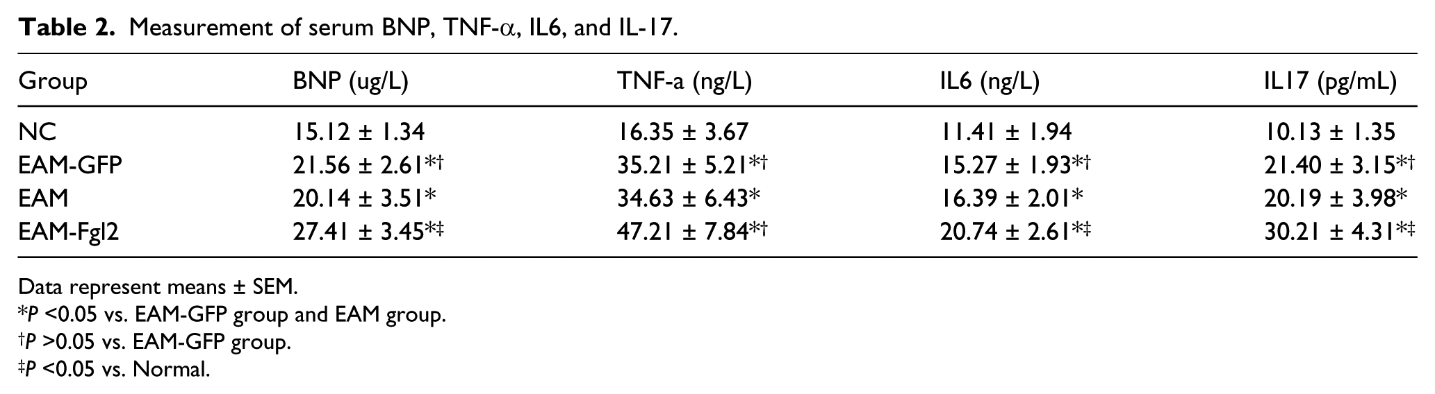
Data represent means ± SEM.
P <0.05 vs. EAM-GFP group and EAM group.
P >0.05 vs. EAM-GFP group.
P <0.05 vs. Normal.
Discussion
Our previous study found that fibrinogen-like protein 2 (Fgl2) gene silencing activates angiopoietin/Tie system and induces myocardial microvascular endothelial cell proliferation and cell migration. Many studies found Fgl2 is involved in immune response. 7 The EAM model rat can be a good tool to innovate new therapeutic approaches for the treatment of myocarditis in humans. In the present study, our data show that Fgl2 overexpression decreased the heart function of EAM model rat, increased the inflammatory cell infiltrations, and increased the level of BNP, TNF-α, IL-6, and IL-17. These results indicate that Fgl2 is a potent target for the treatment of EAM.
BNP is a cardiac neurohormone that is secreted into the plasma from the ventricles in response to ventricular volume expansion and pressure overload. BNP provides an easy method for the early detection of heart failure (HF), and for assessing the severity of HF and the effectiveness of treatment. 8 In this study, we found that Fgl2 overexpression increased the level of BNP, which indicated that Fgl2 increased ventricular volume expansion and pressure overload. In addition, we found that LVDD and LVSD in the EAM-Fgl2 group increased significantly and EF% and FS% in the EAM-Fgl2 group decreased significantly. In conclusion, Fgl2 overexpression deteriorates the heart function of EAM and worsens heart failure.
The study has reported that TNF-α upregulates Fgl2 expression in rat myocardial ischemia/reperfusion injury.
9
We found that Fgl2 overexpression increased the level of TNF-α. To our knowledge, this is the first report. As we know, IL-6,
3
TNF-a, and IL-17 play an important role in immune injury and it is important to improve heart function that decreases inflammatory cytokines.4,5 In this study, we found that Fgl2 overexpression increased the level of TNF-α, IL-6, and IL-17, and which may be the mechanism that causes Fgl2
In summary, we demonstrated that Fgl2 overexpression increased the level of inflammatory cytokines and deteriorate the heart function of EAM. Fgl2 is a potent target to treat myocardial immune injury, which suggests that we can downregulate the expression of Fgl2 and improve the heart function, but it is not clear. The preliminary data, although very encouraging, need to be well discussed and further studies surely continued. It is possible that further studies will provide clues for better understanding of Fgl2 for prevention of treat myocardial immune injury.
Footnotes
Declaration of conflicting interests
The author(s) declared no potential conflicts of interest with respect to the research, authorship, and/or publication of this article.
Funding
This work was supported by a grant from the National Nature Science Foundation of China (No. 30960119, 81260044) and the Technology Support project of Jiangxi province (No. 2010BSA12000).