
Abstract
Hypoplastic amelogenesis imperfecta is a widespread hereditary disease that causes the loss of enamel. The purpose of this study was to investigate the nanoscratch resistance of hypoplastic amelogenesis imperfecta for providing a reference for restorative treatment. Four unerupted third molars from a patient diagnosed with hypoplastic amelogenesis imperfecta and seven unerupted third molars from normal individuals were compared. Atomic force microscopy and energy-dispersive X-ray spectroscopy were used to observe the microstructure and composition of the teeth (enamel and dentin). The nanoscratch tests of teeth (enamel and dentin) were investigated using a nanoscratch tester, scanning electron microscopy, and a stylus profilometer. The results indicated that hypoplastic amelogenesis imperfecta teeth had different microstructures compared to normal teeth. Hypoplastic amelogenesis imperfecta demonstrated a higher composition of organic substance. Meanwhile, the friction coefficient of hypoplastic amelogenesis imperfecta was higher than that of normal teeth, and inferior frictional resistance of hypoplastic amelogenesis imperfecta teeth was observed. The main damaging mechanisms observed in hypoplastic amelogenesis imperfecta under nanoscratch were the combination of delamination, debris, and cracks in enamel with delamination, debris, and plastic deformation in dentin. Our findings suggested that new dental restorative materials should be selected to match the mechanical properties of hypoplastic amelogenesis imperfecta.
Introduction
Amelogenesis imperfecta (AI) is a multiple group of hereditary diseases that affects the enamel of teeth and reduces enamel thickness or decreases mineralization.1,2 Although the exact global incidence of this disease is uncertain, it is estimated that from 1 in 2000 up to 1 in 18,000 people are affected by it.3,4 AI may be differentiated into three main groups: hypoplastic, hypocalcified, and hypomature based on clinical presentation of defects. 5 Compared with normal teeth, hypoplastic AI disturbs the formation of enamel which may cause tooth hypersensitivity and acutely affects facial appearance, oral function, and psychological status of hypoplastic AI patients. 6
Current hypoplastic AI research is focused on etiology and restorative treatment. Although most researchers have focused on the genetic variations of hypoplastic AI,7,8 a genetic therapeutic schedule has not yet been developed. Restorative treatment is the most common approach for hypoplastic AI patients. General methods include all-ceramic crowns, porcelain fused to metal crowns, and so on.1,9 In treatment, the choice of restoration materials is very important for these special patients. The restoration materials, applied consistently with the mechanical properties and tribological characteristics of hypoplastic AI, can protect existing teeth and prolong the life of the prosthesis.10,11 Many previous studies presented the mechanical behaviors of enamel related to enamel’s microstructures.12,13 Significant microstructural differences have been found between hypoplastic AI and normal enamel. Shore et al. 14 discovered the irregular arrangement of constituent crystals within prisms and the difficulty in distinguishing crystal boundaries in AI enamel. Batina et al. 15 used atomic force microscopy (AFM) to learn that AI enamel is porous, with smaller crystal grains than normal human teeth. As pointed out by Wright et al., 16 the enamel prism structure of hypoplastic AI was abnormal and poorly formed. Dentin, a mineralized structure, can not only support the enamel but also prevent the enamel from fracture, 17 and in many cases of AI, dentin is frequently exposed to oral environment after the loss of enamel. 6 Therefore, observing the microstructure and mechanical properties of dentin is also essential. However, research on the tribological characteristics of hypoplastic AI is insufficient at present and should be explored further. Therefore, this study aims to observe the nanoscratch resistance of hypoplastic AI teeth (enamel and dentin) in terms of microstructure and composition by AFM, energy-dispersive X-ray spectroscopy (EDX) and nanoscratch tester.
Materials and methods
Specimen preparation
Four freshly extracted, unerupted caries-free hypoplastic AI third molars were obtained from a 23-year-old female patient in West China Hospital of Stomatology after this study was approved by the local ethical committee, and the patient signed an informed consent. Seven unerupted third molars were collected from other normal individuals and served as controls. These third molars were incubated in water containing 0.05% thymol at 4 °C immediately after extraction. The molars were sectioned parallel to the long axis of the teeth by water-cooled diamond saw (Minitom; Struers, Copenhagen, Denmark). Then the sections were embedded in self-cured resin, with the section surfaces facing up. Subsequently, all samples were polished with abrasive paper from 320 to 4000. The entire polishing process was done under cooling water, and the polishing time of the samples was controlled. After 10 min of ultrasonic treatment (AS515PBD-i ultrasonic cleaner, Tianjin Automatic science instrument company, Tianjin, China) to get rid of surface debris, all polished samples were finally prepared. All the tests in the enamel and dentin were conducted near the dentinoenamel junction.
Microhardness test
The surface microhardness (SMH) of a hypoplastic AI tooth (enamel and dentin) and a sound tooth (enamel and dentin) were measured by a Knoop microhardness indenter (Duramin-1/-2; Struers). A 50 g load was applied for each indentation for 5 s in enamel, while a load of 25 g for 5 s was used in dentin.
Microstructure analysis
All samples were scanned using AFM (SPM-9600; Shimadzu, Kyoto, Japan), with the working mechanism described previously. 18
Nanoscratch testing
To measure the microfriction level of hypoplastic AI, nanoscratching tests were conducted using a nanoscratch machine (CSEM Instruments, Peseux, Switzerland) with a conical diamond tip with a radius of 2 μm. All experiments were conducted at room temperature. Progressive load varied from 0 to 50 mN, and constant load modes were selected. The values for constant load were 20 and 40 mN, with scratch lengths of 200 μm. The remnant widths of the grooves caused by constant load were measured by an Ambios XP-2 stylus profilometer (Ambios Technology, Inc., Santa Cruz, CA).
Scanning electron microscope and EDX analysis
All the morphologies of hypoplastic AI samples were carefully observed via scanning electron microscope SEM (INSPECT, Brno, Czech Republic) to demonstrate damage features. EDX (INCA, OXFORD Instruments, UK) was used to evaluate the concentrations of carbonate (C), oxygen (O), phosphorus (P), and calcium (Ca) of the hypoplastic AI (enamel and dentin) and normal teeth (enamel and dentin).
Statistical analysis
Data were analyzed using the Statistical Package for the Social Sciences (SPSS) Version 13.0 software (SPSS, Inc., Chicago, IL). T-test was used to evaluate the difference in data obtained between hypoplastic AI teeth and normal teeth. P < 0.05 was considered statistically significant.
Results
Microhardness results
Figure 1 shows that hypoplastic AI teeth (enamel and dentin) had statistically significant lower SMH value compared to normal teeth (enamel and dentin). The SMH decreased by 10.9% and 9.8% in the enamel and dentin, respectively.

Surface microhardness analysis of normal teeth versus AI teeth: (a) enamel and (b) dentin.
AFM results
The AFM images of hypoplastic AI teeth (enamel and dentin) and normal teeth (enamel and dentin) are presented in Figure 2. Normal enamel has regular and clear crystals, relatively intact, while hypoplastic AI enamel has a higher porosity surface with larger crystals, apparently randomly ordered loosely (see the arrow). Obvious crystals in uniform grain size and some randomly distributed dentin tubules can be seen in normal dentin. In contrast, hypoplastic AI dentin reflected a smooth surface with obscure grain (see the arrow).
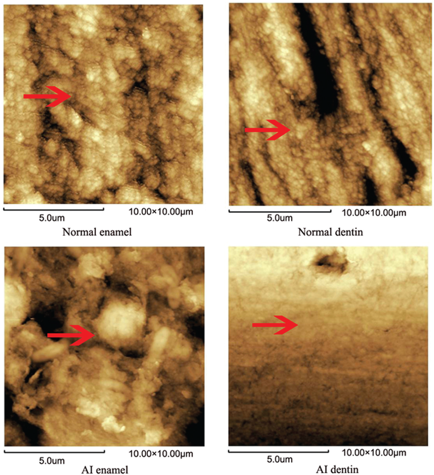
AFM images of normal teeth (enamel and dentin) and AI teeth (enamel and dentin).
EDX results
As shown in Figure 3(a), the content of C in hypoplastic AI enamel increased threefold. The content of P and Ca was reduced by 20% compared with that of normal enamel. In the hypoplastic AI dentin (Figure 3(b)), there was a 14% increase in the content of C, while the content of P was reduced by 8% and the content of Ca was reduced by 5%. All the data obtained above show statistically significant difference and demonstrate the higher composition of organic substance of hypoplastic AI.

Comparison between the elemental compositions of normal versus AI tissues: (a) enamel and (b) dentin.
SEM images of scratch
The typical morphologies of normal enamel and hypoplastic AI enamel after nanoscratch test under ramping loads from 0 to 50 mN are presented in Figures 4 and 5, respectively. The widths of the scratches of normal enamel and hypoplastic AI enamel increased steadily with the imposed load. On the surface of sound enamel (Figure 4), only plastic deformation formed at the beginning of the scratch test, and a little debris piled up on the edges of trace. No cracks were observed on the scratch traces.
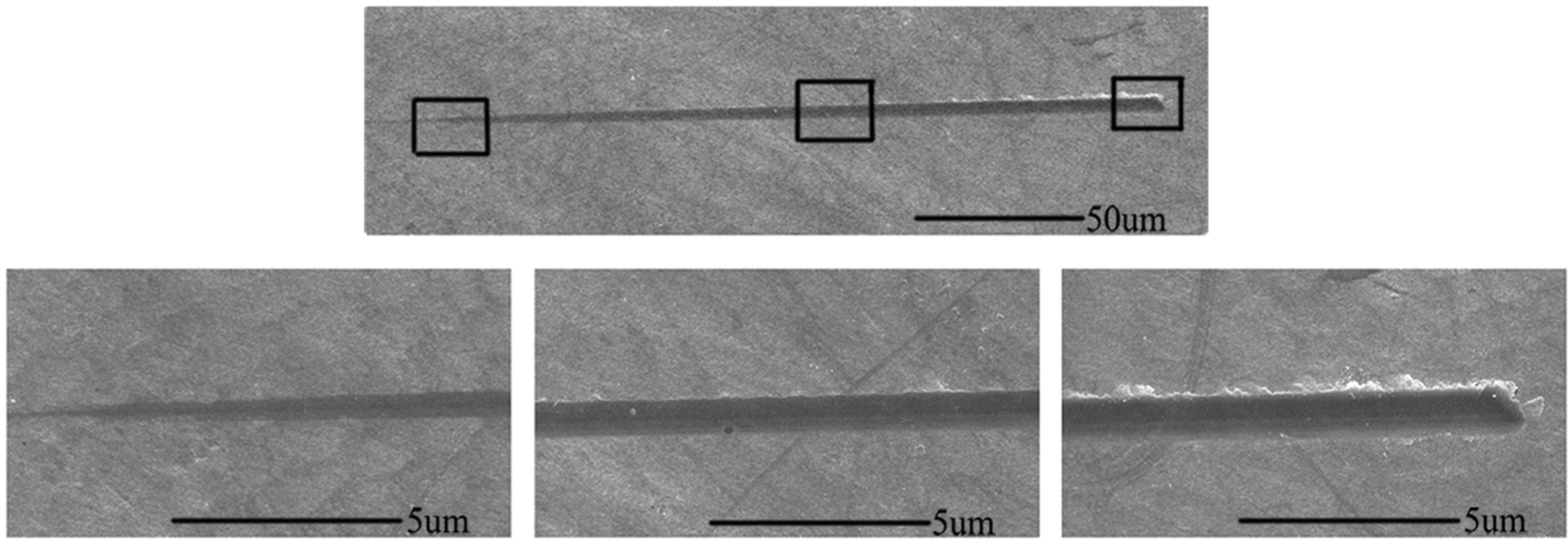
SEM images of nanoscratches of normal enamel with progressive load.

SEM images of nanoscratches of AI enamel with progressive load.
The scratch of hypoplastic AI enamel can be seen in Figure 5. Slight debris formed when the load was about 10 mN. A small amount of delamination occurred on the edge of traces as the load increased to about 25 mN. Meanwhile, more debris and microcracks formed in the scratch trace when the load reached about 30 mN. Some obvious cracks propagated outside scratch trace at the ultimate load. Increasing amounts of debris piled up on the trace with the further increase in load. Debris of hypoplastic AI enamel appeared earlier, and there was more debris in the AI enamel scratch trace compared to normal enamel.
As shown in Figure 6, the scratch trace of normal dentin was too slight to be seen when the load was lower than 10 mN. Very little debris was observed at the load of 50 mN. The typical morphologies of hypoplastic AI dentin surface under the progressive load are presented in Figure 7. At the beginning of the scratch test, the trace was too slight to be seen. When the load increased to 25 mN, a small amount of debris piled up on the edge of the trace. Meanwhile, a small amount of delamination appeared and the width of the scratch increased accordingly. More debris formed at the end of the traces. However, the debris was not as much as that in the hypoplastic AI enamel.

SEM images of nanoscratches of normal dentin with progressive load.

SEM images of nanoscratches of AI dentin with progressive load.
Comparison of friction coefficients
The friction coefficients of normal teeth (enamel and dentin) and hypoplastic AI (enamel and dentin) with progressive loads are presented in Figure 8. The friction coefficient increased slowly with the load. Both the values of hypoplastic AI enamel and dentin were higher compared to the normal enamel and dentin under the same load, respectively.

Friction coefficients of normal teeth and AI teeth with progressive load.
Remnant widths
The remnant widths of the scratch traces on hypoplastic AI (enamel and dentin) and normal teeth (enamel and dentin) at 20 and 40 mN are shown in Figure 9(a) and (b), respectively. All the width of scratch trace in hypoplastic AI teeth (enamel and dentin) and normal teeth (enamel and dentin) increased when the loads go up. The remnant width of hypoplastic AI (enamel and dentin) exhibits a statistically significant increase compared with normal teeth (enamel and dentin).

Remnant width of scratches of normal and hypoplastic AI teeth at 20 and 40 mN: (a) enamel and (b) dentin.
Discussion
In the experiment, hypoplastic AI samples were selected from the same individual. The structures of hypoplastic AI teeth drawn from AFM images showed that hypoplastic AI enamel had a higher porosity surface, which was similar to the results obtained by earlier researchers. 15 Some previous studies also showed that hypoplastic AI teeth have a composition distinguishable from that of normal teeth, in particular, an obvious decrease in calcium concentration. 14 In this study, a reduction in calcium was observed from EDX results. Therefore, it can be concluded that the samples in our study did represent hypoplastic AI patient. All the data were received without individual differences; thus, replication of the experiment is likely to substantiate evidence of good stability and repeatability.
Normal enamel manifests good wear resistance in the mouth due to the unique aligned “prism-shaped” rods. 19 Earlier studies indicated that the mechanical and tribological properties of enamel should be related to its microstructure and chemical composition. When this special structure of enamel changes, the wear property of enamel changes.10,11,20 Hypoplastic AI enamel loses the normal structure because of an erroneous enamel formation mechanism in the hypoplastic AI patient.1,2 The AFM pictures also exhibited the different surface structure of hypoplastic AI enamel (Figure 2). The grains of the hypoplastic AI enamel were randomly aligned and loosely packed, which was completely different from the normal human enamel. This result is identical to the results obtained by Batina et al. 15 Previous studies have reported that normal enamel exhibited debris or an accumulation of material along scratch traces.17,19 Therefore, hypoplastic AI enamel not only demonstrated obvious delamination and debris but also presented apparent cracks in the trace and propagated outside trace. It should be noted that normal dentin showed plastic deformation. 21 In this article, hypoplastic AI dentin was a smooth surface with obscure grain. Therefore, AI dentin presented obvious delamination and debris. In addition, there was less debris in hypoplastic AI dentin compared to hypoplastic AI enamel.
The SMH is a typical feature of the mechanical properties of enamel. The change in microhardness can reflect the different compositions of tooth. Panighi and Sell 22 have revealed that there was a positive correlation between the hardness and the mineral content of the tooth. In this study, AI teeth had lower microhardness, and the EDX results above suggested that the composition of hypoplastic AI enamel and dentin manifested obvious differences compared with that of normal human teeth. There were fewer inorganic materials in hypoplastic AI enamel and dentin. Several studies have shown that the loss of inorganic materials may induce enamel with more susceptibility to abrasion and attrition. 23 Therefore, under friction, hypoplastic AI teeth demonstrated inferior wear resistance to normal teeth; thus, the width of scratch increased.
Based on our knowledge, this study is the first to report on the nanoscratch resistance of hypoplastic AI teeth (enamel and dentin) with microstructure and composition analysis. The results obtained above can provide a useful reference for selecting the better match of restoration material consistently with inferior hypoplastic AI teeth to reduce the attrition of opposite teeth and prolong the life of the prosthesis. But an in vivo study (including clinical experiment) is also necessary to define the properties of the hypoplastic AI enamel and dentin for better treatment. Moreover, a more suitable restorative material for hypoplastic AI patients, in consideration of unique wear resistance of hypoplastic AI teeth, must be identified.
Conclusion
Based on the findings on the nanoscratch resistance of hypoplastic AI teeth, the main conclusions can be made as follows.
Changes in structure and composition of hypoplastic AI teeth (enamel and dentin) may induce relatively weaker wear resistance in hypoplastic AI teeth than that of sound teeth;
The main wear mechanisms of hypoplastic AI teeth were the combination of delamination, debris, and cracks in enamel with delamination, debris, and plastic deformation in dentin.
Footnotes
Academic Editor: Ruey-Jen Yang
Declaration of conflicting interests
The authors declare that there is no conflict of interest.
Funding
This work was supported by the State Key Laboratory of Oral Diseases, Sichuan University, China (SKLODSCU20130044) and the Doctoral Fund of the Ministry of Education of China (20110181110056).