
Abstract

Introduction
James Parkinson discovered Parkinson’s disease (PD), which is one of the most common neurodegenerative disorders. 1 A neurodegenerative disorder is a condition that affects the central nervous system, causing continuous loss of neurons in the spinal cord and peripheral nerves. 2 Neuronal degeneration and cell death are primarily responsible for clinical manifestations of various age-related neurological disorders. 3 PD is a progressive neurodegenerative disorder that affects mainly older adults. The symptoms include bradykinesia, muscle rigidity, tremors (at rest), shuffled gait, and inadequate control over voluntary movements. 4 Previous research has estimated the future prevalence of Parkinson’s disease based on population aging. 5 The etiology of this disease is unknown, and it is affected by multiple factors, including environmental toxins and genetic susceptibilities. 6 However, PD sometimes originates from viral encephalitis or pathological loss associated with cerebral ischemia. Moreover, certain drugs like reserpine and chlorpromazine can induce PD-like symptoms.
Neurotoxins such as MPTP and rotenone (herbicide) are involved in the pathogenesis of PD. Additionally, genetic mutations, including those in α-synuclein, parkin, and leucine-rich repeat kinase 2 (LRRK2), contribute to the development of PD. Typically, symptoms initiate from 1 end of the body and slowly progress to the opposite side over several years. This results in a gradual loss of passive limb movement accompanied by stiffness in the axial and limb regions, with cogwheel rigidity. 7 The pathological characteristics of this disorder include the progressive loss of dopaminergic neurons in the substantia nigra and nerve terminal degeneration in the corpus striatum. Each dopaminergic neuron forms thousands of synaptic contacts in the neostriatum and thus affects the activity of many cells. Rather than participating in specific movements, the dopaminergic system appears to have a tonic, sustaining effect on motor activity. 8 Current treatment regimens for Parkinson’s disease include levodopa preparations (carbidopa-levodopa), dopamine agonists (pramipexole, ropinirole), monoamine oxidase B inhibitors (selegiline, safinamide, zonisamide, and rasagiline), as well as catechol-o-methyl transferase inhibitors (entacapone and tolcapone). However, none of these treatments halt the progression of the disease and are also associated with adverse effects such as nausea, hypotension, sedation, hallucinations, and psychiatric complications. 8
There are several controversies, particularly regarding treatment options. Over the past 20 years, there has been an ongoing debate about the optimal initial therapeutic agent for Parkinsonism. Whether levodopa is superior to dopamine agonists remains a point of contention. Levodopa is associated with various complications, including the on-off phenomenon and long-term effects of dopaminergic therapy, such as tremors, fluctuations, dyskinesia, and psychiatric disorders. Some researchers have even suggested that prolonged levodopa treatment might exacerbate disease progression. 9 Dopamine agonists are medications that primarily target dopamine levels but do not address the core cause of the disease. Recent therapies focus on inhibiting disease progression. Natural therapies have been used to reduce oxidative stress and ROS production. Few novel therapies have been introduced to target α-synuclein, neuroinflammation, and improvement of mitochondrial function. 10
Alternative therapy for Parkinson’s disease includes various exercises and physical activities, which are also crucial for inhibiting the functional progression of the disease. 11 Long-term complications associated with recent treatments may include fluctuations in response, dyskinesias, toxicity, and reduced efficacy. 12 Thus, newer modalities are continually warranted for effectively alleviating disease symptoms, improving quality of life, and halting disease progression. As discussed, dopaminergic neuron degradation begins with mitochondrial dysfunction and oxidative stress. Recently, herbal remedies enriched with phytochemicals have been proven to manage metabolic and neurodegenerative disorders, including PD.13-15 Many herbal extracts are currently being researched for this purpose and have shown effectiveness. Various advanced formulations have also been developed to more effectively target these ailments. 16 These medicinal plants possess various protective actions that help mitigate neurodegeneration’s damaging effects. Some of the herbal extract used includes Curcuma longa, 17 Datura inoxia mil, 18 Myrica esulenta 19 Prunus armeniaca, 20 and Breynia cernua, 21 different parts (roots, tubers, leaves) of these herbal extracts are used in Parkinson like symptoms in animal models. 19 Thalictrum foetidum L. belongs to the family Ranunculaceae and is traditionally used to treat inflammation, endometrial cancer, numbness, joint pain, and trauma. 22 It is commonly known as “Meadow rue,” this plant is referred to by various names in different languages. In the Gilgit-Baltistan region of Pakistan, it is called “Halizee.” 23 Traditionally, it has been used in the Indian medicinal system to treat ulcers, piles, skin infections, burns, colds, coughs, joint pains, and jaundice. 24 The Tuber of this plant is used for wounds, uterine tumors, swellings, and nervous disorders. 22 The Haloperidol-induced cataleptic model is one of the classical models used to induce Parkinson-like symptoms in rodents. This is a rational model because the cataleptic symptoms produced in rodents closely resemble human Parkinson’s disease symptoms. This model is easy to induce without overt toxicity and completion time, which is useful for evaluating the protective effects of various extracts.21,25 This study aimed to explore the therapeutic potential of Thalictrum foetidum L. for Parkinson-like symptoms using a haloperidol-induced animal model. It also provided a rationale for its folkloric use in neurodegenerative disorders and to investigate the underlying mechanism.
Material and Methods
Plant Collection and Identification
Thalictrum foetidum L. was collected from the district of Gilgit Baltistan. Dr Qasim Ali, a famous botanist from the Department of Botany, Government College University Faisalabad, identified the plant. The authentication number 378-bot-23 was issued.
Plant Extract Preparation
Tubers of Thalictrum foetidum L. were cleaned to remove dust and then broken into smaller pieces to facilitate grinding into a powder using a grinder. The resulting coarse powder was mixed with water and ethanol in a 30:70 ratio. The mixture was macerated by vigorous shaking for 7 days. The ethanol was evaporated from the solution using a rotatory evaporator. The remaining residue was dried using a lyophilizer and collected in an amber-colored glass bottle.
Phytochemical Analysis
Phytochemical analysis was carried out using high-performance liquid chromatography (HPLC).
High-Performance Liquid Chromatography (HPLC) Analysis
HPLC was used to determine the phenolic and flavonoid content of Thalictrum foetidum L. The extract was analyzed using an HPLC system (Model = 1269 infinity II, equipped with a quaternary pump, Variable Wavelength Detector [VWD], degreaser [1200 series)], a Zorbax SB Eclipse-C18 column [5 μm, Agilent Technologies, USA]). Several peaks were observed on the chromatogram.
Evaluation of Total Phenolic Content (TPC)
We mixed 0.2 mL of Folin-Ciocalteu’s phenol reagent with sample and standard solutions in separate test tubes. Sodium bicarbonate 1 mL (15%) was added to each test tube. These solutions were kept at room temperature for 2 h. Different concentrations of the solutions were used to generate a linear regression equation. The absorbance of each solution was measured at a wavelength of 760 nm. The standard solution contained all test tube components except the analyte. Gallic acid was used as a reference for constructing the standard curve. Using this standard curve, the sample was calculated in mg per gram of plant extract using linear regression as described by equation no. (1).
26
Estimation of Total Flavonoid Content (TFC)
A mixture of 0.2 mL of sample and standard solutions was prepared in separate test tubes with reagents: 0.1 mL of 10% aluminum nitrate, 0.1 mL of 1 M potassium acetate solution, and 4.6 mL of distilled water. The resulting mixture was incubated at ambient temperature for 45 min. The standard solution in the test tube contained all components, excluding the analyte, with quercetin (QTN) as a reference for constructing the standard curve. Standard curves were generated from the sample solutions at various concentrations, with absorbance readings taken at a wavelength of 415 nm. Total flavonoid content (TFC) was expressed as mg of quercetin equivalents per gram (QEqs/g) of extract, calculated using equation no. (2) derived from the standard curve.
26

DPPH Radical Scavenging Assay
The antioxidant efficacy was assessed using different concentrations of the sample. A 0.05% DPPH solution in methanol was prepared, and 200 μL of this solution was mixed with 80 μL of the sample in 4 mL of methanol. The mixture was incubated in the dark for 30 min. Absorbance was measured at 517 nm using a UV spectrophotometer. The percentage of DPPH reduction by the extract was calculated using the formula provided in equation no. (3).
27
Experimental Animal
Thirty healthy albino mice of both sexes, each weighing between 20 and 25 g, were selected for the study. These mice were obtained and housed in polypropylene cages within an animal facility that maintained appropriate conditions, including a 12-h light/dark cycle and a room temperature of 25°C ± 2. After 1 week of standard feeding and unrestricted access to water, the mice were divided into 6 distinct groups. The study began when ethical approval was granted (Ref. No. GCUF/ERC/257) by the Ethical Review Committee of Government College University Faisalabad. The study adhered to guidelines set by the Institute of Laboratory Animal Resources, the Commission on Life Sciences, and the National Institutes of Health (NIH).
Assessment of Anti-Parkinson Activity
Induction of Disease
Except for the normal control group, all mice were injected with haloperidol (1 mg/kg) once daily via intraperitoneal injection (i.p.) for 21 days to induce Parkinson-like symptoms. Haloperidol was administered 1 h before the initiation of the AETF treatment. 28
Study Design
Study Protocols and Dosing Schedule.

Behavioral Estimation
Cataleptic Test
Catalepsy assessment involved observing mice for their reaction to external stimuli and muscle rigidity following haloperidol administration. Mice were placed on a wooden bar and elevated between 3 and 9 cm using their front limbs. The time taken for the mice to correct their posture was recorded as a measure of catalepsy. The catalepsy episode concluded when the mice either climbed the bar or contacted the floor using their front limbs. Observations were conducted at 30, 60, 90, and 120 min after haloperidol administration. All evaluations took place in a calm environment with a temperature maintained between 23 and 25°C, and each observation period was limited to 5 min. 29
Narrow Beam Walk Test
The primary purpose of this test was to assess mice’s balancing ability and strength. The mice underwent training to navigate a slim, stationary beam. The time taken for the rodents to traverse the narrow beam from 1 end to the other was recorded during the assessment. This duration was used to evaluate motor coordination and balance capabilities. The test aimed to assess the impact of experimental conditions on the mice’s ability to maneuver and sustain equilibrium on the narrow beam. 29
Open Field Test
In the open field test, we checked the mobility and exploratory behavior of the mice. The test was conducted in a square-shaped wooden box measuring 100 cm in length and 45 cm in height. The enclosure was constructed from white-painted plywood and partitioned into 25 segments, facilitating convenient observation and analysis. The mice were placed in the center of the box and allowed to move freely for 15 min. During this period, various behaviors were recorded, including the number of squares crossed in the central and peripheral regions of the box, the total number of crossings, the time spent stretching or engaging in specific activities, instances of defecation, freezing behavior, and different postures assumed by the mice. Ethanol was used to clean the enclosure between trials to ensure accurate observations of each mouse’s behavior in subsequent trials. The documented behaviors provided valuable insights into the mice’s locomotor and exploratory tendencies in response to the experimental conditions. 30
Wire Hanging Test
This test was performed to measure neuromuscular stretches. Apparatus was set as described by Tillerson and Miller 31 It is a non-invasive, cost-effective, and straightforward tool for assessing rodents’ grip strength and neuromuscular coordination. Before the experiment, mice were trained to hold the wire. During the test, the mice were placed on the wire, and the latency time (the time taken to fall from the wire to the surface) was measured up to a maximum of 120 s. 32
Hole Board Test
A hole board test was conducted to analyze the mice’s behavior. The apparatus consisted of a square board measuring 30 cm in length and 30 cm in width, with 16 evenly spaced holes. During a 120-s session per animal, we observed specific behaviors, including focused activities (edge sniffing and head dipping), horizontal actions (like walking and immobile sniffing), vertical movements (such as climbing and rearing), and instances of immobility. 33
Y-Maze Test
The Y-maze test was used to check short-term memory through spontaneous alternations. The Y-maze comprises a central triangular area with 3 equidistant arms: A, B, and C. During the test, a mouse was placed in the central area, facing 1 of the arms. An entry into an arm was recorded when all 4 mouse paws were within the arm. Behavioral sequences, such as ABC, BCA, or CAB (excluding CAC), were noted to identify spontaneous behavior changes, reflecting short-term working memory. The level of spontaneous alternation was measured by calculating the total number of arm entries made by the mouse during the test. The Y-maze test evaluated spatial working memory and short-term memory in animal models, providing valuable insights into cognitive functions and memory-related behaviors.
34
We have used equation no. (4) to calculate the percentage (%) of spontaneous alterations.
Biochemical Evaluation
Preparation of Tissue Homogenate
Tissue homogenates from all organs were prepared individually using an automated tissue homogenizer with 0.1 M phosphate buffer (pH 7.4) at a concentration of 10% (w/v). Protein content was quantified using the Lowry method, with serum albumin as the standard. Absorbance was measured with a UV-Vis spectrophotometer at 660 nm wavelength. This procedure enabled the determination of protein concentrations in the tissue homogenates, essential for various biochemical and molecular analyses. 35
Evaluation of Acetylcholinesterase Activity (ACHE)
For this procedure, a mixture of 2.6 mL phosphate buffer (pH 8) was combined with 100 μL of 2,4-di-thio-bis-nitrobenzoic acid (DTNB) (0.1 M. Additionally, 20 μL of acetylthiocholine iodide was added to the tissue homogenate. This reaction between DTNB and acetylthiocholine iodide triggered a yellow color. Subsequently, the mixture’s absorbance was measured at 412 nm, with readings taken at 2-min intervals over 10 min. Acetylcholinesterase (AChE) activity was calculated using the absorbance changes over time. AChE activity provided valuable insights into tissue enzymatic function and helped in understanding its role in various biological processes. 36
Estimation of Catalases (CAT) Activity
In this test, 1.95 mL of phosphate buffer (50 mM; pH 7), 0.05 mL of tissue homogenate, and 1 mL of hydrogen peroxide (30 mM) were mixed. The mixture’s absorbance was measured at 240 nm. CAT activity was evaluated in μmoles of H2O2 oxidized per minute per mg of protein, as specified in equation (5).
ε = Extinction coefficient of hydrogen peroxide.
V = Volume of the reaction mixture (in mL).
t = Reaction time (in minutes) 37
Estimation of Superoxide Dismutase (SOD) Activity
A 3 mL mixture was prepared, consisting of 2.8 mL of 0.1 M phosphate potassium buffer (pH 7.4) was prepared to determine SOD activity. To this mixture, 0.1 mL of tissue homogenate and 0.1 mL of pyrogallol solution were added. We measured the absorbance at 325 nm by using Formula No. (6).
38

Malondialdehyde Level Estimation (MDA)
MDA level is estimated according to the established protocol already published and using equation no. (7).
21

Reduced Glutathione (GSH) Activity Estimation
Trichloroacetic acid (TCA) is mixed with 1 mL of tissue homogenate. After centrifugation, the supernatant is collected and mixed with 4 mL of phosphate buffer solution and 0.5 mL of 5,5′-Dithiobis (2-nitrobenzoic acid) (DTNB) reagent. After incubating the mixture, the absorbance is measured at 412 nm. This assay measures the activity of GSH in tissue homogenate. GSH activity is calculated using the change in absorbance at 412 nm and equation no. (8).39,40
Estimation of Dopamine and Nor-Adrenaline Levels
This procedure mixed a 0.2 mL aqueous phase sample with 0.1 mL of sodium acetate/ethylenediaminetetraacetic acid buffer (pH 6.9). By thoroughly mixing, 0.1 mL of Na2SO3 solution is added for 1.5 min of oxidation. The solution is then heated at 100°C for 6 min. Both neurotransmitter levels are measured at 350 nm, and formula No. (9) was used for its calculation.
41

Estimation of NF-κB Associated Inflammatory Cytokine (TNF-α and IL-6) Levels
Inflammatory cytokines were measured using ELISA (Enzyme-Linked Immunosorbent Assay) kits purchased from Elabscience, Texas, USA. The assays were conducted according to the manufacturer’s protocols.
Statistical Analysis
Statistical analysis was performed using GraphPad Prism version 8.0.2. Data are presented as mean ± SEM. One-way and two-way Analysis of Variance (ANOVA) were used to assess differences among the groups.
Results
Characterization of Plant Extract
Analysis of Plant Extract by High-Performance Liquid Chromatography (HPLC)
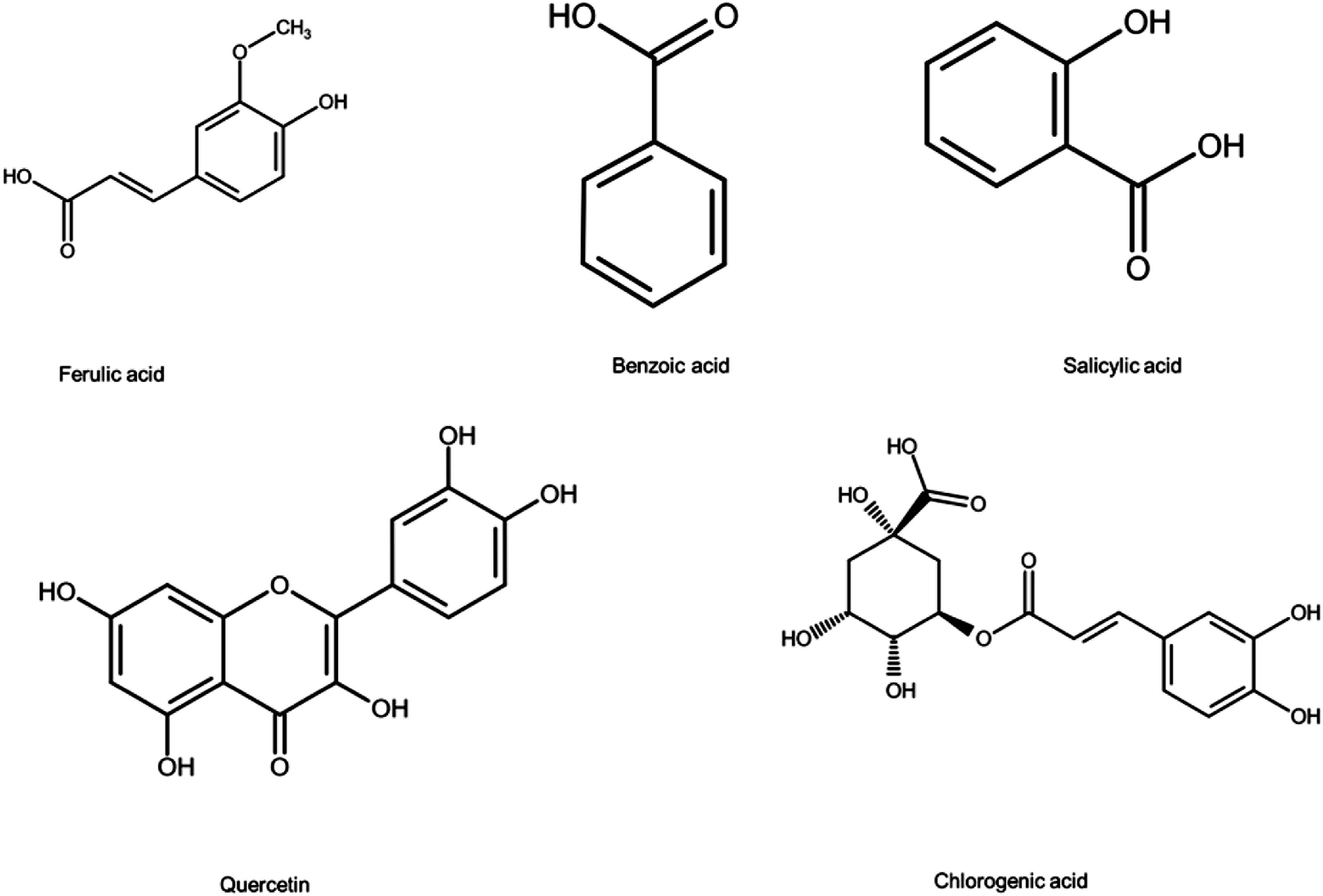
HPLC analysis revealed the presence of various phytoconstituents such as chlorogenic acid 188.26 (mg/kg), ferulic acid 1.5266 (mg/kg), salicylic acid 34.48 (mg/kg), and benzoic acid 2.177 (mg/kg). The flavonoid content found in the aqueous ethanolic extract of Thalictrum foetidum (AETF) was quercetin 47.47 (mg/kg). Phytoconstituents found in AETF are shown in Figure 1 and Table 2, and their chemical structures are shown in Figure 2. HPLC Chromatogram showing phytochemicals found in aqueous ethanolic extract of Thalictrum foetidum (AETF). Flavonoid and Phenolic Contents Found in Aqueous Ethanolic Extract of Thalictrum foetidum (AETF). Structures of various phyto-constituents detected in aqueous ethanolic extract of Thalictrum foetidum (AETF) by HPLC.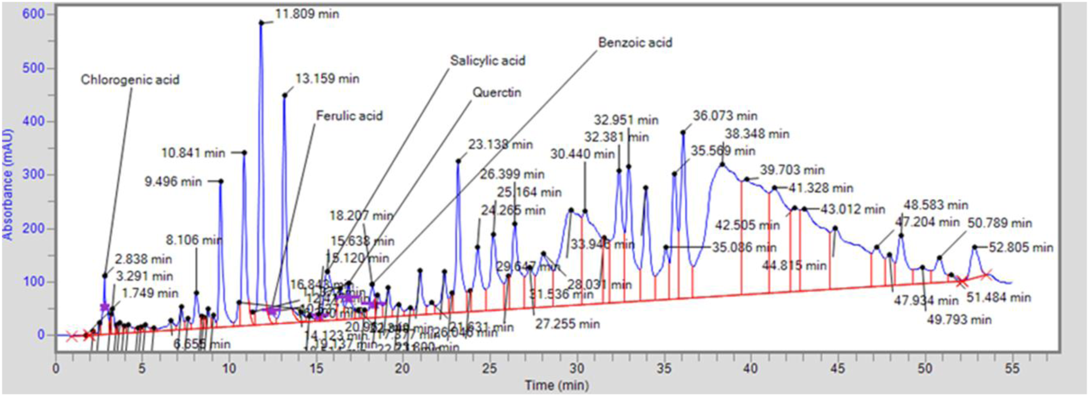

DPPH Radical Scavenging Activity
DPPH Radical Scavenging Potential.

AA, Ascorbic Acid; DPPH: 2, 2 - diphenyl picrylhydrazyl.
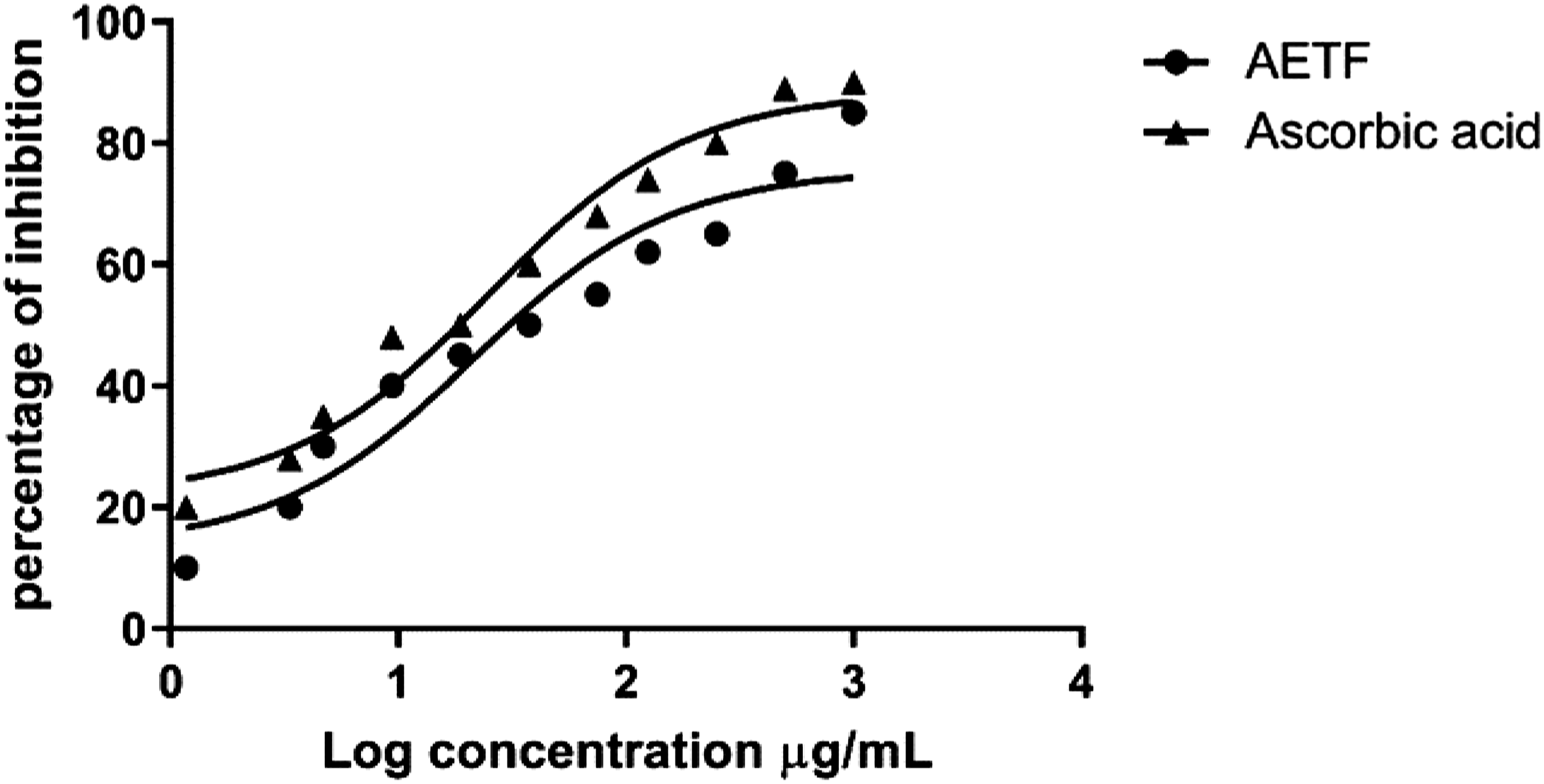
DPPH radical scavenging activity of aqueous ethanolic extract of Thalictrum foetidum (AETF).
Evaluation of Anti-Parkinson Activity
Behavioral Studies
Measurement of Catalepsy
The catalepsy test is used to evaluate muscle rigidity and immobility. All animals were assessed for cataleptic response on specific days (7th, 14th, and 21st) and at fixed intervals (30, 60, 90, and 120 min). Responses were recorded and analyzed after each trial. Higher cataleptic scores indicated that the animals remained in a fixed position for longer periods, potentially due to increased muscle rigidity. The disease control group showed a significant increase in cataleptic scores (P < .001) compared to the normal control group. AETF reduced muscle rigidity and immobility, significantly lowering cataleptic scores (P < .001). AETF effects were dose-dependent, with the most pronounced effects observed at the highest dose. Similar findings were noted with levodopa/carbidopa. Results are shown in Figure 4. The Effect of an aqueous ethanolic extract of Thalictrum foetidum on the cataleptic score. Data were presented as mean ± SEM (n = 6) and were analyzed using a two-way analysis of variance (ANOVA) followed by the Bonferroni multiple comparison test. Symbols were defined as follows: α indicates a significant difference from the normal control group; β indicates a significant difference from the disease group; δ indicates a significant difference from the AETF 200 mg/kg group. Differences were considered significant at P < .05.
Narrow Beam Walk Test
The narrow beam walk test is used to assess motor function, balance, and coordination in mice. The group treated with haloperidol showed significantly (P < .05) longer delay times and more foot mistakes than the normal control group. On the other hand, the group treated with levodopa and carbidopa demonstrated a noticeable improvement, with a shorter time to cross the beam compared to the normal control group. AETF treatment exhibited significantly (P < .05) less time to cross the beam with fewer foot slips. These effects were concentration-dependent, with the most pronounced effects observed at the highest concentration of AETF 600 mg/kg (Figure 5). The effect of an aqueous ethanolic extract of Thalictrum foetidum on the time latency of a narrow beam walk test. Data were presented as mean ± SEM (n = 6) and were analyzed using a one-way analysis of variance (ANOVA) followed by the Bonferroni multiple comparison test. Symbols were defined as follows: α indicates a significant difference from the normal control group; β indicates a significant difference from the disease group; δ indicates a significant difference from the AETF 200 mg/kg group. Differences were considered significant at P < .05.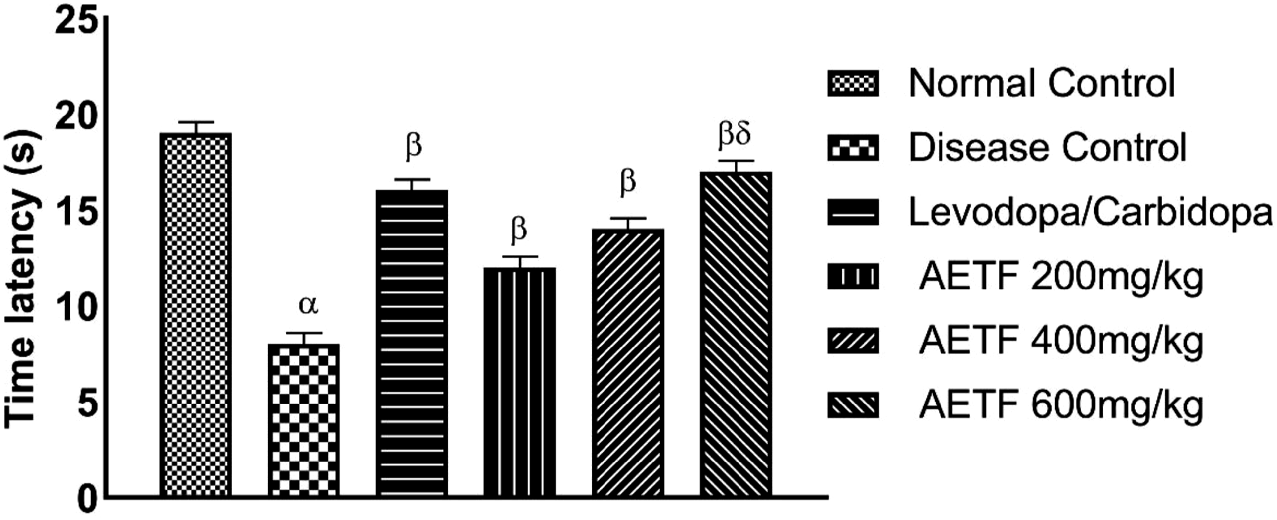
Open Field Test
The open-field test measures exploratory behavior in mice. This test evaluates parameters such as the number of lines crossed, the frequency of rearing, and grooming. Haloperidol significantly decreased (P < .001) the number of squares crossed, indicating reduced locomotor activities. In contrast, treatment with levodopa/carbidopa increased the number of squares crossed compared to the normal control group. AETF 600 mg/kg showed pronounced effects, with mice crossing more squares. This locomotor activity of mice was dose-dependent, with the most pronounced effects observed at this concentration of AETF. Exploratory behavior was assessed based on the number of central explorations. Mice in the disease group showed the lowest number of central explorations, while those treated with AETF at 600 mg/kg concentration showed the highest number Thus, AETF treatment, along with levodopa/carbidopa, reduced depressive behavior and improved exploratory behavior. Results are shown in Figure 6(A) and (B). Effect of aqueous ethanolic extract of Thalictrum foetidum on the Open Field Test. (A) No. of squares crossed (B) No. of central explorations. Data were presented as mean ± SEM (n = 6) and were analyzed using a one-way analysis of variance (ANOVA) followed by the Bonferroni multiple comparison test. Symbols were defined as follows: α indicates a significant difference from the normal control group; β indicates a significant difference from the disease group; δ indicates a significant difference from the AETF 200 mg/kg group. Differences were considered significant at P < .05.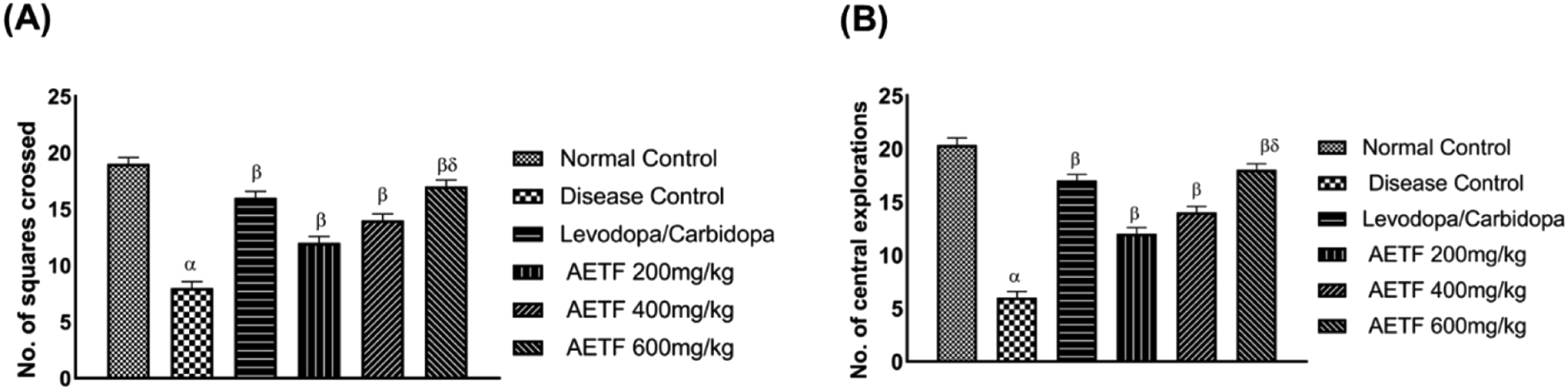
Wire Hanging Test
The wire-hanging test is used to assess the motor coordination of experimental animals. The animals were placed on an inverted cage top, and the time they stayed on the wire before falling off was recorded. In the disease control group, the wire-hanging time of the mice was significantly reduced (P < .001) compared to the normal control group. However, treatment with AETF significantly increased the wire hanging time of the mice in a dose-dependent manner compared to the disease control group. Similar results were also observed with levodopa/carbidopa treatment (Figure 7). Effect of aqueous ethanolic extract of Thalictrum foetidum on the wire hanging test. Data were presented as mean ± SEM (n = 6) and were analyzed using a one-way analysis of variance (ANOVA) followed by the Bonferroni multiple comparison test. Symbols were defined as follows: α indicates a significant difference from the normal control group; β indicates a significant difference from the disease group; δ indicates a significant difference from the AETF 200 mg/kg group. Differences were considered significant at P < .05.
Hole Board Test
Hole board tests are commonly used to assess an animal’s curiosity, anxiety, neophilia, and stress. This test typically measures head dips and edge sniffs, which indicate focused exploratory behavior, reduced anxiety and stress, and increased neophilia and curiosity. Additionally, vertical explorations, such as climbing and rearing, are evaluated. Our study found that disease induction negatively affected exploratory behaviors, as evidenced by a significant (P < .001) reduction in the number of head dips and edge sniffs, as well as significant (P < .001) decreases in climbing and rearing behavior. AETF at 200 mg/kg, 400 mg/kg, and 600 mg showed improvements in behavioral changes, including increased curiosity and exploration (neophilia) and reductions in anxiety and aggression. The most significant effects were observed with the highest dose of AETF (600 mg), resulting in increased head dips, head sniffs, climbing, and rearing behavior. The results are displayed in Figure 8(A) and (B). Effect of aqueous ethanolic extract of Thalictrum foetidum on the hole board test (A) focused and (B) horizontal exploratory behaviors. Data were presented as mean ± SEM (n = 6) and were analyzed using a one-way analysis of variance (ANOVA) followed by the Bonferroni multiple comparison test. Symbols were defined as follows: α indicates a significant difference from the normal control group; β indicates a significant difference from the disease group; δ indicates a significant difference from the AETF 200 mg/kg group. Differences were considered significant at P < .05.
Elevated Y-Maze Test
The test is typically conducted to evaluate how a disease and its treatment affect mice’s recognition ability and working memory. Compared to the normal control group, there was a significant decrease (P < .001) in behavior (recognition ability and working memory) after the mice were injected with haloperidol. This decrease was evident from the reduction in arm entries and the number of triads crossed by the mice during the elevated Y-maze test. AETF concentrations (200, 400, and 600 mg/kg) improved the behavior changes induced by haloperidol, with significant improvements observed in the number of arm entries and the number of triads crossed (P < .05, P < .01, and P < .001, respectively). Similar effects were observed in the proportion of spontaneous alternation behavior, with significant improvements at these doses. The most significant effects (P < .001) on spontaneous alteration behaviors were observed at AETF 600 mg/kg results are shown in Figure 9(A)–(C). Effect of aqueous ethanolic extract of Thalictrum foetidum on elevated Y-maze test (A) No. of arm entries (B) No. of triads crossed (C) spontaneous alterations (%). Data were presented as mean ± SEM (n = 6) and were analyzed using a one-way analysis of variance (ANOVA) followed by the bonferroni multiple comparison test. Symbols were defined as follows: α indicates a significant difference from the normal control group; β indicates a significant difference from the disease group; δ indicates a significant difference from the AETF 200 mg/kg group. Differences were considered significant at P < .05.
Estimation of Neurotransmitter Levels in the Brain
Measurement of Dopamine and Noradrenaline Levels
The study analyzed dopamine and noradrenaline levels in the brain tissues of control and experimental mice. Administration of haloperidol resulted in a substantial decrease (P < .001) in the levels of both neurotransmitters in the disease group. In contrast, the treatment groups showed significantly increased dopamine and norepinephrine levels. Additionally, treatment with Thalictrum foetidum demonstrated a dose-dependent ameliorative effect, with the most significant increase observed at the highest concentration (600 mg/kg). Results are shown in Figure 10(A) and (B). Effect of aqueous ethanolic extract of Thalictrum foetidum on (A) dopamine and (B) nor-adrenaline. Data were represented as ± SEM (n = 6) and compared by using a one-way analysis of variance (ANOVA) followed by the bonferroni multiple comparison test. α: showed a significant difference from the normal control group. β: showed a significant difference from the disease group. δ: showed a significant difference from the AETF 200 mg/kg. The difference is considered significant if P < .05.
Measurement of Acetyl Cholinesterase (AChE) Activity
The disease control group showed a significant increase in AChE levels compared to the normal control group. Treatment with AETF resulted in a significant, dose-dependent reduction in AChE levels, with the most prominent effect observed at the 600 mg/kg concentration of AETF. The results are shown in Figure 11. Effect of aqueous ethanolic extract of Thalictrum foetidum on acetylcholinesterase (AchE) level. Data were represented as ± SEM (n = 6) and compared by using a one-way analysis of variance (ANOVA) followed by the bonferroni multiple comparison test. α: showed a significant difference from the normal control group. β: showed a significant difference from the disease group. δ: showed a significant difference from the AETF 200 mg/kg. The difference is considered significant if P < .05.
Role of Aqueous Ethanolic Extract of Thalictrum foetidum on Oxidative Stress Markers (SOD, CAT, GSH and MDA)
Based on previous research, it is well-established that the degradation of dopaminergic neurons is a significant factor in the disease. Reactive oxygen species (ROS) are produced in dopaminergic neurons through various pathways, including the action of dopamine itself. Several defective gene products, such as α-synuclein, parkin, and LRRK2, contribute to complex pathological processes, worsening oxidative stress and ROS levels. 42 ROS regulates critical pathways that modulate cellular responses to growth hormones and cytokines, including the mitogen-activated protein kinase (MAPK) and phosphoinositide 3-kinase (PI3K) pathways. For instance, several enzymes in the MAPK and PI3K pathways, which are crucial for mediating cellular responses to growth hormones and cytokines, are directly regulated by ROS. 43 Oxidative stress and ROS generation are countered by key enzymatic antioxidants, including superoxide dismutase (SOD), catalase (CAT), and glutathione (GSH).
In this study, we assessed the antioxidant markers in brain tissues. The results showed a significant (P < .001) reduction in the level SOD, CAT, and GSH in the disease group compared to controls. Treatment with AETF significantly elevated their levels in a dose-dependent manner. The lowest concentration of AETF (200 mg/kg) had a non-significant effect, while the highest concentration (600 mg/kg) produced the most significant impact. Additionally, we measured malondialdehyde (MDA), a marker of ROS generation and lipid peroxidation. MDA is a polyunsaturated fatty acid peroxidation byproduct and indicates ROS levels and neurodegeneration. Rodents in the disease group have significantly (P < .001) higher concentrations of MDA. Treatment with AETF at concentrations (400 and 600 mg/kg) significantly decreased MDA levels, with dose-dependent effects. All the results are shown in Figure 12(A)–(D). Effect of aqueous ethanolic extract of Thalictrum foetidum on (A) SOD (B) CAT (C) GSH and (D) MDA levels. Data were represented as ± SEM (n = 6) and compared by using a one-way analysis of variance (ANOVA) followed by the bonferroni multiple comparison test. α: showed a significant difference from the normal control group. β: showed a significant difference from the disease group. δ: showed a significant difference from the AETF 200 mg/kg. The difference is considered significant if P < .05. SOD: superoxide dismutase; CAT: catalase; GSH: glutathione; MDA: malondialdehyde.
Role of NF-κB Initiated Inflammatory Cytokine Pathway in Neurodegeneration Associated With Parkinsons Disease
NF-κB is primarily expressed in neuroglial cells and triggers an inflammatory signaling cascade involving microglial cells, which may contribute to the degeneration of dopaminergic neurons. TNF-α and IL-6 are key cytokines generated during this inflammatory cascade and are characteristic of the neurodegeneration associated with PD. The study results indicated that TNF-α and IL-6 levels were increased in the disease group compared to the normal control group (P < .001). A significant dose-dependent effect was observed in the AETF treatment group relative to the disease control group; administration of AETF at a dose of 600 mg/kg resulted in a marked reduction in TNF-α and IL-6 levels (Figure 13(A) and (B)). Effect of aqueous ethanolic extract of Thalictrum foetidum on NF-κB associated cytokine (A) TNF-α and (B) IL-6. Data were represented as ± SEM (n = 6) and compared by using a one-way analysis of variance (ANOVA) followed by the bonferroni multiple comparison test. α: showed a significant difference from the normal control group. β: showed a significant difference from the disease group. δ: showed a significant difference from the AETF 200 mg/kg. The difference is considered significant if P < .05.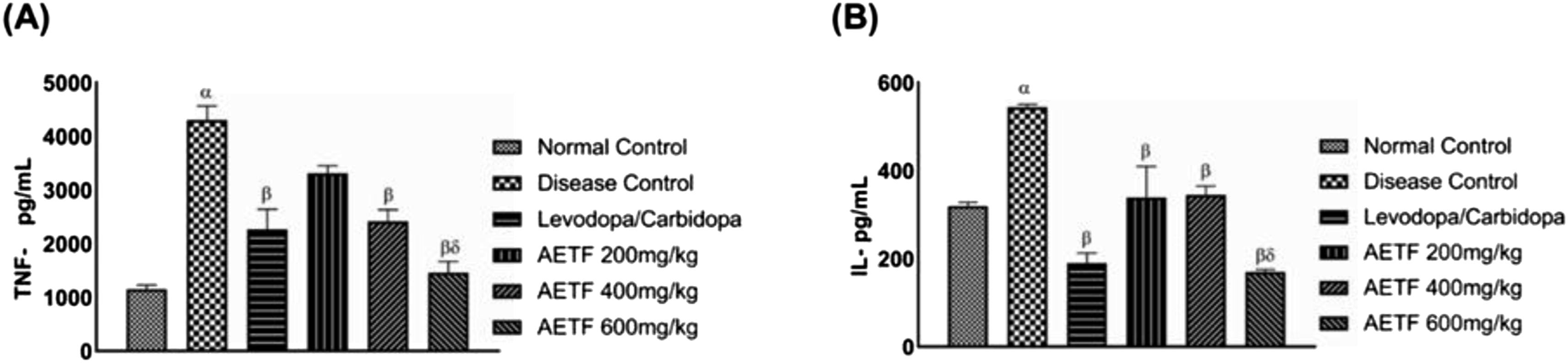
Histopathological Evaluation of Brain Tissue for the Presence of Protein Aggregation
Histopathological examination of brain tissue from the disease control group revealed the presence of neurofibrillary tangles (NFT), Lewy bodies (LB), and vacuolated cytoplasm. In contrast, the normal control group showed no vacuolated cytoplasm, NFT, or Lewy bodies. Mice treated with AETF (200 mg/kg) showed neurofibrillary tangles and Lewy bodies, but in fewer numbers than observed in the disease control group. Mice receiving AETF (400 mg/kg) had only a few Lewy bodies and no neurofibrillary tangles. The histopathological analysis of mice treated with AETF at higher concentrations demonstrated normal histology with no evidence of protein aggregates (Figure 14(A)–(E)). Effect of aqueous ethanolic extract of Thalictrum foetidum on histopathology of mice brain tissues: (A) brain section from normal control group displaying normal histology of brain tissues (B) brain tissues from PD mice showing numerous Lewy bodies, vacuolization, and neurofibrillary tangles. (C) PD mice treated with levodopa/carbidopa containing reduced no. of lewy bodies (D) (D) histopathology of brain tissues treated with AETF 200 mg/kg showed fewer lewy bodies and fewer abnormal protein aggregates. (E) Brain tissues from PD mice treated with AETF 400 mg/kg showed only 1 Lewy body and no neurofibrillary tangles. (F) AETF 600 mg/kg was devoid of visible protein aggregates. INF: infiltration, LB: lewy bodies: V: vacuolization, NFT: neurofibrillary tangles, P: plaques. All the pictures were taken at 40×.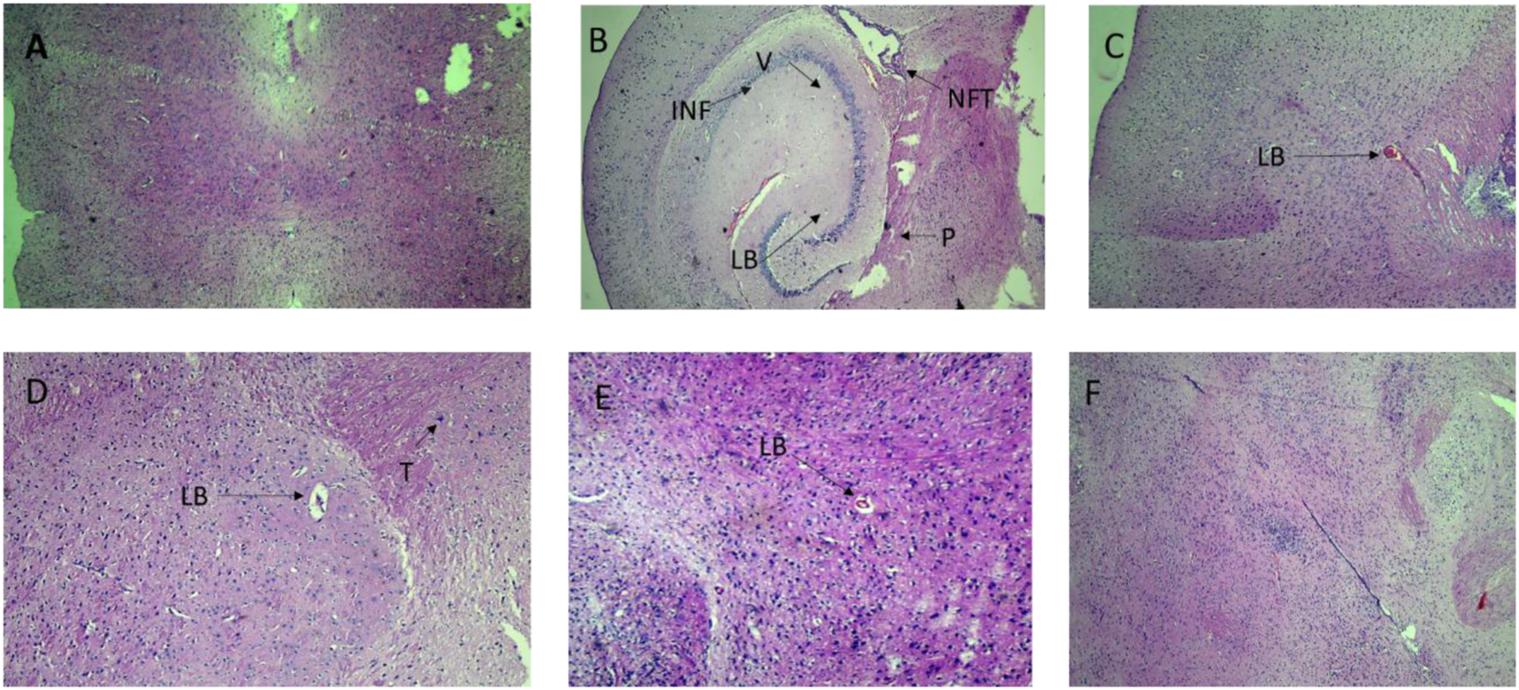
Discussion
Parkinson’s disease (PD) is a complex neurodegenerative disorder that generally progresses with age and is characterized by the degeneration of dopaminergic neurons in the substantia nigra and the corpus striatum. PD is one of the most common and disabling neurodegenerative disorders affecting the elderly. Its pathology is multifaceted, with various etiological and pathological abnormalities contributing to its onset. Genetic abnormalities, such as mutations involving α-synuclein and parkin, play a significant role in its pathogenesis by developing aberrant protein aggregates in the brain. Additionally, exposure to neurotoxins like rotenone, which is used as an herbicide, and MPTP further worsens dopaminergic neuronal degradation. Neurodegeneration and functional impairments associated with PD are influenced by oxidative stress, ROS production, and neuroinflammation, with prevalence increasing with age. Current treatments for PD primarily offer symptomatic relief rather than slowing disease progression. However, none of these treatments are ideal, and each comes with potential toxicity. Therefore, developing treatment modalities that could halt or reverse the neurodegeneration of dopaminergic neurons would be a significant advancement.
Recent research has proven that neuroprotection and inhibition of oxidative processes in neurons may play a crucial role in mitigating neurodegeneration. Natural compounds derived from organic sources have gained attention due to the presence of phenols and flavonoids, which act as natural antioxidants. 13 These compounds have the potential to counter oxidative stress, reduce neuroinflammation, and improve motor and cognitive functions.44,45 Thalictrum foetidum is a famous medicinal plant rich in flavonoids and phenols and has a traditional potential to be used for neurodegenerative disorders. HPLC analysis revealed the presence of phytochemicals such as chlorogenic acid, ferulic acid, salicylic acid, benzoic acid, and quercetin. The DPPH radical scavenging assay demonstrated that the aqueous ethanolic extract of Thalictrum foetidum (AETF) exhibited strong radical scavenging activity, with an IC50 value of 21.50 μg/mL, compared to ascorbic acid, which had an IC50 value of 25.05 μg/mL (Figure 3). This antioxidant activity is attributed to the phytoconstituents in AETF, which possess inherent capabilities to scavenge free radicals produced by oxidative stress in neuronal tissue. These findings aligned with previously published data. 44
In vivo antiparkinsonian activity is assessed using animal models that exhibit Parkinson-like symptoms. Various chemicals such as haloperidol, MPTP, and paraquat are used to develop these symptoms. 28 In this study, haloperidol (1 mg/kg) was administered for 21 days to induce Parkinson-like symptoms in mice, including catalepsy, motor function abnormalities, hypokinesis, and postural instability. Behavioral analysis was carried out through various tests such as cataleptic score measurement, narrow beam walk test, wire hanging test, hole board, and elevated Y-maze test. The cataleptic test evaluates motor coordination and the ability of rodents to correct their posture when subjected to an externally imposed awkward position. Time is taken to correct the posture, serve as a measure of catalepsy. Mice treated with haloperidol showed significantly higher cataleptic scores (P < .001) compared to the normal control group, indicating increased muscular rigidity and motor incoordination. AETF significantly reduced cataleptic scores in a dose-dependent manner, with the greatest reduction observed at the highest concentration (AETF 600 mg/kg). These results suggested that AETF ameliorates haloperidol-induced behavioral abnormalities in rodents (Figure 4). Our findings are consistent with previously published data. 21
The narrow beam walk test assesses an animal’s balance and lower limb performance. 46 Rodents in the haloperidol group showed foot slips and delays in crossing the beam, likely due to impaired balance and motor coordination due to haloperidol-induced damage. AETF was effective at higher concentrations (AETF 400 mg/kg and 600 mg/kg), significantly reducing the time latency to cross the beam (Figure 5). The open field test measures anxiety levels, locomotor activity, and exploration ability in experimental animals. The disease-induced group showed a significant decrease in locomotor activity and exploration (P < .001). 46 In contrast, AETF treatment improved exploration and reduced anxiety in a dose-dependent manner (Figure 6). Similar findings were reported by Uzma Saleem and colleagues, who demonstrated that a curcuminoid formulation increased exploration and reduced anxiety in the open-field test. 47
In the wire-hanging test, AETF-treated mice spent more time on the wire than the normal control group, indicating improved motor function and endurance (Figure 7). The hole board test was also conducted to evaluate the animals’ curiosity, anxiety, neophilia, and stress. Our results revealed that AETF significantly increased head dips, edge sniffs, climbing, and rearing behaviors in a dose-dependent manner, indicating reduced anxiety and increased curiosity (Figure 8(A) and (B)). Mice recognition ability was assessed using the elevated Y-maze test, which measures the number of arm entries and triads crossed. Treatment with AETF increased the number of arm entries and triads crossed in a dose-dependent manner (P < .05, P < .01, and P < .001). Similar improvements were observed in spontaneous alteration behavior (Figure 9). These findings are consistent with previously published data. 48
Symptoms of Parkinson’s disease (PD) typically manifest when dopamine levels fall to approximately 40% of normal. We assessed dopamine levels in brain tissues and found a significant decrease in the disease group (P < .001) (Figure 10). Treatment with AETF restored dopamine levels in a dose-dependent manner. Similarly, reduced noradrenaline levels were also restored with AETF treatment (Figure 10). Increased acetylcholinesterase (AChE) activity is also associated with PD, and cholinergic antagonists are often used as adjunct therapies. We observed elevated AChE levels in the disease-induced group, but AETF treatment significantly reduced AChE activity dose-dependently (Figure 11). Histopathological examination of brain tissues from the haloperidol group revealed the presence of Lewy bodies (LB), vacuolation (V), neurofibrillary tangles (NFT), and plaques (P), which are characteristic of PD. Treatment with AETF reduced these abnormal protein aggregates, demonstrating neuroprotective effects and attenuation of neuronal degradation (Figure 13).
The production of reactive oxygen species (ROS) primarily occurs in brain tissues, which consume about 20% of the body’s oxygen supply. This high oxygen demand contributes to the generation of ROS. Neurons and glial cells are key sources of ROS, with their production beginning from several pathways, including the electron transport chain, monoamine oxidase (MAO), nitric oxide (NO), and NADPH oxidase. ROS production plays a significant role in the degradation of dopaminergic neurons. 42 In addition to ROS generated from dopamine metabolism, low levels of glutathione (GSH) and other antioxidants, such as catalase (CAT) and superoxide dismutase (SOD), contribute to oxidative stress. High levels of calcium and iron in the substantia nigra pars compacta further worsen this process. Moreover, lipid peroxidation induced by elevated levels of polyunsaturated fatty acids leads to additional neurotoxic effects. These combined activities result in neuroinflammation and neurotoxicity. 43 Therefore, it can be conferred that inhibition of oxidative stress, neuroinflammation, and enhancement of neuronal protection might be an effective way to halt the disease progression and to ameliorate the pathogenicity and symptomatology associated with PD. Similar findings have been reported in previous research, which indicates that medicinal plants can mitigate oxidative stress in metabolic disorders.44,49
AETF is rich in polyphenols such as chlorogenic, salicylic, benzoic, ferulic, and flavonoids like quercetin. Recent studies have demonstrated that ferulic acid (FA) reduced neuronal inflammation and improved behavioral impairments in the 1-methyl-4-phenyl-1,2,3,6-tetrahydropyridine (MPTP) Parkinson mice model. FA is widely recognized as a potent antioxidant that protects against oxidative damage by lowering ROS levels and enhancing the activity of cellular antioxidant enzymes. 50 Our study findings also aligned with previous research, showing significant improvements in superoxide dismutase (SOD), catalase (CAT), and glutathione (GSH) levels, along with reduced malondialdehyde (MDA) levels, a marker of lipid peroxidation (Figure 11). The antioxidant and anti-inflammatory properties of FA are likely responsible for its protective effects. 50
Salicylic acid is another phytochemical found in AETF. It is a well-known antioxidant with strong free radical scavenging properties. It has been shown to provide neuroprotection against MPTP-induced neurotoxicity. Salicylic acid’s ability to scavenge free radicals contributes significantly to its neuroprotective effects. Furthermore, salicylic acid has been shown to inhibit MPTP’s neurotoxic effects on the brain’s enzymatic defense system, which includes superoxide dismutase, glutathione peroxidase, and catalase. 51 Additionally, salicylic acid treatment has been reported to enhance phenylalanine ammonia-lyase (PAL) activity, hydrophilic total antioxidant activity (H-TAA), and phenolic content in apricot fruit by regulating H2O2 metabolism during postharvest storage. 52
Another phytoconstituent found in AETF was Benzoic acid, which improved behavioral and biochemical changes and mitigated oxidative stress in a ROS-induced PD model. Benzoic acid also restored liver, kidney, and blood markers, suggesting reduced pathological changes. It has various applications in neurological disorders such as multiple sclerosis. 53 Chlorogenic acid (CGA), a polyphenol in various medicinal plants, has been reported to possess potential anti-PD effects. It has been shown to delay the development of Parkinson’s disease in Caenorhabditis elegans through autophagy induction. 54 Quercetin, a flavonoid, has strong antioxidant capabilities. It can potentially reduce rotenone-induced neurotoxicity, alleviate endoplasmic reticulum (ER) stress-induced apoptosis, and mitigate oxidative stress. In PD models, quercetin can induce autophagy and modulate the neuronal microenvironment, which may help prevent neuronal death. Therefore, quercetin might serve as a chemoprotective agent against the development of PD. 55 The DPPH test measures the ability of the stable free radical 2,2-diphenyl-1-picrylhydrazyl to react with hydrogen donors. All samples showed increased radical-scavenging activities with higher concentrations. 56
Tumor necrosis factor alpha (TNF-α) is a key proinflammatory cytokine involved in neuroinflammation by increasing the expression of the BACE1 gene (bata site amyloid precursor protein cleaving enzyme 1). BACE1 contributes to the accumulation of amyloid-beta and tau hyperphosphorylation, both of which are pathological hallmarks of Parkinson’s disease. Research has shown that anti-inflammatory agents targeting TNF-α can offer therapeutic benefits for cognitive impairments associated with Parkinson’s disease. 57 Neurodegenerative disorders are associated with elevated levels of proinflammatory cytokines such as interleukins IL-1α, IL-1β and IL-6, which are found near amyloid plaques and contribute to neuroinflammation 58 Our study found that TNF-α and IL-6 levels were significantly increased in the disease group compared to the normal control group (P < .001). Treatment with AETF decreased these levels dose-dependently (Figure 12).
A limitation of our study was the limitation of resources, which prevented us from extending our research to the molecular level. We planned to expand the project by incorporating additional quantitative PCR (qPCR) data and evaluating the expression of genes related to the disease. Additionally, we intend to collect more data on dopamine neuron degradation and assess the impact of treatments on halting disease progression.
Conclusion
In conclusion, the aqueous ethanolic extract of Thalictrum foetidum (AETF) has improved the potential to alleviate Parkinson’s disease symptoms in animal models. AETF appears to protect against the degradation of dopaminergic neurons. Its neuroprotective effects are likely due to its ability to mitigate oxidative stress in dopaminergic neurons and reduce the expression of inflammatory cytokines such as TNF-α and IL-6, which are activated through the NF-κB inflammatory pathway. These findings warrant further investigation using a detailed molecular approach to confirm and elucidate the underlying mechanisms
Footnotes
Acknowledgments
The authors are grateful to Dr Hafiz Muhammad Qasim, Assistant Professor, Department of Applied Linguistics, Government College University Faisalabad, for providing English language editing services at no cost.
Author Contributions
Concept and design, J.H, X.U, MF, L.H.; Data Acquisition, analysis, and interpretation, J.H, X.U, Z.C, M.F, A.J.; intellectual content development, H.M.A.R, R.S, M.A, S.R; final version approval and integrity, J.H, X.U, M.F, L.H, H.M.A.R, R.S, S.R.
Declaration of Conflicting Interests
The author(s) declared no potential conflicts of interest with respect to the research, authorship, and/or publication of this article.
Funding
The author(s) received no financial support for the research, authorship, and/or publication of this article.