
Abstract
The patient was contaminated with multiple radionuclides 38 years ago due to an accident. To investigate the effects of radionuclide contamination on humans, he has been followed up by examinations for many years. Long-term effects gradually emerge in these years. Lung cancer was diagnosed by medical examinations. Besides, chronic gastritis with intestinal metaplasia was indicated by gastroscopic biopsies, while colorectal polyps found by colonoscopy. All 13 colorectal polyps were removed, and radical surgery for lung cancer was performed. Fortunately, pathological examinations indicated that it was early lung cancer. The ground glass nodule (GGN) in left lung identified during the follow-up will be resected when needed. It is speculated that multiple manifestations of the patient may be related to radiation, and different lesions in the organs may be related to systemic adaptive response. However, longer follow-up is needed due to a lack of effective and direct evidence. This work is expected to provide experiences for similar patients’ treatment and follow-up.
Introduction
For more than a century, a large number of experimental and epidemiological studies have been carried out on the biological effects of ionizing radiation on humans. 1,2 In this case, the patient had accidental burns combined with multiple radionuclides contamination 38 years ago. He has been followed up since the accident. 3,4
Once radiation accidents occur, different scenarios may appear. Patients exposed to radiation will suffer damages of organs and psychological distress at different levels. 5
Besides the clinical treatments, follow-up plays important roles in the study of the long-term radiobiological effects. A lot of accidental patients were followed up for a long time. For example, the mother and fetus exposed in the radiation accident in Xinzhou of China have been followed up for 16 years. 6 Clean-up workers of the Chernobyl accident have been followed up for more than 30 years. 7 Various studies of radiobiological effects on exposed persons were conducted in the Fukushima Daiichi nuclear accident. 8,9
It is widely recognized that the radionuclide contamination causes damage effects on the victim’s tissues and organs in a long time. So, it is necessary and valuable to implement medical follow-up for the patient. The valuable information acquired in this work is important for the future study of the radiation-induced, long-term biological effects.
Materials and Methods
General Information
The patient “Z” was born in 1945. He had been diagnosed with hypertension since 15 years ago but was not affected by diabetes, hepatitis, heart disease, pneumonia, tuberculosis, asthma or HIV, and so on. Appendix was removed because of appendicitis in 1976. He does not smoke and has moderate drinking. His father died from gastric carcinoma at the age of 83, and his mother died a natural death at the age of 93. He was a mechanic maintenance worker in a radioactive plant from May 1972 to February 1979. He often worked in radiological hot zone before the accident. After the accident, he was transferred to administration post and retired in 2005. He had a healthy son before the accident. One month before the accident, he inhaled PuO2 (plutonium dioxide) during his work, and plutonium value in urine was 0.037 Bq/24 h. CaNa3-Diethylenetriaminepentaacetic acid (CaNa3-DTPA) was used 3 times to remove radionuclides at that time.
Accident
On February 22, 1972, when the worker “Z” was cutting pipes containing strong radioactive residues using electric welding gun
Early Medical Treatment Post Accident
One hour after the accident, 2% new bromogeramine was used to clean the wound, then the 4% EDTA decontamination solution was applied to wash the wound, and the exposed area was decontaminated. The wound of the extremities was bandaged with aseptic dressing. Two hours after the accident, the patient was intramuscularly injected with 4 mL 10% CaNa3-DTPA and 50 mg dolatin. The patient’s hair was burned and curled and at the same time a palm size of skin in the left head, face, and neck became brown. Except the nasal skin with a diameter of 1.5 cm, all the skin of face, eyelids, lips, ears, and jaw got brown. There were 2 flushes and some blisters in the upper neck and upper anterior chest with a size of palm. The skin of left wrist dorsal became brown with a size of 3 × 4.5 cm. Lumpy eschar was scattered in about one-third area of the left calf, while the peripheral skin was damaged. The burned area accounted for about 15% of the total body surface area, which was assessed as first to second degree burns. The patient was hospitalized in the Affiliated Hospital of China Radiation Protection Institute. Measures such as decontamination of the body skin and wound as well as radionuclide absorption blocking and removal were taken. Comprehensive treatment was implemented to control infection and protect important organs. After a long time, the wound healed and the patient recovered well. There were multiple scars, pigmentation, and depigmentation around the skin of lips, head, neck, chest, left forearm, and right lower limbs. He was diagnosed as chronic radiation skin injury.
Dose Estimation Post Accident
Surface contamination detector was used to estimate contamination of the patient during hospitalization in another hospital. The injured scab and hair contaminated by radionuclides were collected for radiochemical analysis and physical measurement. The results showed that the samples contained 144Ce-144Pr, 106Ru-106Rh, 137Cs, 95Zr-95Nb, 90Sr-90Y, 239Pu, and 238U. The wound was the main way by which radionuclides entered the body. After successful decontamination, sodium iodide NaI(TI) whole-body counters and physical analysis of urine and feces were used to estimate the dose, the total committed effective dose of 7 kinds of radionuclides was estimated to be 0.7 Sv. The committed equivalent dose of bone surface was 14 Sv, and the equivalent dose of liver was 3 Sv. The absorption dose of the skin from β-ray was estimated to be 10.44 Gy by the chemoradiation analysis results of the injured scab. The absorption dose of skin was estimated to be 9.36 Gy by the surface contamination survey meter. The results from 2 methods were nearly the same.
Medical Examination
Medical follow-up includes: observation of clinical manifestations, peripheral blood cell analysis, examination of liver and kidney functions, analysis of blood glucose, blood lipid and immune function, analysis of lymphocyte chromosome aberration, examination of thyroid function, imaging examination, tumor marker examination, and gastrointestinal endoscopy. Cattell’s 16 personality factor test, self-rating anxiety scale, and self-rating depression scale were used to assess psychological status of the patient.
Results
Clinical Manifestation
The patient had good sleep and appetite; however, he had a dry mouth sometimes. Physical examination showed that there were multiple scars, pigmentation, and depigmentation around the skin of lips, head, neck, chest, left forearm, and right lower extremities. The patient’s skin of head, neck, chest, and back showed scattered verrucous eminences in dark brown without pain and itch.
Laboratory Examination
We have reviewed the abnormal changes in our hospital between 2000 and 2009 (Table 1, the data for 2005 were absent since he didn’t come for physical examination due to his private reason). An abnormal echo in the right lobe of the thyroid gland was found in 2003. Total thyroid ectomy was carried out in 2004. Postoperative pathological analysis showed nodular goiter with focal lymphocytic follicle formation. The patient had been taking thyroxine all the time since the surgery. Thyroid function test indicated a little higher level of thyroid-stimulating hormone.
Abnormal Changes in Follow-Up in Our Hospital Between 2000 and 2009.a

a “+” indicated the results were abnormal, ”−” indicated the results were within normal range. “NA” indicated the examination was not conducted.
In 2017, the numbers of white blood cells, hemoglobin, and platelets detected by blood cell analysis were normal. Chromosome aberrations were analyzed in 338 lymphocytes from peripheral blood and 2 acentric fragments were found. There was no dicentric chromosome, translocation, or inversion. Functions of liver and kidney as well as fasting blood glucose level were normal. The level of immunoglobulin A was a bit higher than normal value (the measurement value was 5.41 g/L while the normal range was 0.700∼4.000 g/L). The levels of IgG and IgM were normal. B lymphocyte ratio was a bit lower than normal value, with the measurement value of 4.8% while the normal range was 5% to 13%.
In August 2017, gastroscopy showed that the mucosa of the gastric antrum was spotted in red and white, mainly white, and the surface was rough. The scattered eminence lesions were found with local hyperemia and edema. A biopsy of the gastric antrum was performed, and the pathological analysis showed that superficial mucous chronic inflammation with intestinal metaplasia in local glandular (Figure 1).
H&E staining of biopsy sections from gastric antrum. Microscopic observations under an Olympus BX41 with 40× (A) or 100× magnifications (B). H&E indicates haemotoxylin and eosin.
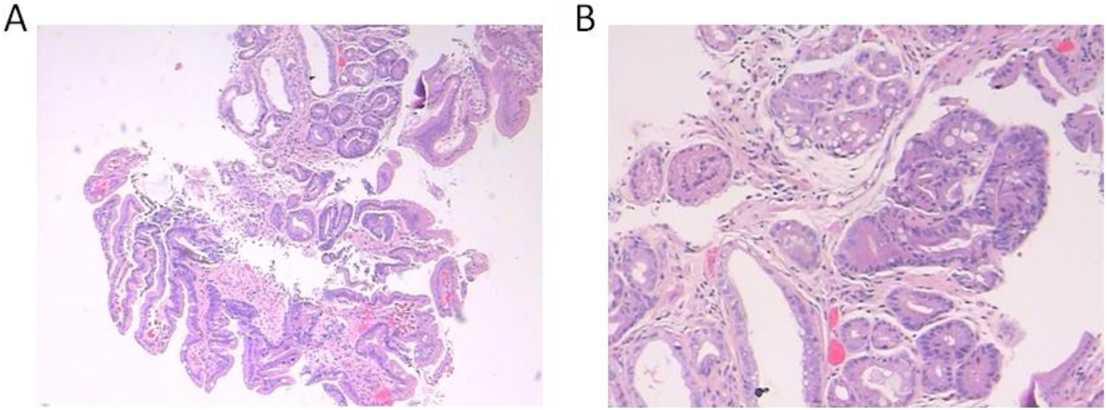
The colonoscopy showed that 13 polyps of different sizes were found in the colorectum. The smaller polyps were removed by biopsy forceps, and the larger ones were clamped with a clip. They were diagnosed as colorectal mucosa polyps through the pathological analysis (Figure 2).

H&E staining of biopsy sections from colorectal polyps. Microscopic observations under an Olympus BX41 with ×40 (A) or ×100 magnifications (B). H&E indicates haemotoxylin and eosin.
Bone density examination revealed osteoporosis. Twenty-eight years after the accident, compression fractures happened in the patient’s third lumbar vertebra accompanied with osteoporosis. Carcinoembryonic antigen (CEA) is a glycoprotein acting as an antigen to cause an immune response in patients. It is a broad-spectrum tumor marker that reflects the presence of multiple tumors. The level of CEA was higher (8.57 ng/mL) in 2017, while 5.50 ng/mL in 2015 and 6.80 ng/mL in 2016.
Computed tomography (CT) scan showed a tubercle in superior lobe of right lung and local pleura was dragged, conforming to the characteristics of lung cancer. Computed tomography scan also showed an irregular GGN in the upper lobe of the left lung, with a size of 20 mm × 24 mm, which was suspected to be early-stage cancer (Figure 3).

Chest CT scanning result. CT scanning indicated a tubercle in superior lobe of right lung with local pleura invasion (A) and GGN in the upper lobe of the left lung (B). CT indicates computed tomography; GGN, ground glass nodule; H&E, haemotoxylin and eosin.
Psychological Assessment
The patient was in good mental health, without anxiety and depression. The 16PF scale showed that the patient was characterized with good intelligence, emotional stability, humility, obedience, responsibility, and so on. But sometimes he showed excessive self-criticism.
Medical Treatment
On August 19, 2017, radical resection of right upper pulmonary carcinoma by video-assisted thoracic surgery was conducted. Anti-infection and hemostasis were carried out after the operation, and the patient recovered well. Histopathologic analysis revealed infiltrating adenocarcinoma with moderate differentiation in right upper lung, which has invaded the pleura without neural and vascular invasion (Figure 4).

H&E staining of the lung adenocarcinoma sections. Microscopic observation under an Olympus BX41 with ×40 (A) or ×100 magnification (B). H&E indicates haemotoxylin and eosin.
Further high-throughput gene sequencing and the single-nucleotide polymorphism (SNP) analysis showed that epidermal growth factor receptor (EGFR) mutation (p.L858R) in exon21 was positive with a frequency of 34.62%. Results of genetic risk-related gene mutations indicated that no pathogenic mutation was found (Table 2). Follow-up examination is kept being carried out for the upper lobe of the left lung, and necessary operation will be conducted in due time.
Results of Genetic Risk Related Gene Mutations.a

a “Benign” indicated this type of mutation didn’t increase the risk of cancer, ”Uncertain” indicated the relationship between this type of mutation and the risk of cancer is not clear. “Risk factor” indicated this type of mutation may increase the risk of cancer. “Pathogenic mutation” means it will increase the incidence of the cancer.
Discussion
Internal contamination of radionuclides as internal sources to produce sustained radiation to the human body and then the internal radiation occurs. Internal exposure causes changes first, then damages, and finally harms to organism, which are called internal radiation effects. Radionuclides selectively enter different tissues and organs by inhaling, feeding, skin, or wound. As many as 7 kinds of radionuclides entered the body of the patient. The distribution, deposition, and excretion of these nuclides are different from each other depending on different energy, toxicity, and half-life period, so their toxicological effects also vary. Multiple radionuclides may have complex effects. Once the long half-life radionuclides such as 137Cs, 90Sr, 239Pu, and 238U enter the human body, the damage effects may appear gradually as time goes on. However, the lesions found in this medical follow-up could not be attributed to the effects of radiation damage.
239Plutonium, 90strontium, and 95zirconium are all bony nuclides. Among them, 239 plutonium is the most toxic. Due to the lack of clinical and pathological features of malignant tumors induced by ionizing radiation, it is difficult to identify whether the malignant tumor is caused by radiation based on clinical analysis result of the individual case. We can only judge whether the tumor has a certain relation with the irradiation through epidemiology analysis. There are many studies on the carcinogenic effect of plutonium all over the world, mainly in the observation of the dose and the long-term carcinogenic effect by radiation in the nuclear industry. 10 The risk of lung cancer from occupational plutonium exposure was analyzed in Mayak and Sellafield staff. The 2 cohorts are the most informative in the world with plutonium urine monitoring programs. There was a linear relationship between cumulative internal plutonium lung dose and lung cancer mortality and incidence in the cohort. 11
A case–control study at the Rocky Flats Plant in Colorado from 1951 to 1989 showed that workers whose cumulative internal lung doses of more than 400 mSv had bigger lung cancer risk. 12 However, it has been reported that low-level radiation dose may help prevent cancer mortality among nuclear workers. 13 The patient overexposed in this accident has been unevenly exposed to bone, liver, and skin with relatively very high doses. However, this patient only has lung cancer but without cancinogenesis in other organs. It might be due to adaptive response induced by his previous small dose exposures. Although the overall death rate from cancer in workers monitored for plutonium exposure was also significantly low relative to national rates (SMR = 89, 581 deaths; 2P = .005). 14 A single-center retrospective study in Sichuan, China, showed significant increases in the number of patients with lung adnenocarcinoma from 1995 to 2015. 15 In summary, it is hard to say the patient’s incidence of lung cancer was related to the radionuclide contamination.
Radiation effects are influenced by environmental, dietary, and other carcinogens stresses. So, it is hard to say lung cancer of the patient was induced by radiation. Although most sporadic cases with lung cancer are related to environmental factors, genetic susceptibility may also play an important role, and a number of lung cancer associated with SNPs have been identified although many remain to be found. 16 Both genetic background and environmental factors play a role in the carcinogenesis among which ionizing radiation is an important factor. The mechanism underlying radiation-induced carcinogenesis is not yet clear. Genomic instability is thought to play a major role in cancer development after irradiation. 17 Although gene mutation is supposed to play an important role in the carcinogenesis, the number of germline mutations related with familial lung cancer is small. Through whole-genome sequencing of lung cancer specimens, more mutations that may associate to lung cancer are likely to be found. The mutations in genes other than EGFR may be of greater significance. 18 For example, the BRCA2 mutation may affect nearly 0.1 to 2% of the population. 19 The patient in our study has EGFR mutation, but there were no other pathogenic mutations.
The current standard of China 20 clearly claimed that the construction of the dose–effect relationship of occupational radiological tumors must be based on the dose, the sex of the patient, the age of both exposure, and the onset of the disease. The diseases appropriate to radioactivity etiological diagnosis include external irradiation induced lung cancer. But due to the lack of the external radiation dose data, it is impossible to analyze the etiological probability of radioactive tumor. Referring to the international approach, the case involved 7 kinds of radionuclides, and the dose of each specific nuclide is difficult to be estimated precisely, so the probability analysis of the radiation-induced tumor is also difficult. The appropriate assessment of the dose of the patient by dose reconstruction will be helpful to assess the dose–effect relationship and to help people get reasonable accidental compensation.
Spontaneous fractures and osteoporosis in the patient may be related to the 90Sr contamination. 106Ru is a pure beta radiation source. Its daughter 106Rh is a beta and gamma radiation source. 106Ru deposits mainly in the kidney and skeleton and produces acute radiation damage to the body. However, the main chronic effects of 106Ru are intestinal mucosa degeneration, colorectal polyps. But there are many causes of polyps, including chronic intestinal inflammation, aging, abnormal development of immature embryos, low intake of fiber foods, genetic factors, and so on. 21,22 Endoscopic examination revealed gastritis, intestinal metaplasia, and multiple colorectal polyps. However, whether they associated with radionuclide contamination in vivo needs further study.
In recent years, the pollution accident of radionuclide contamination has been greatly reduced due to the improvement of radiation protection measures and the enhancement of personnel’s awareness of radiation protection. Almost no similar accident was reported, and most of research focused on workers in nuclear plants. Thus, the case of this study is important. In this case, tumor marker CEA in the blood of the patient was found to be higher 2 years ago. Because the patient was afraid of medical radiation, further investigation was not carried out. How often and which kind of medical examinations should be carried out still need to be clarified. We draw lessons and experience from abroad research about medical follow-up of similar accidents. Twenty-six workers in Los Alamos nuclear weapons factories in the United States overtook 239Pu, for which a long-term medical follow-up was conducted. 23 The use of new imaging technologies make patients exposed to high levels of the ionizing radiation during diagnosis and treatment, which has attracted increasing concerns from both public and medical professionals. For example, the risk of CT scan on human health was controversial and has been discussed in many studies. 24 While no direct evidence exists demonstrating the risk of secondary cancer with radiation exposure, a lot of evidence suggests dose and age play important roles in the increase in malignancy risk induced by ionizing radiation. 25 So, if the patient was found abnormal during the follow-up, necessary medical examinations should be conducted, without considering medical radiation exposure.
In any case, we need to pay attention to these accident victims and develop an individualized follow-up program based on the effects. Although this case didn’t present a clear dose–effect relationship, it may provide some ideas for future research as well as some experience in medical treatment and follow-up of this kind of patients.
Footnotes
Acknowledgment
Authors thank the help given by Mr Longjiang Xu in providing pathological data.
Declaration of Conflicting Interests
The author(s) declared no potential conflicts of interest with respect to the research, authorship, and/or publication of this article.
Funding
The author(s) disclosed receipt of the following financial support for the research, authorship, and/or publication of this article: This work was supported by the National Natural Science Foundations of China (No. 11405235, 81702806) and Co-construction State Key Laboratory of Radiation Medicine and Protection (No. BM201727).