
Abstract
Polymer blending is a promising method to provide nanofibers with improved properties and minimal defects. Recently, gelatin-chitosan-based nanofibers have attracted great attention due to their biocompatible properties which have made them a great candidate for biomedical applications. However, current methods for fabricating gelatin–chitosan nanofibers require the use of corrosive and toxic solvents and cross-linking agents. In the present research, gum tragacanth followed by thermal annealing is employed to improve the properties of nanofibers. The morphology of the electrospun blend nanofibers was characterized using a scanning electron microscope, while the miscibility and thermal behavior of the blends were determined using a Fourier transform-infrared spectrometer/attenuated total reflectance. The optimum results were achieved in blend gelatin–chitosan–gum tragacanth in the ratio 7:3:1, which resulted in nanofibers with a mean diameter of 115.8 ± 10.66 nm. Antibacterial tests were conducted against Staphylococcus aureus and Escherichia coli bacteria. Biodegradability of blend nanofibers was also investigated. It is proved that thermal annealing in presence of gum tragacanth could be a suitable candidate for the stability of nanofibers followed by omitting toxic cross-linkers.
Introduction
Nanofibers from biocompatible polymers have demonstrated prospective applications in biomedicine, including wound dressings, 1 drug delivery, tissue engineering scaffolds, and much more. The unique properties of nanofibers, such as high surface-to-volume ratio, 2 insignificantly small pore size, 3 high oxygen-permeable porosity, and ease of fabrication have made them such practical materials.
Electrospinning is one of the most flexible and simple techniques that allow for the production of fibers with diameters ranging from tens of nanometers to several micrometers.4,5 The electrospun nanofiber morphology is affected by the material being used and processing parameters. 6
Chitosan, a linear, semi-crystalline polymer and an N-deacylated product of chitin, is a naturally biocompatible, biodegradable, non-toxic, and bio-adsorbable polysaccharide and the second most abundant biopolymer behind cellulose. Due to the aforementioned properties, chitosan is used in medical fields such as tissue engineering and wound dressing.7,8 The content of free amino groups in the polysaccharide, defined as the degree of deacetylation (DD), can be employed to differentiate between chitin and chitosan.9,10 Chitosan also contains free amino groups which make it a positively charged polyelectrolyte in a pH lower than 6; this contributes to its higher solubility in comparison to chitin. However, this property makes chitosan solutions highly viscous, and protonated –NH2 groups restrict the formation of the continuous fibers, complicating its electrospinnability by producing beads rather than neat fibers. 11 The dynamic interactions between polymer chains, such as entanglement, hydrophobic interaction, and hydrogen bonding, are essential for reducing the fiber diameter to the nanoscale level and maintaining continuous fiber formation.
To overcome chitosan electrospinnability challenges, polymer blending is suggested, more specifically with a polyelectrolyte for its ability to be negatively charged due to pH conditions.12–15 Hence, gelatin, a natural biopolymer produced by hydrolytic extraction of animal collagen can be considered an alternative for blending with chitosan. Depending on the pretreatments on collagen, there are two types of gelatin: type A and type B. If collagen undergoes acidic pretreatments, gelatin type A is extracted and if alkaline pretreatments are processed, type B is obtained. This natural polypeptide, unlike synthetic polymers that are mainly non-ionic, is a polyelectrolyte with many ionizable groups due to its carboxyl and amide functional groups. 16 Collagen molecules are mainly stabilized by intra- and inter-chain hydrogen bonding. 17 Gelatin is also known to have no antigenicity, and it is more economical than collagen, making it an attractive component for the fabrication of prostheses and incorporation into drug delivery systems, and wound healing materials.18–20
Several research studies on electrospinning of gelatin and chitosan blend nanofibers indicate that in order to overcome the high viscosity of both polymers and omit toxic solvents such as trifluoroacetic acid (TFA), hexafluoroisopropanol (HFIP), and TFA/DCM (dichloromethane), a third polymer can be utilized as a suitable alternative.21–26 On the other hand, to reduce water solubility of gelatin, electrospun nanofibers should be cross-linked with some methods, such as UV radiation and chemical cross-linking, in which the main material being used is glutaraldehyde and thermal cross-linking has been used in several research studies. 15
Gum tragacanth (GT), a natural polymer, is also known for its excellent biological properties such as biodegradability, biocompatibility, antibacterial, and wound healing activity. GT is a dried exudation obtained from the stems and branches of Asiatic species of Astragalus. They are materials of high molecular weight, which are soluble in water, or which can be at least dispersed therein. GT is a complex, heterogeneous, and anionic carbohydrate with prominent structural stability to heat, acidity, and aging.5,27,28 Tragacanthin, galacturonic acid part of tragacanth which is water soluble and is a neutral, branched with a high molecular weight which gives highly viscous solutions, and bassorin, the other part of tragacanth, is a complex of methoxylated acids that are insoluble in water and swells to form a gel or viscous solution.8,9
GT has been used as an emulsifier, hydrogel membranes, and pharmaceutical for burn wound healing applications.29–31 It has also been reported that presence of gum arabic in gelatin and chitosan blend can decrease the solution viscosity and cause ease of electrospinning of blend nanofibers, but there is no report for gelatin–chitosan–gum tragacanth to our knowledge. 22
As mentioned earlier, common methods for electrospinning of gelatin and chitosan nanofibers require the use of toxic and corrosive solvents and cross-linking agents. The main objective of this work was to merge all chitosan, gelatin, and GT properties together by conducting a study on the possibility of electrospinning their blends by which the final nanofibers could be suggested as a great candidate to be used in wound dressing and drug delivery applications. It is worth mentioning that in most works done in this field, polyvinyl alcohol (PVA) has also been used in the blend to facilitate electrospinning, which was tried to avoid in this work. This purpose was achieved by using aqueous acetic acid as a solvent and thermal cross-linking instead of chemical one. The viscosity-decrease effect of GT was witnessed to ease of electrospinning of blend nanofibers and the resulted membrane.
The morphology and miscibility and thermal behavior of nanofibers were examined using scanning electron microscope (SEM), Fourier transform-infrared spectrometer/attenuated total reflectance (FTIR/ATR), and differential scanning calorimetry (DSC) techniques. Biodegradability and antibacterial properties of blend nanofibers were also investigated.
Materials
All the polymers used in this research are biodegradable including medium molecular weight chitosan powder with DD = 75%–85% obtained from Sigma-Aldrich, USA, gelatin from bovine skin type B was obtained from Sigma-Aldrich, Germany. GT used in this study was a high-quality ribbon type and was collected from the stems of floccosus species of Astragalus bushes, grown in the central regions of Iran (Figure 1). Solvents which have been used are glacial acetic acid (AcOH) from Chem-Lab, Belgium. Phosphate buffer saline (PBS) was obtained from Medicago, Sweden.

The schematic of gum tragacanth preparation from the Iranian plant to a natural polymer. Plants, dried gum tragacanth, gum tragacanth powder, and dissolved gum tragacanth are shown.
Electrospinning procedures
Chitosan, gelatin, and GT were used to prepare an electrospinning solution as demonstrated in Table 1. Chitosan 3wt% in 60% acetic acid and gelatin 20wt% in 80% acetic acid were dissolved separately.32,33 Raw GT was ground into fine powder, and 0.5wt%, 1wt%, and 1.5wt% were added to the chitosan–gelatin solution and stirred for 2 h. Blend solutions of chitosan–GT and gelatin were prepared with a different weight ratio of Gel–CH–GT at 60°C (Table 1). Polymers were fed into a 20-mL syringe, and a needle gauge 21 was used as a nozzle in the electrospinning unit with a rotating collector. The applied voltage was 20 kV, and tip-to-collector distances and the flow rate were fixed at 150 mm and 0.008 mm min−1, respectively. Figure 2 shows the steps for preparing polymer solutions and electrospinning of nanofibers, using a rotating collector at the rotating speed of 10 m min−1.
Electrospinning and solution parameters of nanofiber samples.


Schematic diagram of the solution preparing and electrospinning process.
Scanning electron microscopy
The morphology of the electrospun webs was observed by a Bal-Tech SCD 005 SEM, with accelerated voltages of 18–20 kV and magnification of 2500×. Before observation under SEM, all the samples were sputter coated with a gold layer. The diameters of the 10 fibers of each sample were determined by analyzing SEM images with snapshot software.
Fourier transform-infrared spectroscopy/attenuated total reflectance
The characterization of the chemical structure of the nanofiber samples was done by FTIR/ATR technique (Tensor 27, Bruker). All data were recorded by means of a ZnSe internal reflective element in the range of 500–4000 cm−1.
Cross-linking of nanofiber mats
Thermal treatment
As an alternative to chemical cross-linking agents, thermal annealing treatment of samples was carried out in a vacuum oven at 60°C and 120°C for 120 min and then cooled down at room temperature, and the initial dry weight of each mat (w1) was measured.
Stability of nanofiber mats
The nanofiber mats were immersed in PBS at pH = 7.4 with shaking at 37°C. After 24 h and 7 days of incubation, the nanofiber samples were dried overnight and the residual weight (w2) was measured. The remaining weight percentage was used to investigate the stability of nanofiber mats and calculated as follows

Differential scanning calorimetry
A DSC (TA Instrument 2010, USA) was used to evaluate the thermal properties of nanofiber webs. All samples were first dried, and about 4 mg of each sample was loaded in an aluminum pan and sealed and then heated at a flow rate of 10°C min−1 from −50°C to +200°C in a nitrogen environment (one reverse cycle).
Water contact angle measurements
Contact angle measurements were performed according to the international standard method no. ASTM D724-99. Deionized water was dropped onto sample, and a picture of the drop was captured using a video camera (SSC-DC318P, Sony Co., Amirkabir University of Technology). The contact angles could be calculated by software through analyzing the shape of the drop (Image J).
Antibacterial assessment
In order to investigate the antibacterial effect of samples, a suspension of Escherichia coli (ATCC 11303) and Staphylococcus aureus (ATCC 6538) was prepared from fresh colonies on tryptic soy agar (TSA), and then, the nanofiber mats were added, followed by incubation at 35°C for 24 h. Antibacterial activity was calculated according to the following equation

where A and B are the bacterial colonies before and after shaking, respectively.
Results and discussion
Scanning electron microscopy
Morphology and diameter of electrospun nanofibers are influenced by many parameters, such as electrospinning and solution conditions. In this research, electrospinning parameters such as voltage, working distance, and flow rate were kept constant for all samples; hence, the viscosity and role of each polymer in the solutions could determine the morphology of prepared nanofibers. Due to the high molecular weight, high viscosity, and lack of entanglements of chitosan and GT, the mentioned two polymers could not form nanofibers in pure form, instead some droplets appeared on the collector (Figure 3(a) and (c)). Despite chitosan and GT, gelatin formed a uniform and beadless structure with the average diameter of 256.59 ± 42.63 nm (Figure 3(b)). Gelatin–chitosan with the weight ratio of 7:3 formed nanofibers with an average diameter of 70.45 ± 16.73 nm (Figure 3(d)). 32 During electrospinning, along with the formation of beads and droplets, the solution became highly viscous and could not flow through a syringe needle and blocked it several times. Figure 3(e) to (g) shows SEM images of gelatin–chitosan–GT nanofibers with a weight ratio of 7:3:0.5, 7:3:1, and 7:3:1.5, respectively. When the content of GT increases from 0.5wt% to 1wt%, the average diameter of nanofibers decreases from 121.65 ± 29.84 nm to 115.8 ± 10.66 nm, and more uniform and defect-free nanofibers were produced in the sample with the GT content of 1wt% in comparison with a sample with 0.5wt%. However, the blend included 1.5 weight ratio of GT in gelatin–chitosan–GT; nanofibers with the average diameter of 98.05 ± 13.44 nm were electrospun (Figure 3(g)). Nanofiber breakage, unevenness, and beads appeared due to an increase in viscosity, and brittleness of the blend was caused by an excessive amount of GT. Considering these results, a sample with the weight ratio of 7:3:1 of gelatin–chitosan–gum is considered optimum, and the annealing procedure was applied to it according to the method mentioned in the experimental part. It can be seen that after annealing, the average diameter of gelatin–chitosan–gum with the weight ratio of 7:3:1 mentioned polymers decreases to 109.65 ± 36.41 nm (Figure 3(h)).

SEM image of (a) chitosan, (b) gelatin, (c) gum tragacanth, (d) gelatin–chitosan nanofibers, (e) gelatin–chitosan–gum tragacanth 0.5% nanofibers, (f) gelatin–chitosan–gum tragacanth 1% nanofibers, (g) gelatin–chitosan–gum tragacanth 1.5% nanofibers, and (h) gelatin–chitosan–gum tragacanth 1% nanofibers after heat.
As it can be seen in Figure 4(a), there is no significant change in nanofibers heated below Tg at 60°C. Following annealing over Tg (determined to be ~ 90°C obtained from DSC results), inter-fiber bonding can form, which is pointed by circles in (Figure 4(b)). During thermal annealing, temperature accelerates the inter-diffusion of polymer macromolecules at the contact point of nanofibers and causes fiber–fiber fusions and even a slight decrease in the average diameter (Figure 5). Considering the fact that nanofibers completely dry when deposited onto the collector, annealing procedure produces webbing effect within the nanofiber web and produces a merging of nanofibers, which can improve the stability of the formed nanofibers, needed for the final application in biomedical fields. It is known that the motions of polymer macromolecules are restrained below Tg and polymer chains become more flexible over Tg, which cause merging and accelerating interfusion of fibers. 34
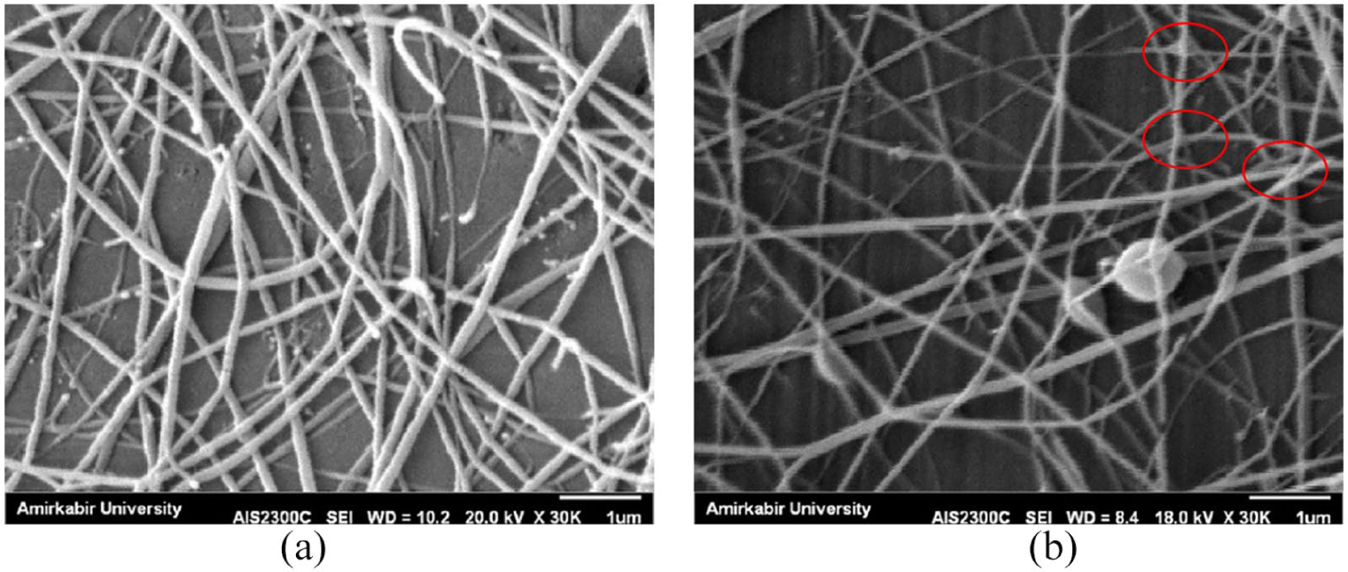
SEM image of (a) gelatin–chitosan–gum tragacanth and (b) gelatin–chitosan–gum tragacanth annealed at 120°C.

Schematic of possible cross-linking mechanism for thermal annealing treatment.
On the other hand, due to the swelling property of GT, there is some merging on overlap points, by which annealing can be more effective in GT-included samples rather than in gelatin–chitosan ones.
Fourier transform-infrared spectroscopy/attenuated total reflectance
Figure 6 shows the FTIR spectra of (a) gelatin, (b) chitosan, (c) GT, (d) gelatin–chitosan nanofibers, (e) gelatin–chitosan–GT nanofibers, (f) gelatin–chitosan–GT blend after being heated. There are many similar peaks observed in FTIR/ATR spectra, due to common functional groups found in chitosan, gelatin, and GT.

FTIR spectra of electrospun nanofibers of (a) gelatin, (b) chitosan, (c) gum tragacanth (d) gelatin–chitosan nanofibers, (e) gelatin–chitosan–gum tragacanth nanofibers, and (f) gelatin–chitosan–gum tragacanth after annealing.
For instance, all the samples exhibited a broadband around 3350 cm−1and a weak peak at 2922 cm−1, which are assigned to the groups of O–H and CH2, respectively. There are some peculiar peaks which appear for pure chitosan and gelatin to explain their special properties. The appearance of signal around 1740 cm−1 and a peak at 1555 cm−1 confirms the existence of amide and amine groups. The absorbance peak at 1066 cm−1 is prominent in pure chitosan and corresponds to the feature –C–O–C– bonds in Figure 6(b). As it can be seen in the spectra of the gelatin sample, the bands at 1640, 1540, and 1240 cm−1 corresponded to the stretching of amide I, N–H bending of amide II, and N–H bending of amide III, respectively. 34
The existence of these peaks for chitosan and gelatin is obvious and apparent evidence for a perfect combination of these two polymers and ionic interaction between positively charged chitosan (Figure 6(d)) and negatively charged gelatin. In other words, FTIR/ATR verifies the chemical structure and proves the possibility of miscibility and unaffected structures of chitosan and gelatin after the electrospinning process.
Figure 6(c) represents FTIR/ATR spectra of GT. The peaks at 1397, 1495, and 1747 cm−1 are related to asymmetric and symmetric carboxylate and carbonyl stretching, respectively. 29
Despite the high similarity of Gel–CH and Gel–CH–GT, their significant difference is in the intensity ratio of amide I and amide II after annealing. Also, the peak of the OH band is broader than that of Gel–CH–GT before annealing, which could be related to hydrogen bonding. The difference in ratio could be related to intensified inter- and intra-molecular interaction induced by annealing, in which it could be expected that thermodynamic movement of macromolecular chains intensifies under high temperature.29,33,34
Along with promoting ionic complexation between gelatin (carboxylate (–COO−) ions) and chitosan (ammonium (–NH3+) ions), heat treatment may produce the formation of hydrogen bonding between hydroxyl groups in GT and a carboxyl group in gelatin.
Stability of nanofibrous mats in aqueous solution
In order to investigate improvement in stability of thermally treated nanofibers, the nanofiber mats of Gel–CH, un-annealed Gel–CH–GT, and annealed Gel–CH–GT at 120°C were soaked in PBS buffer for 24 h and 7 days, and the remaining weight percentage of the samples was used to represent its stability. Gel–CH, un-annealed Gel–CH–GT, and annealed Gel–CH–GT at 60°C lost about 30% and 15% of their initial weight, respectively, after 1 day in PBS and lost about 80% and 70% of their initial weight after 7 days, respectively. However, in the case of the nanofibrous mats annealed at 120°C, remaining weights after 24 h and 7 days were about 70% and 45% of the initial weight, respectively. This is due to improvement effect of annealing on inter-fiber bonding, the conjunction of oppositely charged polymers, and also hydrogen bonding between macromolecules.
Differential scanning calorimetry
DSC is found to be an appropriate tool which is exclusively used to investigate thermal and morphological properties of blend nanofibers. As it is shown in thermal analysis of chitosan, gelatin, and GT (Figure 7(a) to (c)), a broad endothermic peak corresponds to dehydration of chitosan, gelatin, and GT starting at almost 20°C and ending at 150°C. The endothermic peak for all samples appeared at around 90°C (Tg) and is associated with hydrogen-bonded water in nanofibrous samples.

DSC thermograms of electrospun nanofibers of (a) chitosan, (b) gelatin, (c) gum tragacanth, (d) gelatin–chitosan–gum tragacanth, and (e) gelatin–chitosan–gum tragacanth annealed at 120°C.
Moreover, theoretically, a completely miscible blend should result in the formation of a single-phase transition between the transition temperatures of the chitosan, gelatin, and GT. In addition, no secondary peak was observed in the Gel–CH–GT sample, suggesting good miscibility of all the blend samples and the absence of phase separation (Figure 7(d)). Good miscibility is related to inter-molecular interaction between oppositely charged polymers. The highest peak position shift is in nanofibers heated at 120°C, which implies steadiness of nanofibers during heating.21,34
Water contact angle measurements
The surface hydrophilic property plays an important role in biomedical application. To investigate the influence of blending polymers on the surface hydrophilic property of electrospinning gelatin–chitosan–GT nanofibers, the water contact angle (WCA) measurement was done and shown in Figure 8. As it can be seen, gelatin–chitosan–GT nanofibers showed a contact angle of about 35° which is less than 90°, indicating that the surface is hydrophilic. 32

Water contact angle (WCA) measurements of gum tragacanth–chitosan–gum tragacanth.
Antibacterial properties
The antibacterial property of nanofibrous mat plays an important role in its application as a wound dressing. All bacteria in colony-forming units on agar plates for pure gelatin sample survived entirely. However, for pure chitosan, chitosan–gelatin, and gelatin–chitosan–GT nanofibers, there is a higher grade of suppression of bacterial colonies (Table 2). This is due to the interaction between a negatively charged bacterial cell surface and positively charged chitosan. On the other hand, the antibacterial effect of chitosan is more effective against Staphylococcus aureus in comparison with Escherichia coli due to stronger binding and lysis on gram-positive bacterial cell walls.
Anti-bacterial activity of nanofibers with gram-positive Staphylococcus aureus and gram-negative Escherichia coli.

It is assumed that the
Conclusion
Nanofibrous membranes of the chitosan–gelatin–GT blend were electrospun using acetic acid as solvent and annealing as cross-linker. Incorporation of GT has been found to improve electrospinnability and morphological properties of the gelatin–chitosan blend. The effect of the blend weight ratio and solution concentration on the morphology of nanofibers was also investigated using SEM, FTIR/ATR, and DSC. Optimum condition was identified as 7:3:1 weight ratio of gelatin–chitosan–GT, which resulted in decrease of the fiber diameter to 115.8 ± 10.66 nm. Annealing processing at 120°C caused better stability of nanofibers; hence, thermal annealing can be used as a suitable method instead of using chemical and harmful cross-linkers. Due to the mentioned properties and optimizing the nanofiber properties, resulted nanofiber mats can be used in biomedical applications such as wound dressing and tissue engineering scaffolds.
Footnotes
Declaration of conflicting interests
The author(s) declared no potential conflicts of interest with respect to the research, authorship, and/or publication of this article.
Funding
The author(s) received no financial support for the research, authorship, and/or publication of this article.