
Abstract
Spirometry-induced bronchoconstriction (SIB) refers to the paradoxical narrowing of the airways triggered by deep inhalation (DI) during spirometry testing. This response is atypical but can occur in subjects with heightened airway sensitivity, such as those with asthma. The present case report described a male patient who developed SBI in response to DI during spirometric maneuvers. A 34-year-old male patient with personal (i.e., rhinitis) and family allergy consulted our pulmonary department for recurrent wheezing dyspnea associated with exertional dyspnea level 2 according to the modified Medical Research Council dyspnea scale. Asthma diagnosis was suspected and a spirometry test was required. During five consecutive spirometric efforts, the patient developed a SIB with forced expiratory volume in one second (FEV1) decreases (in mL and % of initial value [%Initial]) of 50 mL (1%Initial), 270 mL (8%Initial), 480 mL (14%Initial), and 600 mL (18%Initial), respectively. Following the spirometry tests, the patient presented symptoms such as wheezing, shortness of breath, and chest tightness. The bronchodilator test (i.e., 400 µg of short-acting bronchodilator) was clinically significant with an increase of FEV1 by 1,260 mL (40%Predicted) (FEV1 passed from 2.80 L [65%Predicted] to 4.06 L [95%Predicted]). An alleviation of symptoms was noted, which supports the diagnosis of SIB. Clinicians should exercise caution when asking asthmatic patients to repeatedly perform DI for spirometry testing.
Keywords
Introduction
Spirometry-induced bronchoconstriction (SIB) refers to an unusual airway narrowing triggered by the forced expiratory maneuvers performed during spirometry testing (Haynes, 2015; V. C. Moore, 2012; Orehek et al., 1980; Suzuki et al., 1990). Although uncommon, this response can occur in subjects with heightened airway sensitivity, such as those with asthma (Haynes, 2015; Marthan & Woolcock, 1989; V. C. Moore, 2012; Orehek et al., 1980, 1981; Suzuki et al., 1990). Both deep inhalation (DI) and forceful exhalation (FE), essential components of spirometry, have the potential to provoke bronchoconstriction in susceptible subjects (Haynes, 2015; Marthan & Woolcock, 1989; V. C. Moore, 2012; Orehek et al., 1980, 1981; Suzuki et al., 1990).
There are only a few documented cases of SIB in the literature. A PubMed search conducted on January 30, 2025, using the following keywords ((((bronchoconstriction-induced deep inspirations[Title/Abstract]) OR (Spirometry-induced bronchoconstriction[Title/Abstract])) OR (Inspiration-induced bronchoconstriction[Title/Abstract])) OR ((deep inspiration[Title/Abstract])) AND (bronchoconstriction[Title/Abstract])) retrieved 112 papers, with only one comparable case report published in 2015 (Haynes, 2015). That case described a patient who experienced significant bronchoconstriction following DI during spirometry testing (Haynes, 2015).
The present case report described a male patient who developed SBI in response to DI during spirometric maneuvers.
Observation
This case report was prepared following the “CAse REports” guidelines (Gagnier et al., 2013).
Clinical Findings
A 33-year-old former smoker (10 pack-years) was referred to the outpatient pulmonary clinic for recurrent wheezing and exertional dyspnea (level 2 according to the modified Medical Research Council dyspnea scale; Fletcher et al., 1959), which had been progressing for 6 months. His medical history included allergic rhinitis and a shrimp allergy, both present for 5 years. In addition, the patient had a family history of allergy, with his mother and brother also suffering from allergic rhinitis. Physical examination and laboratory tests were unremarkable. Given the suspicion of asthma, the patient was referred to the physiology and functional exploration department for spirometry, along with a bronchodilator (BD) test or a methacholine challenge test.
Timeline
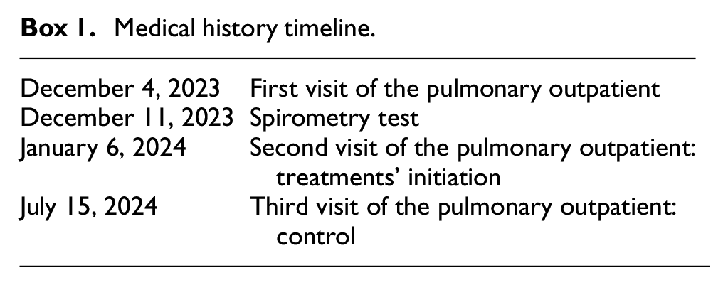
The patient’s medical history is summarized in Box 1.
Medical history timeline.
Diagnostic Assessment
The patient’s decimal age (33.48 years) and anthropometric measurements (height: 176 cm, weight: 73 kg, body mass index: 23.6 kg/m2) were recorded as per departmental protocol. Spirometry was performed according to the latest international guidelines (Graham et al., 2019) using a calibrated spirometer (Medisoft, Sorinnes, Belgium) with a nose clip. The four phases of the forced vital capacity (FVC) maneuver—maximal inspiration, “blast” of expiration, sustained complete FE for up to 15 s, and rapid inspiration to full lung capacity—were explained and demonstrated by an experienced technician (Graham et al., 2019).
During the FVC maneuvers, the patient exhibited signs of SIB, evidenced by a progressive decline in forced expiratory volume in 1 s (FEV1) over five consecutive efforts (Table 1, Figure 1). By the fourth and fifth attempts, FEV1 had decreased by 480 mL (14% of initial value [%Initial]) and 600 mL (18%Initial), respectively. The patient also developed symptoms of wheezing, shortness of breath, and chest tightness. A diagnosis of SIB was confirmed based on an FEV1 drop of more than 10%Initial (Parsons et al., 2013), prompting the cessation of FVC maneuvers. In addition, the emergence of progressively more concave expiratory flow-volume loops strengthened the SIB diagnosis (Figure 1). The five spirometric attempts met international acceptability criteria (Graham et al., 2019); though repeatability standards were not satisfied due to worsening SIB. Following a discussion between the technician performing the spirometry and the interpreting physicians (MA and HBS in the authors’ list), equipment malfunction and suboptimal patient effort were ruled out as potential causes (Haynes, 2015).
Decreases of FEV1 and FVC Over the Five Expiratory Efforts.

Note. FEV1 = Forced expiratory volume in 1 s. FVC = Forced vital capacity.
Changes in: Absolute value (mL) = Effort x value − Effort1 value; %Initial (%) = (Effort x value minus Effort1 value)/Effort1 value.

Superimposed flow-volume loops and volume-time curves from spirometry effort 1 to effort 5. Effort 5 has lower flows and volumes compared with effort 1.
The pre-BD data from all efforts and the technologist’s suspicion of SIB were shared with the interpreting physician (HBS in the authors’ list). Due to baseline airway obstruction, the methacholine challenge test was canceled in accordance with laboratory protocol, which contraindicates the test in cases of low FEV1 at 65%Predicted (Plantier et al., 2018) (Table 2). Consequently, 400 µg of a short-acting BD was administered (Graham et al., 2019; Stanojevic et al., 2022), and spirometry was repeated 15 min later (Table 2, Figure 2). The BD test was clinically significant with an FEV1 increase of 1,260 mL (i.e., FEV1 improved from 2.80 L [65%Predicted] to 4.06 L [95%Predicted]), accompanied by symptom relief, confirming the diagnosis of SIB.
Spirometric Data Pre- and Post-Administration of 400 µg of Short-Acting Bronchodilator.

Note. FVC = Forced vital capacity. LLN = Lower limit of normal. ULN = Upper limit of normal.
Absolute change = post-bronchodilator value minus pre-bronchodilator value.
%Predicted change = %Predicted post-bronchodilator minus %Predicted pre-bronchodilator.
Predicted values and z-scores were derived from the 2012 global lung function initiative norms (Caucasian group).

Pre- and post-bronchodilator flow-volume loops and volume-time curves.
The Global Lung Function Initiative 2012 spirometric norms for Caucasians were applied, and test results were interpreted based on the latest international guidelines (Barkous et al., 2024; Ben Saad, 2022; Quanjer et al., 2012; Stanojevic et al., 2022) (Table 2, Figure 2). Typically, spirometry reports the best pre-BD effort as the one with the highest sum of FVC and FEV1 (Graham et al., 2019; Haynes, 2015) (i.e., effort 2 in this case, where “FVC + FEV1” = 8.57 L [Table 1]). Given the progressive bronchoconstriction, effort 5, with the lowest FEV1 (Tables 1 and 2), was chosen as the pre-BD value to more accurately reflect the patient’s latest ventilatory status (Haynes, 2015). The state of ventilation that accompanied effort 2 no longer existed, and comparing the post-BD data to a then-nonexistent milieu might be misleading and potentially affect test interpretation (Haynes, 2015).
Spirometry results (Table 2, Figure 2) indicated a moderate, pure obstructive ventilatory defect (i.e., low FEV1/FVC z-score = −3.13, low FEV1z-score = −2.73, and normal FVC z-score = −0.79). The BD test was clinically significant with an FEV1 increase of 30%Predicted (i.e., exceeding the recommended threshold of 10%Predicted; Stanojevic et al., 2022), and the post-BD FEV1/FVC ratio normalized at 0.81 (i.e., z-score = −0.13). Pre- and post-BD flow-volume loops are superimposed in Figure 2.
Even if the highest FEV1 value from the pre-BD efforts had been reported as the best pre-BD value (i.e., FEV1 = 3.40 L from effort 1, Table 1), a clinically significant increase in FEV1 following BD administration would have been reported (i.e., 15%Predicted increase: [(4.06 – 3.40)/4.29], Table 2).
Therapeutic Intervention
The patient was referred to his pulmonologist, who confirmed the diagnosis of bronchial asthma. Treatment was initiated with a combination of Budesonide-Formoterol 400/12 µg, prescribed as one inhalation twice daily, with an additional dose as needed, in accordance with the international guidelines (GINA, 2024).
Follow-up and Outcome
At a six-month follow-up, the patient reported significant clinical improvement, with complete resolution of dyspnea.
Discussion
This case report provides four key takeaways: (a) DI can trigger bronchoconstriction during FVC maneuvers; (b) Careful assessment is necessary to distinguish SIB from equipment failure or suboptimal patient effort; (c) Reporting the highest FEV1 as the “best value” after bronchoconstriction occurs may be misleading, and instead, the lowest FEV1 should also be considered for accurate interpretation; and (d) Clinicians should exercise caution when requesting repeated DI maneuvers, particularly in acutely ill asthma patients undergoing diagnostic procedures (Haynes, 2015).
Our case aligns with a previously reported case by Haynes (Haynes, 2015), describing a 49-year-old male with a history of childhood asthma who experienced increasing wheezing and chest tightness post-exercise. During spirometry, this patient exhibited coughing, mild chest tightness, and a progressive decline in FVC and FEV1 from the first effort [FVC = 5.05 l (92%Predicted), FEV1 = 3.28 l (76%Predicted)] to the seventh effort [FVC = 4.36 l (79%Predicted), FEV1 = 2.54 l (60%Predicted)] (Haynes, 2015). The respective decreases in FVC and FEV1 between efforts 1 and 7 were 14% and 22%, respectively (Haynes, 2015). Following BD administration (2.5 mg of albuterol and 0.5 mg of ipratropium), the patient exhibited a clinically significant response, with post-BD FVC [5.85 L (106%Predicted), an increase of 1,490 mL (27%Predicted) from effort 7] and post-BD FEV1 [4.06 L (94%Predicted), an increase of 1,520 mL (34%Predicted) from effort 7]] (Haynes, 2015).
Clinicians performing and interpreting spirometry tests should be aware that DI could influence airway function, either facilitating bronchodilation or triggering bronchoconstriction (Burns & Gibson, 2002; Cockcroft & Davis, 2006; Haynes, 2015; Lim et al., 1989). Overlooking these effects could lead to errors in spirometry interpretation, potentially attributing bronchoconstriction to equipment malfunction or poor patient effort, which may result in false-negative test outcomes (Allen et al., 2005; Cockcroft & Davis, 2006; Haynes, 2015). The primary risk of a false-negative diagnostic test is the failure to detect a disease/condition when it is actually present, leading to delays in treatment, worsening of the condition, and potential harm to the patient (Schneider et al., 2009). For instance, an asthma patient may exhibit bronchoconstriction solely due to DI during spirometry, leading to underdiagnosis (Haynes, 2015).
Generally, SIB is attributed more to DI than to FE during the expiratory phase of spirometry (Haynes, 2015; Kamio et al., 1990; B. J. Moore et al., 1997; Suzuki et al., 1990). On one hand, Moore et al. (B. J. Moore et al., 1997) demonstrated no significant difference in methacholine challenge test responses between protocols that included repeated FEs to residual volume (without DI) and those excluding both DI and FE. On the other hand, Suzuki et al. (1990) reported a decline in specific airway conductance after an FE maneuver from functional residual capacity; however, this effect was less pronounced compared to the bronchoconstriction triggered by DI. Their study suggested that SIB in asthmatics may result not only from DI reaching total lung capacity but also from FE reaching RV, with the bronchoconstriction likely driven by increased parasympathetic activity (Suzuki et al., 1990).
Possible Mechanisms of SIB
In healthy subjects or those with mild asthma, DI typically induces bronchodilation or has little impact, whereas some asthmatic patients experience transient but significant bronchoconstriction following DI (Burns & Gibson, 2002; Lim et al., 1989; Suzuki et al., 1990). Interestingly, while patients with mild asthma have limited BD responses, those with severe asthma often exhibit pronounced bronchoconstriction after DI (Lim et al., 1989). The mechanisms underlying DI-induced bronchoconstriction (i.e., SIB) remain unclear, but multiple factors are believed to contribute (Brown et al., 2001; Golnabi et al., 2014; Haynes, 2015; Samee et al., 2003; Tgavalekos et al., 2007; Venegas et al., 2005). Marthan et al. (1989), proposed that bronchoconstriction could be explained by ion exchange across airway smooth muscle cell membranes. The myogenic response is regulated by calcium ion influx through voltage-dependent calcium (VDC) channels, and this response can be suppressed by VDC antagonists (Marthan & Woolcock, 1989). Their findings indicate that airway stretching during DI causes membrane depolarization in airway smooth muscle cells (Marthan & Woolcock, 1989). Notably, administering a calcium channel antagonist sublingually significantly reduces the DI-induced decrease in specific airway conductance in asthmatic patients (Marthan & Woolcock, 1989). An alternative hypothesis proposed by Fish et al. (1981) suggests the presence of an intrinsic mechanism regulating bronchomotor tone through active lung volume changes, which may be impaired in asthma. Another proposed mechanism involves lung tissue hysteresis, which leads to reduced lung recoil pressure and weaker mechanical forces exerted on the airway walls (Brusasco & Pellegrino, 1995; Burns & Gibson, 2002). This results in a balance of hysteresis between the airways and lung parenchyma after DI (Brusasco & Pellegrino, 1995; Burns & Gibson, 2002). Asthmatic airway hyperreactivity may be attributed more to reduced relaxation capacity rather than increased airway smooth muscle contractility (Fish et al., 1981).
According to Lim et al. (1989), “the more severe the obstruction, the greater the constrictor effect of a DI.” The bronchoconstrictive effect of a DI is closely linked to the degree of inflammation in the obstructive process (Lim et al., 1989). In asthmatic patients, the response to a DI may serve as a functional marker of the primary mechanism underlying airway obstruction (Lim et al., 1989). In asthmatic airways, which are characterized by an increased number of blood vessels and a leaky, low-pressure capillary bed, fluid leakage into the airway wall can occur (Burns & Gibson, 2001). This fluid accumulation thickens the airway wall, reduces airway diameter, and lowers specific airway conductance (Burns & Gibson, 2001). In addition, the excess fluid makes the airway wall stiffer, thereby reducing its compliance. As a result, the airway becomes less likely to collapse during FE (Burns & Gibson, 2001). Even minor fluid accumulation within the airway wall can enhance smooth muscle contraction, leading to significant airway narrowing (Brown et al., 1995). Consequently, even small fluid shifts may contribute to the observed bronchoconstriction (Brown et al., 1995). In contrast, in healthy subjects without airway inflammation, this mechanism is either absent or negligible (Brown et al., 1995).
Advances in imaging techniques have greatly enhanced the understanding of bronchial obstruction in asthma (Brown et al., 2001; Golnabi et al., 2014; Haynes, 2015; Samee et al., 2003; Tgavalekos et al., 2007; Venegas et al., 2005). These techniques have demonstrated that asthma responses involve dynamic and heterogeneous patterns of bronchoconstriction and bronchodilation, accompanied by patchy zones of parenchymal hypo-expansion and hyper-expansion, respectively (Brown et al., 2001; Golnabi et al., 2014; Haynes, 2015; Samee et al., 2003; Tgavalekos et al., 2007; Venegas et al., 2005). The constricted airway zones and hypo-expanded parenchymal areas are referred to as ventilation defects (Golnabi et al., 2014; Samee et al., 2003). Due to airway-parenchymal interdependence, the areas surrounding ventilation defects often undergo bronchodilation and relative parenchymal hyper-expansion through radial traction (Haynes, 2015). Repeated DI, particularly when lung volume is limited, may fail to sufficiently expand these ventilation defects (Haynes, 2015). As a result, ventilation may become concentrated in hyper-expanded regions, potentially causing unstable boundary zones of ventilation defects to collapse, leading to the formation of larger and more numerous ventilation defects (Haynes, 2015). Some authors suggest that DI performed under conditions of airway obstruction may generate significant intrathoracic pressure fluctuations, contributing to airway edema and increased airway smooth muscle shortening (Burns & Gibson, 1998, 2002; Fish et al., 1981). Burns and Gibson (1998, 2001, 2002) proposed that negative intrathoracic pressure acting on the airway capillaries promotes airway wall edema and reduces airway caliber. During normal and rapid DI, this mechanism may play a role in bronchoconstriction among asthmatic patients (Burns & Gibson, 1998, 2002; Fish et al., 1981). Similarly, it appears that repeated DI could worsen airflow obstruction due to a reduction in deflation recoil (Lim et al., 1989).
One limitation of this case report is the reliance on FEV1 alone for diagnosing SIB. First, using FEV1 as the sole parameter can be misleading, as some asthmatic patients exhibit a similar response to DI as healthy subjects or those with hay fever (Orehek et al., 1980). A more comprehensive assessment would include static lung volumes or specific resistances, as demonstrated by Haynes (Haynes, 2015). Second, some authors recommended measuring the transient stretch of airway muscle, as this factor is believed to enhance the beneficial effects of DI by reducing the contractile ability of airway smooth muscle (Bosse, 2019; Macklem, 1989).
Conclusion
In healthy subjects, DI transiently dilates the airways, whereas many asthmatic patients experience bronchoconstriction through a mechanism that remains partially understood. The SIB observed after a DI is not only due to stretching and relaxation-induced changes in the airway and lung tissues but also to temporary pressure fluctuations within the chest that affect inflamed airway walls in asthmatic patients. Clinicians should exercise caution when requesting multiple DIs for spirometry tests, peak-flow measurements, and auscultation, as these maneuvers may exacerbate airway obstruction (Haynes, 2015).
Footnotes
Patient Perspective
The patient reported an improvement in his condition and informed the authors that “he feels much better with the treatment, leads a normal life, and hopes not to undergo spirometry again.”
Informed Consent
Written consent was obtained from the patient for the publication of this case report.
Funding
The author(s) received no financial support for the research, authorship, and/or publication of this article.
Declaration of Conflicting Interests
The author(s) declared the following potential conflicts of interest with respect to the research, authorship, and/or publication of this article: The authors wish to disclose that an artificial intelligence tool (ChatGPT 3.5 ephemeral) was utilized to enhance the clarity and coherence of the manuscript writing. The tool was utilized for language refinement purposes only, ensuring the text was clear and coherent without altering the scientific content or generating any new text (Dergaa et al., 2023).