
Abstract
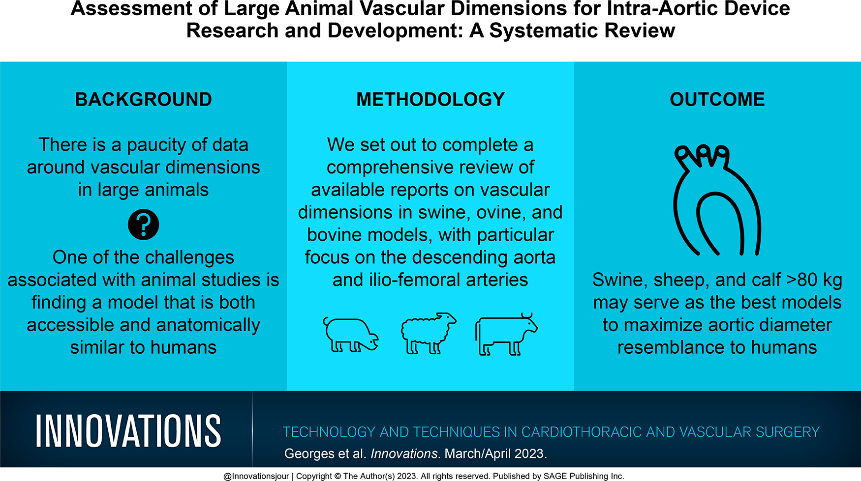
Animal studies are often required to evaluate new cardiovascular medical devices before they reach the market. Moreover, first-generation novel devices including aortic endovascular prostheses and circulatory support devices are often larger than later iterations or tested in a limited range of sizes. One of the challenges in evaluating these devices is finding a model that is both accessible and anatomically similar to humans, as there is a paucity of data on vascular dimensions in large animals. We set out to complete a comprehensive review of available reports on vascular dimensions in swine, ovine, and bovine models, with a particular focus on the descending aorta and ilio-femoral arteries. We searched Embase and MEDLINE databases for reports of descending aorta and peripheral vascular dimension in large animal models. Data from swine, ovine, and bovine models were separated by weight into 3 categories: 40 to 60 kg, 61 to 80 kg, and >80 kg. We also incorporate our computed tomography angiography data from 4 large sheep and 9 calves into this review. Swine, sheep, and calf >80 kg may serve as the best models to maximize aortic diameter resemblance to humans. If device implantation can be achieved in aortas of smaller dimensions, care should be taken to ensure access site suitability such as the common femoral artery in these smaller animals.
Central Message
There is a paucity of data on vascular dimensions in large animals. The results of this comprehensive review of the available literature found that swine, sheep, and calf >80 kg may serve as the closest models to replicate aortic diameter in humans.
Introduction
The development of new cardiovascular medical devices commonly relies on animal studies to evaluate their safety and efficacy before they become available on the market. One of the challenges with animal studies is finding a model that is both readily available and anatomically similar to humans. 1 This is particularly challenging for early generation devices, which are often larger than versions of the devices reaching the market, or complex devices, which can initially be manufactured in a single or limited range of sizes. For the medical device development community, there are no guidelines based on vascular anatomy to determine the appropriate animal model. As such, the animal model may be chosen based on operator and center experience. If the model is suboptimal, multiple animals may be screened. Screened animals that do not receive the device can sometimes be repurposed but may also be terminated at the end of the study. Due to the prior development of coronary and peripheral stents, there are some data on small animal vascular dimensions.2–6 When the size of the device permits the use of small animals, they may be preferred because of their lower cost and housing requirements and because they are generally easier to handle. However, this is not always the case, and there is currently a paucity of data on vascular dimensions in large animals.2–6 The success of endovascular aortic repair7,8 and, more recently, intra-aortic mechanical circulatory support, 9 suggests continued development efforts in this area. Better understanding of large animal vascular dimensions could help improve study efficiency, lower costs, and result in less animal sacrifice.
We set out to complete a comprehensive review of available reports on vascular dimensions in swine, ovine, and bovine models, with a particular focus on the descending aorta and the ilio-femoral arteries. We also incorporated our computed tomography angiography data from 4 large sheep and 9 calves into this review.
Methods
We searched Embase and MEDLINE databases from their inception to January 9, 2023, using the MeSH and text terms shown in the Appendix. Two review authors (G.G. and T.C.) independently read the titles and abstracts of relevant papers retrieved by the search strategy described above to identify potentially suitable studies. Full publications of all potentially relevant studies were retrieved and stored in a Mendeley library. Two review authors (G.G. and T.C.) determined article eligibility independently, using a standardized inclusion form. Only original articles were included. Studies were also excluded if none of the following were present: thoracic descending or abdominal aorta dimensions, aortic arch branches, and femoral or iliac artery dimensions. Ascending aorta measurements were not included in this study as they are better documented owing to transcatheter aortic valve preclinical studies and more accessible measurement via echography. All imaging modalities were accepted. We limited the scope of this review to the 3 most common large animal models: porcine, ovine, and bovine. Studies were excluded if animal weight was not mentioned. We did not apply any language restrictions. We resolved any disagreements by discussion with the third review author (P.V.). The selection process and reasons for exclusion are presented in Figure 1. For each study, we sought the animal cohort size and characteristics (i.e., weight, age, breed) and arterial vasculature dimensions. We recorded data on an electronic data extraction form. When measurements were presented only as a range, the mean value was recorded. We made no further attempt to impute missing values. Animals from all 3 species (swine, ovine, and bovine) were separated by weight into 3 categories: 40 to 60 kg, 61 to 80 kg, and >80 kg. All vascular dimensions are presented as weighted means and standard deviation. Only the weighted means were calculated when no standard deviations were reported, and they could not be calculated from data present in any of the articles for a certain weight and animal model category. The number of animals from which each value was extracted is also presented.
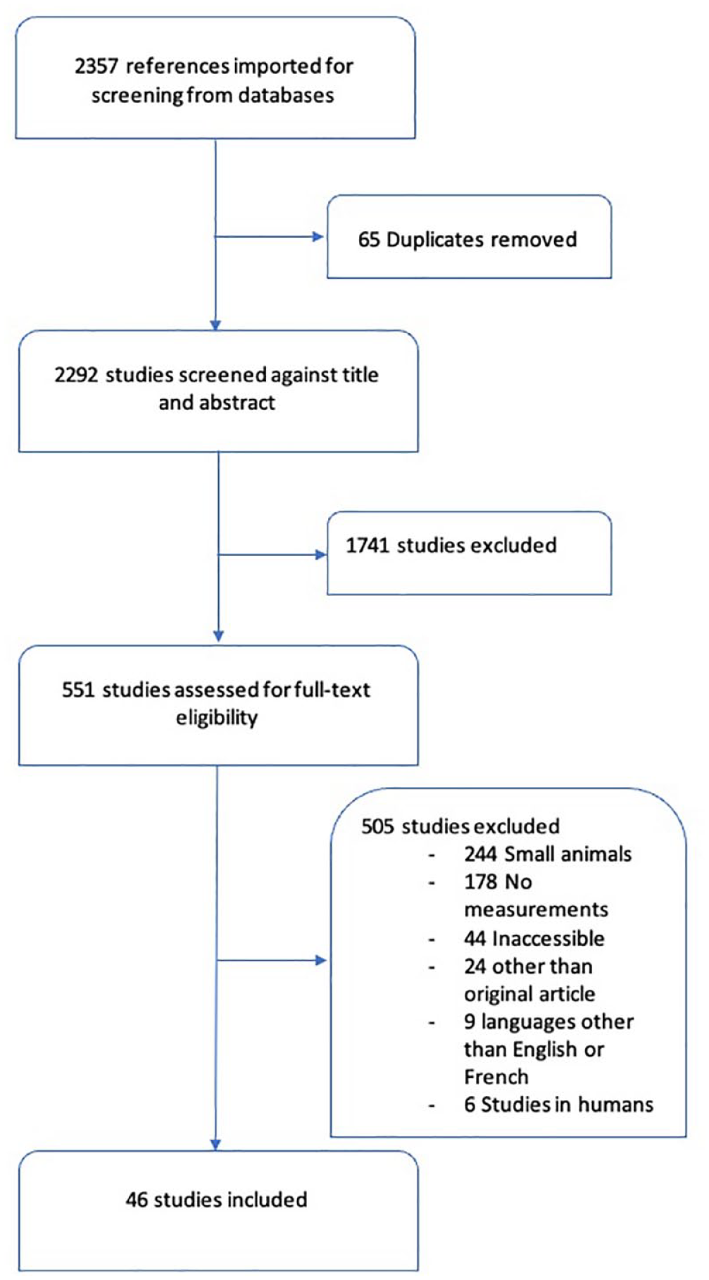
Selection process.
We chose to include data from 4 sheep and 9 calves from our computed tomography angiography library. All animals were part of different acute terminal cardiovascular studies that required preoperative pan-corporeal computed tomography angiography. Permission was granted by the institutional animal welfare committee. Animals were housed under controlled environmental conditions, and there was an acclimatization period of 1 week before the experiment. Briefly, standard anesthesia treatment was provided with a mix of midazolam (0.1 mg/kg) and ketamine (4.0 mg/kg) administered intravenously. The animals were intubated, and anesthesia was maintained with propofol or isoflurane. Examination was performed on a 2 × 128 slice multidetector computed tomography scanner (SOMATOM Definition Flash; Siemens Healthineers, Erlangen, Germany). Images were analyzed using 3Mensio software (Pie Medical Imaging, Maastricht, Netherlands). The results were analyzed with data extracted from the literature review.
Results
The search found 2,357 papers. After duplication removal, this number was reduced to 2,292. We excluded 1,741 articles because they did not meet the study inclusion criteria. A total of 551 papers were selected for full-text review, of which 505 were excluded based on not meeting the study criteria. Of the 46 selected studies, 30, 15, and 1 reported swine, ovine, and bovine data, respectively.2,10–53 In the 1 paper we found describing bovine arterial dimensions, only thoracic aorta measurements were determined to be relevant in this study. We found no study describing aortic arch branch diameters in calves and only 1 study in sheep reporting brachiocephalic trunk diameters. The included studies are presented in Table 1.
Studies Included in the Systematic Review and Animal Characteristics.
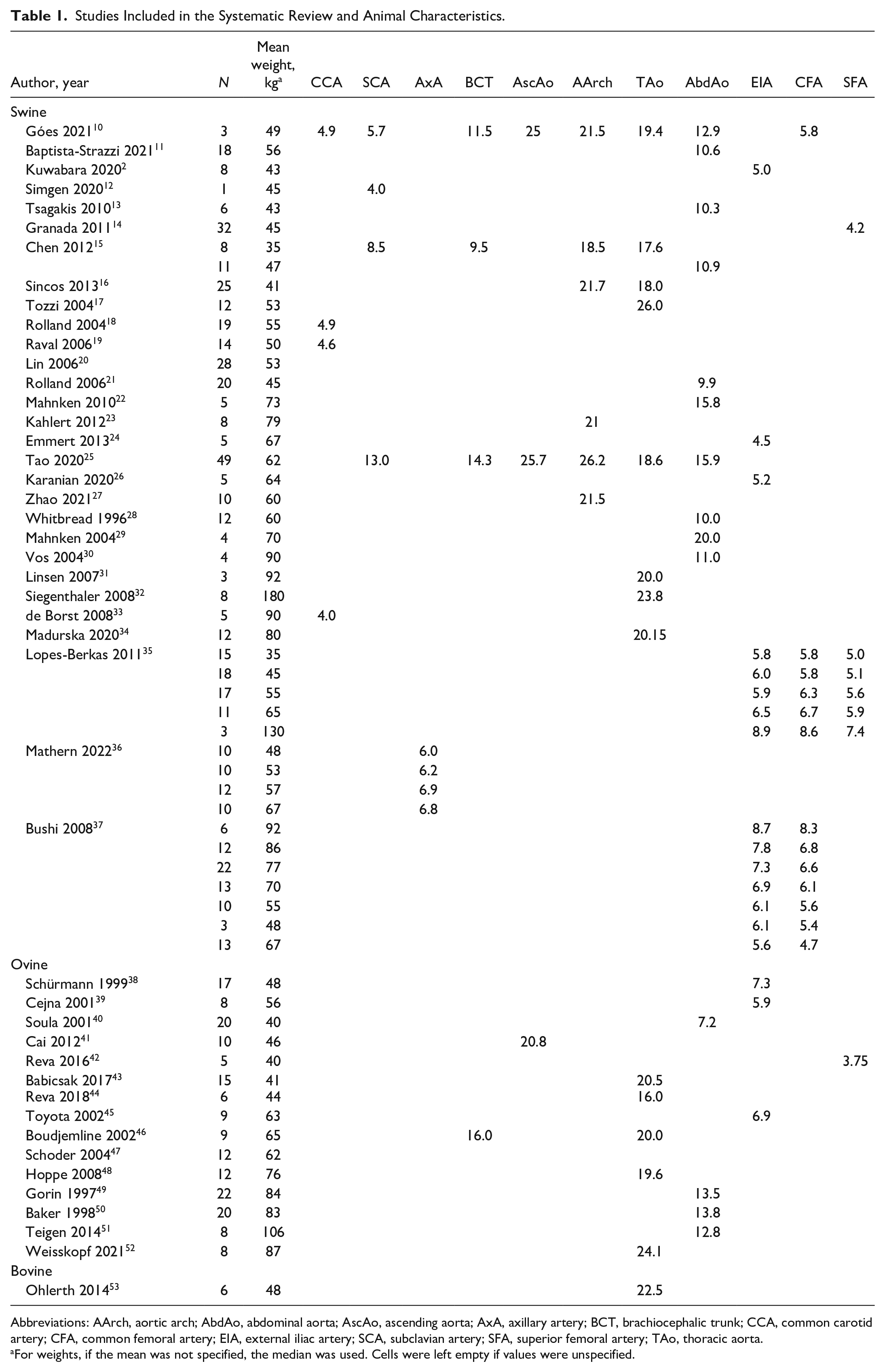
Abbreviations: AArch, aortic arch; AbdAo, abdominal aorta; AscAo, ascending aorta; AxA, axillary artery; BCT, brachiocephalic trunk; CCA, common carotid artery; CFA, common femoral artery; EIA, external iliac artery; SCA, subclavian artery; SFA, superior femoral artery; TAo, thoracic aorta.
For weights, if the mean was not specified, the median was used. Cells were left empty if values were unspecified.
Vascular dimensions by species and animal weight are summarized in Figure 2. Swine 60 to 79 kg were found to have the largest aortic arch diameter (24.9 ± 2.0 mm), while sheep >80 kg had the largest descending thoracic aorta (22.7 ± 1.9 mm), and calves had the largest abdominal aorta (17.4 ± 2.0 mm). Although proximal descending thoracic aorta diameters were similar, overall, all models tended to possess smaller descending thoracic aorta and aortic arch diameters compared with normal human size (aortic arch: 19 to 21 mm, descending thoracic aorta: 24 to 30 mm, abdominal aorta: 14 to 30 mm).54,55
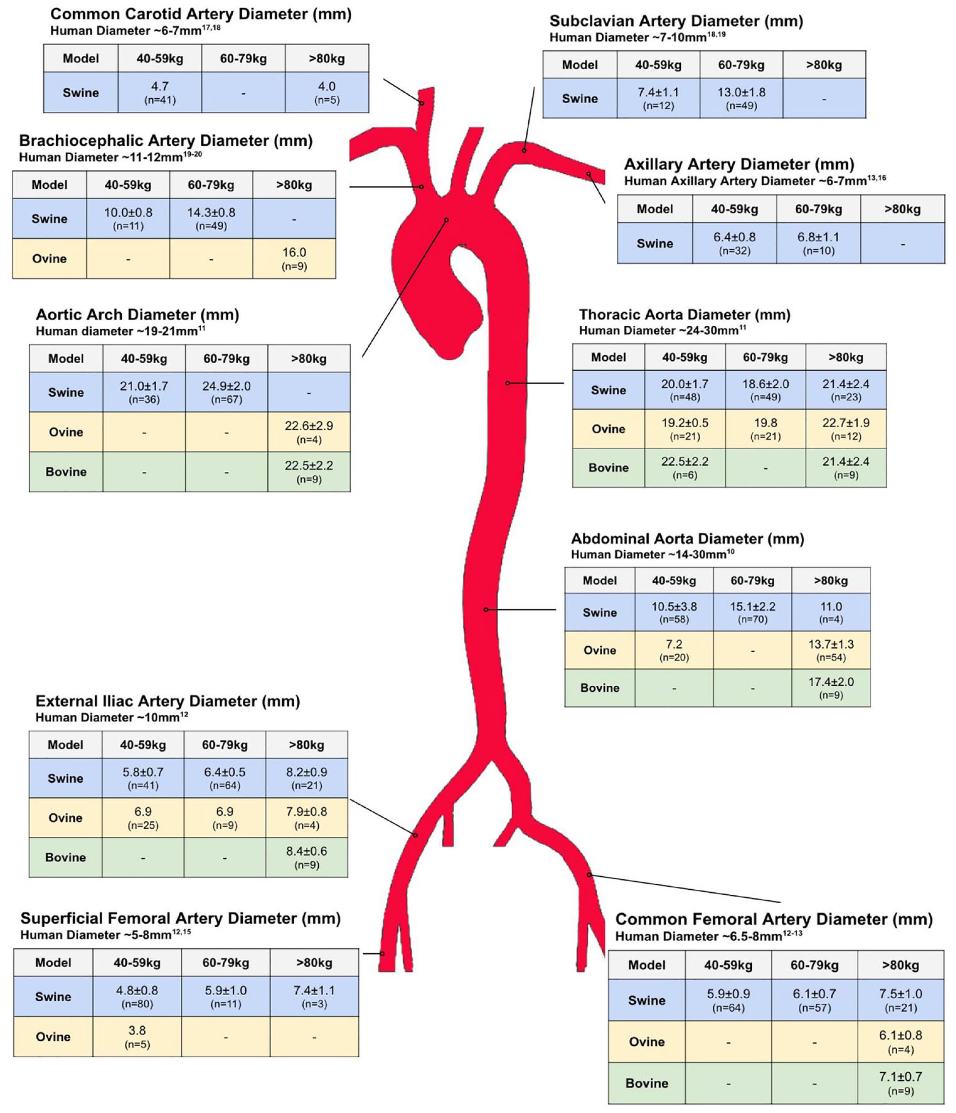
Arterial diameters in pigs, sheep, and calves by weight. Vascular dimensions are presented as weighted means and standard deviations. Only weighted means are presented when no standard deviations were reported and they could not be calculated from data present in any of the articles.
In swine, external iliac, common femoral arteries, and superficial femoral arteries increased with weight. All 3 models had smaller external iliac arteries compared with what is found in humans (~10 mm). 56 The largest common femoral arteries were found in swine >80 kg (7.5 ± 1.0 mm), followed by calves >80 kg (7.1 ± 0.7 mm) and sheep >80 kg (6.1 ± 0.8 mm). Swine and calves >80 kg had the closest common femoral artery diameter to humans, in which mean diameters varied from 6.5 to 8 mm.57,58 Superficial femoral arteries in swine were similar to those in humans (4.8 ± 0.8 to 7.4 ± 1.1 mm in swine 40 to 59 kg and >80 kg vs 5 to 8 mm in humans).56,59
Axillary arteries were comparable in size to those of humans (6 to 7 mm)57,60 in both 40 to 59 kg (6.4 ± 0.8 mm) and 60 to 79 kg swine (6.8 ± 1.1 mm). Common carotid arteries were significantly smaller in swine (~4 mm) than in humans (6 to 7 mm).61,62 Subclavian artery diameter was similar to humans (7 to 10 mm)62,63 in swine 40 to 59 kg (7.4 ± 1.1 mm) and were much larger in swine >80 kg (13 ± 1.8 mm). Brachiocephalic trunk was also most similar to human diameters (11 to 12 mm)63,64 in swine 40 to 59 kg (10.0 ± 0.8 mm) and became larger with increasing weight (14.3 ± 0.8 mm in swine >80 kg).
Discussion
Preclinical in vivo trial success is largely dependent on choosing the right model. Reports of thoraco-abdominal aorta diameter and aortic branch diameters in large animal models are scarce. To our knowledge, this study is the first comprehensive review of the available data on arterial vascular diameters in swine, ovine, and bovine models. We found the 3 species to be potentially suitable for preclinical work, especially in weights over 80 kg. With the development of endovascular aortic prostheses and mechanical circulatory support, refinement in anatomical knowledge of large animal vasculature is mandatory to optimize screening, reduce cost, and avoid unnecessary sacrifice. Endovascular mechanical circulatory support devices are extremely complex to manufacture and are typically available only in a single size.9,65,66 It may be easier to plan preclinical studies for endovascular aortic prostheses, which come in a range of sizes. In certain cases, only the smaller devices may be implanted in preclinical studies if their larger counterparts are shown to be equivalent in terms of mechanical performance ex vivo.
Our results show that the arterial diameters of swine, sheep, and calves over 80 kg are most similar to those of humans. We therefore suggest the use of larger animals for studies requiring vascular access sites and aortic implantation sites that resemble that of humans. However, it should not be forgotten that bigger, heavier animals may offer a challenge to study personnel in terms of handling and manipulation. These animals may also require more sturdy equipment and may be too heavy for standard equipment such as operating tables. Choosing animals that are fully grown may be advantageous in subacute and chronic studies if implanted devices are to be left in place. Another potential pitfall of choosing larger animals is their corpus length. In our experience with calves, the average distance extending from the common femoral artery to the aorta at the level of the diaphragm was ~55 cm and was ~77 cm from the common femoral artery to the aortic isthmus. In humans, these distances have been evaluated at ~36 cm and ~60 cm, respectively. 67 Thus, in certain cases, material designed for human use may be too short, such as delivery sheaths that typically have a maximum working length of 65 cm. In this case, alternative access sites closer to the implant site may be required.
Some limitations of the current study should be acknowledged. First, animals were categorized by weight but not breed, which may play a role in vascular dimensions. Second, anatomical landmarks to identify the height of measurement of vessels were rarely provided. This may mostly have an impact on thoracic and abdominal aorta dimensions and may account for some variability in the results. More thorough precise documentation of the exact site of measurement in future studies may help improve upon this limitation (i.e., specifying mid-descending aorta or referring to bony landmarks and vessel branches).
When choosing an animal model for cardiovascular trials, other factors that are beyond the scope of this study should be considered. Anatomical particularities other than arterial diameters may influence the choice of model. For example, in contrast to the human aortic arch, cows possess a common brachiocephalic trunk that divides into the subclavian, vertebral, and carotid arteries. Depending on the device being studied, this may influence the level of the implantation site. 1 Mechanical properties of the targeted vessel may also differ between the animal model and that of humans. For example, a study on human and porcine aortic tissue found human aortas to be significantly stiffer compared with their porcine counterpart and highlighted a greater proportion of elastin and less collagen fibers in the porcine samples. 68 If the trial requires heart failure to be induced, susceptibility to arrythmia and coronary artery dominance should also be considered. Although porcine coronary artery distribution is right dominant as in humans, sheep are left dominant.1,69,70 Nonanatomical factors may also play a role in selection of the species, such as potential zoonotic disease transmission and need for prestudy quarantine. 1
Conclusions
This study was meant to provide a reference point for choosing the right animal species and weight range for future research in the field of endovascular aortic devices. Swine, sheep, and calves >80 kg may serve as the best models to maximize aortic diameter resemblance to humans. If device implantation can be achieved in aortas of smaller dimensions, care should be taken to ensure access site suitability, such as the common femoral artery, in these smaller animals.
Supplemental Material
sj-pdf-1-inv-10.1177_15569845231164134 – Supplemental material for Assessment of Large Animal Vascular Dimensions for Intra-Aortic Device Research and Development: A Systematic Review
Supplemental material, sj-pdf-1-inv-10.1177_15569845231164134 for Assessment of Large Animal Vascular Dimensions for Intra-Aortic Device Research and Development: A Systematic Review by Gabriel Georges, Thomas Couture and Pierre Voisine in Innovations: Technology and Techniques in Cardiothoracic and Vascular Surgery
Footnotes
Declaration of Conflicting Interests
The authors declared no potential conflicts of interest with respect to the research, authorship, and/or publication of this article.
Funding
The authors received no financial support for the research, authorship, and/or publication of this article.
Supplemental Material
Supplemental material for this article is available online.
References
Supplementary Material
Please find the following supplemental material available below.
For Open Access articles published under a Creative Commons License, all supplemental material carries the same license as the article it is associated with.
For non-Open Access articles published, all supplemental material carries a non-exclusive license, and permission requests for re-use of supplemental material or any part of supplemental material shall be sent directly to the copyright owner as specified in the copyright notice associated with the article.