
Abstract
Models depicting normal human anatomy have been a feature of Western medical teaching since the Renaissance. Some were collected by McGill University’s medical museum after its founding in 1821. In 1907, a fire in the medical building destroyed virtually the entire collection. Many models were purchased to replenish it during the following sixteen years. The majority of these, as well as those acquired after 1923, were consolidated in the Maude Abbott Medical Museum (MAMM) in 2012. We undertook a review of all models acquired by McGill’s Medical Museum/MAMM since the start of medical faculty calendar records in 1852/1853. A total of 433 were identified, of which 323 are still present. We discuss the uses of this collection and speculate about how 110 models have been “lost.” The review provides an understanding of the use of models in anatomy teaching in one North American university during the past 175 years.
Introduction
Models depicting normal human structure have been a feature of Western anatomy teaching since the Renaissance. 1 At that time, one of the most compelling reasons for their use was the lack of sufficient corpses for dissection because of religious and legal restrictions. Over the years, other advantages became evident, including:
the ability to better depict small anatomical features, such as the middle ear or parts of an embryo;
the lack of offensive odor that was frequently generated during anatomical dissection before the development of modern embalming procedures;
more natural color than organ specimens preserved in alcohol or formaldehyde;
the intimacy provided to the student by touch and three-dimensional manipulation, and the theoretical improved understanding of structure that this entailed;
the relative ease of storage and acquisition for teaching year after year;
the ability to permanently affix numbers or letters indicating specific anatomic features as special learning aides.
The creation and use of models was particularly important in the nineteenth and early twentieth centuries, coinciding with greater importance assigned to anatomy teaching in medical school curricula and increasing medical school enrollment (Alberti 2011; Spencer 2008; Talairach-Vielmas 2014). Although less employed, such use has continued to the present time, sometimes combined with traditional dissection in the anatomy laboratory or more recently developed teaching methods such as radiologic imaging (Fredieu et al. 2015).
Many materials have been used for model construction, including clay, wood, ivory, leather, and rubber. Because of the ease with which it could be worked, relative affordability, and the ability to simulate a life-like appearance by the addition of dyes, wax was the most popular substance in the 1700s, particularly in Italy (Haviland and Parish 1970). Other materials, such as plaster and papier-mâché, followed in the 1800s. Currently, plastic is by far the most widely used.
McGill University’s medical museum began around 1821 at the time of the founding of the Montreal Medical Institution. Its first anatomy professor was John Stephenson. There are no official records of the museum’s creation or its first teaching materials; however, we know that Stephenson used human specimens because a complete skeleton that he mounted in 1826 is present in today’s Maude Abbott Medical Museum (MAMM). Although few details are available, many other skeletal preparations and some anatomical models were added to the anatomy teaching collection throughout the nineteenth century.
In 1907, fire in McGill’s medical building resulted in the destruction of most of its pathological and almost all its anatomical material (Abbott 1910). A new campus building that contained a large amount of space for the medical museum was built in 1909. Many specimens that illustrated pathological abnormalities or normal anatomy were donated between 1907 and 1910 by museums in North America and Europe to assist in the reconstitution of the collections (Abbott 1910). Although most of the anatomical donations were skeletal, some were models. In addition, the Director of the Anatomy Museum at that time, Francis Shepherd, decided to purchase models from European manufacturers to supplement the collection. This was carried out mostly in seven years between 1908 and 1914. A new Anatomy Department Chair, Auckland Geddes, was appointed in 1914. Although he spent almost all his time as Chair in England as part of the War effort, he sent a variety of models from there to McGill to augment its collection.
Entries in the museum logbooks that document the acquisition of these post-fire acquisitions effectively stop in 1923, despite their presence in the MAMM collection today. This situation changed with the official re-establishment and naming of the museum as the MAMM in 2012; models donated by McGill faculty members or private individuals are now accessioned with accompanying descriptive information such as provenance, manufacturer, etc.
An investigation of the material remaining in museums can be useful to understand both the history of anatomy teaching and the sociocultural aspects related to it (Spencer 2008). Knowledge of what is missing from the collection—material that has been “lost”—can also be a subject of interest (Claes and Deblon 2017). We thus decided that it would be worthwhile to:
(1) document the anatomical models acquired by the original McGill Medical Museum and its successor the MAMM;
(2) document the models that remain in the MAMM collection and investigate the reasons for the loss of those that have disappeared; and
(3) review the past and current uses of models, particularly in student anatomy teaching but also in less traditional topics such as the history of medicine, aesthetics (the model as art), and patient education.
Methods
Information concerning the presence and nature of anatomical models acquired by the original museum and by the MAMM was sought in three sources:
(1) Reports of the museum in the McGill University Faculty of Medicine calendars published between the date they first originated (1852/1853) and the medical building fire (1907/1908).
(2) Museum logbook entries between 1908 and 1923. No logbooks are available before 1908; if they did exist, they were presumably destroyed in the fire. A new set of logbooks was started after the fire, the first entry being in 1908. These logbooks included columns labelled “short description” and “source.” The words “model” or “cast” in the former column identified most models. In some cases, these words were not present; instead, the name of a substance such as wax or plaster had been written to suggest that the item was a model. Such entries were corroborated to be models by the word “purchased” (sometimes with a manufacturer’s name) in the “source” column. We documented every entry identified as a model by these indicators for the fifteen-year period from 1908 to 1923, after which new entries effectively ceased, and no new models were accessioned.
(3) The MAMM database that contains information about all models currently held in the museum, including those accessioned in the 1908 to 1923 logbooks, those acquired between 1924 and 2012 but not documented in any identifiable source, and those acquired and accessioned by the MAMM between 2013 and 2025.
Excluded from the dataset were models of prehistoric human and animal bones and associated objects such as flint tool artefacts. Models that illustrate disease (e.g., wax moulages of conditions such as ichthyosis and breast cancer) were also excluded, since these were acquired by and used in the pathology museum, after the anatomy and pathology components of the original medical museum separated in 1876.
We documented all available information about the type of organ or tissue modeled, manufacturer, model substance, provenance, and date of acquisition. Such information was not available for every model. In some such instances, particularly if the model appeared to be part of a series as indicated by consecutive logbook entries, we extrapolated the information from adjacent entries.
Items from the three sources were combined into three groups:
(1) models archived in the University calendars from 1852 to 1907;
(2) models accessioned in Department of Anatomy logbooks between 1908 and 1923; and
(3) models acquired by the Department of Anatomy between 1924 and 2012 and by the MAMM between 2013 and 2024.
Results
1852 to 1907
The earliest documentation of the presence of anatomical models at McGill is in the university calendar of 1853/1854, which stated “that a considerable number of Anatomical and Pathological Preparations in WAX, ordered from Paris, will arrive before the commencement of the Session” (McGill University Faculty of Medicine 1852–1853). No details about these are available and no other entries related to models are present in other calendars until 1857/1858. The latter indicated that the museum had “a selection of casts in wax and in papier-mâché, prepared at the manufactories of Guy and Thibert in Paris” (McGill University Faculty of Medicine 1857–1858). Again, their precise number and the details of their nature are not indicated. However, since the descriptive word “lesion” was associated with them, it is almost certain that they illustrated pathological abnormalities rather than normal anatomy.
No additional mention is made of model acquisition until 1891/1892 when the following are indicated as having been “recently added”:
- a complete set of Steger’s beautiful colored casts taken from the celebrated frozen sections of Professor’s His and Braune of Leipzig,
- a complete set of Steger’s brain sections,
- a set of Prof D J Cunningham’s beautiful casts of the brain in situ, showing the relations of the convolutions to the skull, and
- models of the cerebrospinal and sympathetic nervous system (unspecified manufacturer/modeler).
No entries related to additional models were identified in calendars between 1891/1892 and 1907/1908. None of the objects listed in the calendars are in the current MAMM database and most, if not all, presumably were destroyed in the 1907 fire.
1908 to 1923
During this period, 294 models were identified in the logbooks, of which 184 (62%) are present in the museum today. Thus, of all models acquired between the 1907 fire and 1923, approximately one third have been “lost.”
The majority of these acquisitions (271, 92%) were entered between 1908 and 1914, with peaks in 1909 (57, 19%) and 1914 (156, 53%) (Figure 1). The date 1909 is likely significant because it is the year when the newly reconstructed medical building opened; space for model storage was presumably at a premium, if available at all, until that time. In 1914, the year after Auckland Geddes was appointed Chair of the Anatomy Department, 156 models were acquired, of which twenty-five were donated specifically by him. Presumably this represents his interest in the department’s growth in his first year as its Chair. The marked decrease in accessions after 1914 may reflect his time commitment to the war effort to the detriment of McGill’s anatomy department (despite his remaining as titular Chair until 1919).
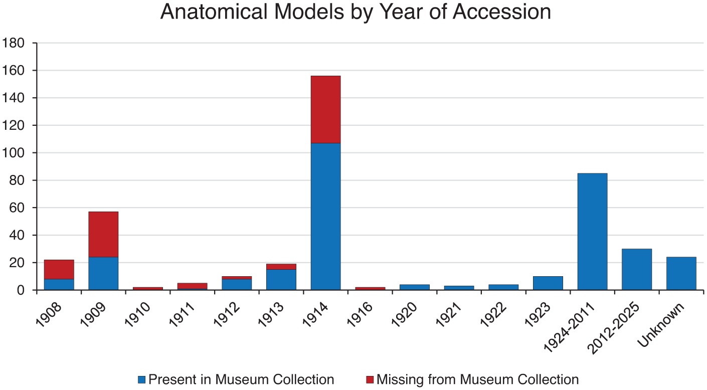
Models acquired by McGill’s Department of Anatomy and the Maude Abbott Medical Museum 1908 to 2025.
Specific features of the models are documented in Figures 2 (manufacturer) and 3 (material). Most consisted of specimens showing embryology (17%), the nervous system (14%), or the head/neck region (10%). The most represented modeler/manufacturer was Adolf Ziegler and son (123 models, 42%). Many of their models were developed in association with the anatomist Wilhelm His and demonstrated normal human embryological/fetal development (Figure 4). This subject lent itself particularly well to model creation because of the minute size of the material under study.
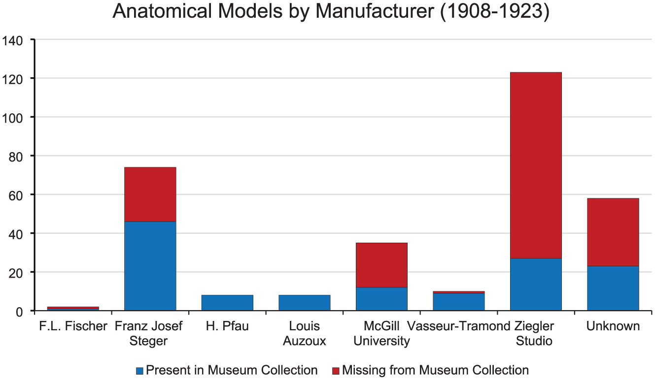
Models acquired 1908 to 1923: manufacturer.
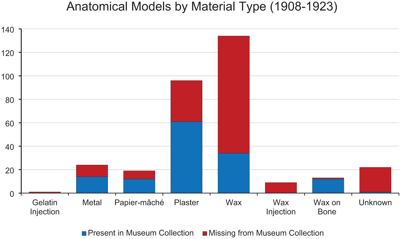
Models acquired 1908 to 1923: material type.
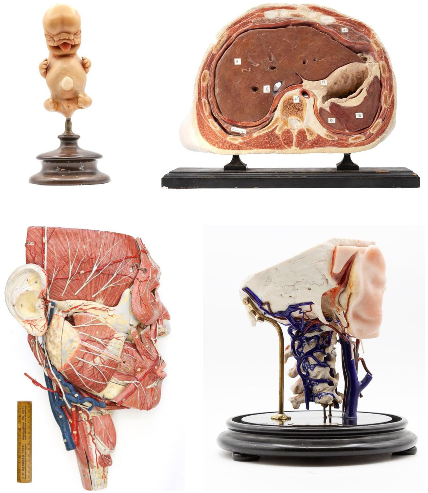
Models purchased 1908 to 1923: (A) Zeigler wax model of human embryo, c1914, (B) Steger cross sectional plaster model of upper abdomen. c1910, (C) Auzoux papier-mâché model of the face, c1920, and (D) Pfau wax-on-bone specimen of the temporal and adjacent bones. Its sequestration in a glass bell jar (not shown) accounts for its excellent preservation, c1910.
Franz Josef Steger, a particularly successful nineteenth century modeler, contributed the next most abundant part of the collection (74 models, 25%). These consisted mostly of representations of gross anatomical features such as muscles, brain, liver, etc. (Figure 4). Many of the Ziegler and Steger models were purchased as series, such as the urogenital system (Ziegler) and the brain (Steger). Ziegler’s models were constructed of wax and Steger’s primarily of plaster, which explains the high proportion of the two modelling substances during this period.
Of the 323 models that are present in the museum today (i.e., documented in the current MAMM database), 184 were accessioned in the museum logbooks between 1908 and 1923. We suspect that twenty-four of the remaining 139 were acquired before 1923 despite not being indicated in the logbooks. This is based on their appearance or similarity to images in published books or articles, and on the collecting activity of the Anatomy Department at that time. This group includes eight Auzoux papier-mâché models (a head, several views of the pelvic organs, and representations of male and female reproductive organs), four Pfau wax-on-bone models of the temporal bone and middle ear structures, and a series of seven painted metal models illustrating stages of development of the inner ear from an unknown manufacturer.
Interestingly, 35 (11%) of models accessioned during this period were created at McGill. Of these, a relatively large number (16) were metal casts of various organs or structures, such as the temporal bone showing the middle/inner ear, the trachea, the right heart ventricle, and the brain ventricular system. Casts of various joints created by injection of an unknown substance (8) or gelatin (one) were also made in the anatomy dissection room during this period; none have survived.
1924 to 2025
Since 1924, 115 models have been acquired, 85 (74%) between 1924 and 2012 and 30 (26%) since the 2012 MAMM inauguration (Figure 1). Manufacturers and model substances are shown in Figures 5 and 6. Organ type/anatomical region differed somewhat from those of 1908 to 1923, the three most common being head/neck (27%), nervous system (19%), and embryology (10%). Examples which exemplify the pre-2012 acquisition period are a set of five plaster casts of face anatomy and a series of seventeen representations of portions of the brain, both made by surgeon Carlton Smith at the University of Toronto in the 1970s/1980s (Davies 2004). Plaster is the most common substance employed in this time period, although plastic is a close second.
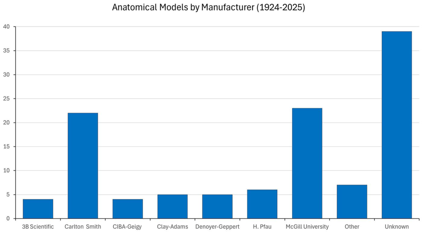
Models acquired 1924 to 2025: manufacturer.
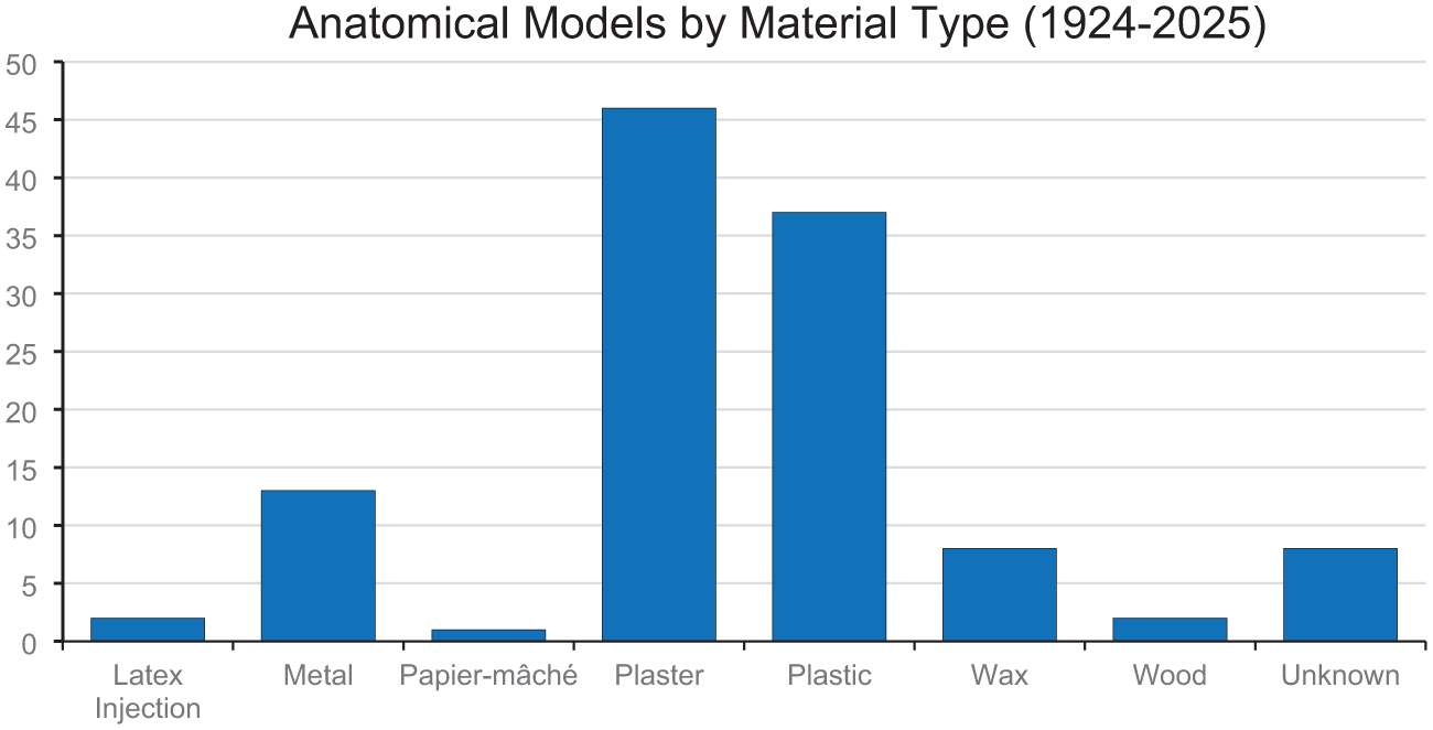
Models acquired 1924 to 2025: material type.
The models acquired by the MAMM since 2013 have several origins. The first, related to the current museum’s current role of preserving the University’s material cultural history, consists of donations of objects “discovered” in a retiring professor’s desk drawer or a cupboard being cleaned as part of a renovation project.
A second source is donations from private individuals whose deceased physician relative used anatomical models in their offices for patient education. These objects differ from the others in that they were presumably used to demonstrate the location of disease and/or potential treatment of it. They include organs such as brain and heart as well as more complex models such as the entire male genitourinary system. All are plastic and many have accompanying pharmaceutical company labels, suggesting that they were given to the physician by the company as a promotional activity. Seven similar models presumably meant for anatomy student teaching, also of plastic but without pharmaceutical labels, were acquired from the University Bookstore as part of a downsizing secondary to a change of location.
A third source consists of seventeen 3D printed plastic models. Since the academic use of 3D printing is relatively recent, all these objects have been acquired since 2018. Some, such as a series of seven hearts, were constructed in the anatomy department and painted by a professional artist as part of an investigation of their possible use in teaching. Others, such as copies a cervical vertebra and a temporal bone, have been created in the MAMM for use in student exercises of close observation and anatomical knowledge.
Discussion
To the best of our knowledge, 433 anatomical models were acquired by McGill’s medical museums, both the original one associated with the Department of Anatomy between 1908 and 2012 and the current MAMM. However, because of lack of documentation the actual number is certainly higher. For example, the anatomist and modeler Dr. Carlton Smith of the University of Toronto developed a set of twenty-four models to accompany his book The Serial Dissection of the Human Brain (Smith 1981). After the death of his wife, Smith is said to have donated two sets of the models to every medical school in Canada in her name (Davies 2004). The MAMM has seventeen of these models; no documentation of their arrival in the anatomy department is available and what happened to the remaining thirty-one models that were presumably sent is not evident.
McGill’s anatomy department did not seem to rely heavily on models for teaching prior to the 1907 fire. According to the calendar entries, none were deemed worthy of mention in the 33-year period from 1857/1858 to 1890/1891. Moreover, after acquisition of material related to nervous system anatomy around 1891/1892, nothing more was documented during the following sixteen years. Despite this, some models seem to have been acquired and presumably used during this time. For example, in 1919, Anatomy Department Professor Francis Shepherd indicated that the former anatomy demonstrator William Fuller had made “beautiful casts of frozen dissections . . . as far back as 1868” (Shepherd 1919). According to Shepherd, one unspecified set of these was in the museum when it was destroyed in the 1907 fire.
Although it has been suggested that the use of models in anatomy teaching decreased in importance relative to “live” body dissection during the 1900s, (Hallam 2016) their acquisition and, by extension, use was still important in at least some centers. That McGill was likely one of these indicated by its having acquired 294 models in the sixteen-year period after fire consumed its pre-1907 collection. Most of these (271) were acquired in the seven years following the fire. Presumably, this was because of the need to replenish the material available for teaching. It is possible that the Anatomy Department had special funding from the medical faculty to replenish its collections. The new Strathcona Medical Building that was constructed in 1909 included a large amount of museum space (almost 15% of the building’s area) which the faculty undoubtedly wished to be used. However, specific details about funding and the amounts paid for individual models are not available and this hypothesis cannot be confirmed.
As might be expected, the nature of the material purchased changed over time. Before 1913, most models were gross specimens such as torsos and pelvises manufactured by the Steger Company. In fact, the seventy-four models by this Company that were purchased and the forty-six that remain in the MAMM appear to comprise one of the larger such collections (Cornwall and Smith 2014). In 1914, most newly acquired models (113) came from Ziegler and illustrated embryological development. The majority of these were non-human, including complete series illustrating the development of Amphioxus (25 models) and the frog (25). The reason for this change in the focus of the collection towards developmental anatomy is not clear. However, although it may have been simply one of logistics—the budget allowing only so many purchases per year—it may also indicate an increased emphasis on embryology teaching.
What happened to the Museum’s 110 specimens acquired between 1908 and 1923 that have been “lost”? Undoubtedly, there are several reasons. Repetitive use by cohorts of students resulting in enough damage for the specimen to be no longer considered useful for teaching is perhaps the most likely. Breakage, particularly of the more delicate specimens, also must have occurred. Many such damaged models were likely discarded (although a few discovered in storage have survived). The observation that some wax-on-bone models of the temporal bone/inner ear by Pfau are much better preserved in both color and construction than other similar size and type of objects is probably explained by the fact that they have been preserved in glass bell jars, safe from “prying” student’s hands (Figure 4).
It has also likely that some items have been discarded because of a belief that they were too “old fashioned” for teaching (Maerker 2015). One retired McGill Anatomy Department professor we interviewed recalled a decision to stop using information from animal development studies in the teaching of human embryology. This made some Ziegler model series—such as those of amphioxus and the frog indicated above—irrelevant. It is not known if these were discarded, donated elsewhere, sold, or suffered some other fate. Whatever the process, none of these models are present in the MAMM today.
On the other hand, the fact that some series of human embryological material remain suggests that they were considered valuable for teaching in some way. Such importance is also illustrated by what appears to be the replacement of some lost models. For example, thirteen of the original sixteen specimens in one Ziegler human embryo wax series are extant in our collection. However, three of these are plaster replicas virtually identical to the original except that the construction material is not wax. Although we do not have written confirmation, it is possible that these were purchased or perhaps constructed locally to replace the damaged or lost originals.
Talairach-Vielmas (2014) has emphasized the precarious nature of anatomical model collections, particularly with respect to inadequate storage space, poor maintenance, limited funding, movement within or between institutions, and—perhaps most importantly—lack of interest. This would appear to be particularly relevant for delicate and wax models. An example of this at the MAMM was the 1998 discovery of a collection of wax moulages (not included in the figures for this article because they demonstrated pathology) haphazardly stored in a large cardboard box on the top shelf of a poorly ventilated and non-temperature-controlled basement storage room. Some of the moulages were wrapped in newspaper dated 1973, indicating that they had been unused for over twenty-five years. The wood base of some moulages had impacted the demonstrative wax face of their neighbor resulting in deformity. These moulages had presumably been stored in the box to keep them “safe.” However, the poor storage conditions, perhaps combined with movement of the box from time to time, resulted in significant damage. Apart from the injury itself, it is easy to understand how the entire collection might have been discarded if the box had been opened by someone who considered the damage sufficient to render the specimens “useless.”
Although the use of models for anatomy teaching has undoubtedly decreased significantly since the 1900s, (Hopwood and Ziegler 2002) they were certainly used at McGill throughout the twentieth century. This can be seen in photographs in which they are being manipulated and are evident in easily accessible storage cabinets in the 1940s (Figure 7). Moreover, new models were acquired into the late 1900s, such as a series of Smith plaster face dissections for teaching surgeons in training from the 1980s to the early 2000s. Finally, models are still used to illustrate anatomical features difficult to appreciate in routine student dissections. A collection of approximately thirty-five plastic models, most depicting the larynx, inner ear or heart, is stored on shelves near the entrance to the dissection laboratory. These have not been accessioned by the museum and are instead actively used for demonstrating aspects of anatomy which cannot be appreciated by routine student dissection—the rationale behind the acquisition of most models that have been accessioned by the MAMM.
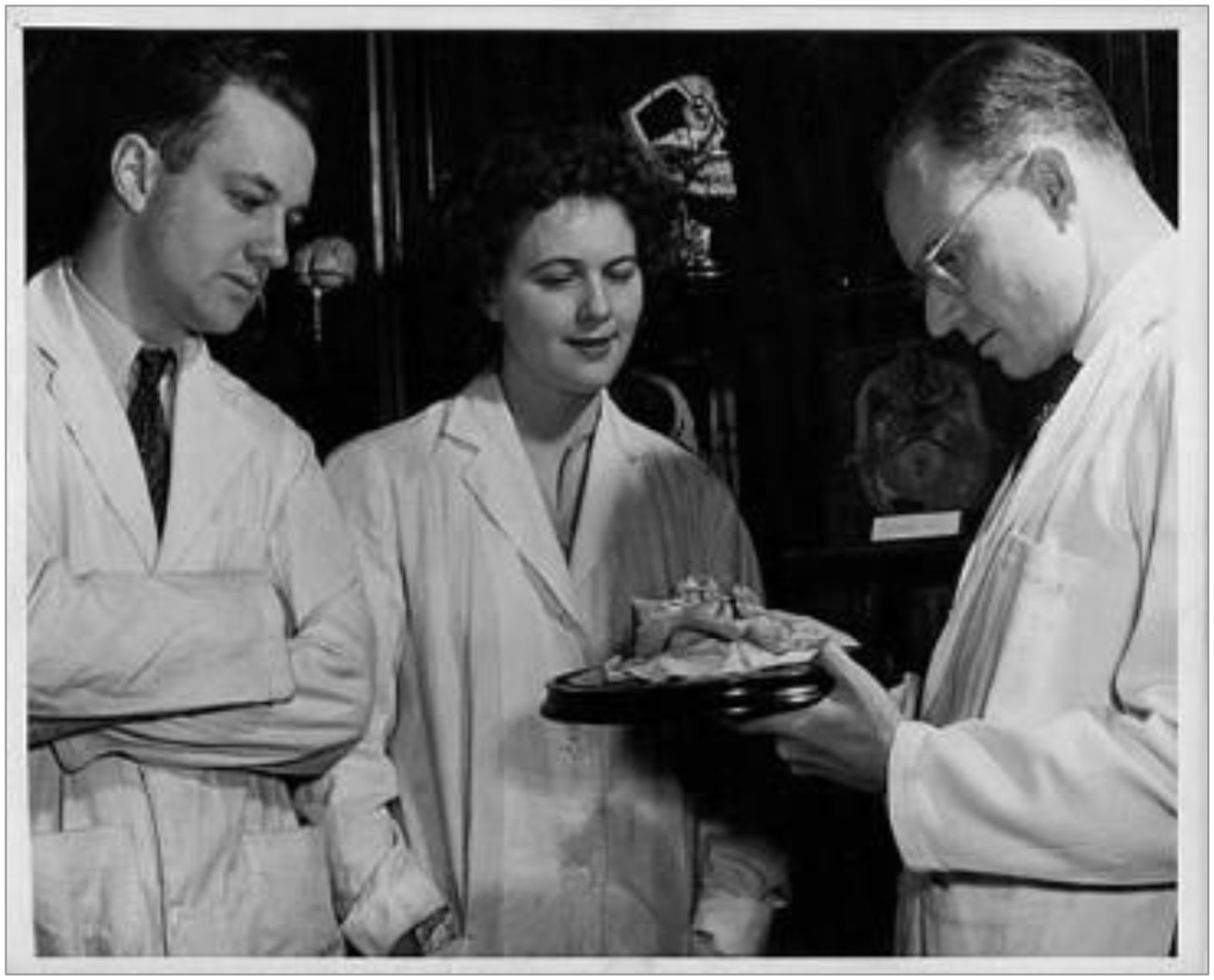
McGill anatomy professor Martin Banfill demonstrating a Tramond wax-on-bone model of the nerves of the eye to two students in a photograph dated 1946. A Tramond hemi-cranium can be seen at the top of a storage cabinet behind. Both models are still in the MAMM collection.
Although the primary use of museum models has been for teaching anatomy to medical and other healthcare students, historically they have also been exhibited to the public, for both educational and entertainment purposes (Talairach-Vielmas 2014). It could be argued that the “specimens” in the recent Body Worlds exhibits are the best-known modern-day manifestation of the latter (Moore and Brown 2007). It has also been suggested that anatomical models have been used to illustrate and presumably advocate for eugenic theory and to elaborate on some psychological theories (Spencer 2008). There is no evidence that any of McGill’s anatomical models were used for these purposes.
Although the great majority of models in the current MAMM collection were used for student teaching and originated in the Anatomy Department before 2012, two additional “categories” of model have been acquired by the MAMM since that date and used for “non-traditional” purposes. One of these is a small collection of models developed by pharmaceutical companies for the purpose of drug marketing and given to physicians for patient education. These have been donated by deceased physician’s relatives and were accessioned mainly to fulfill the MAMM’s mission of documenting the history of medicine via material objects. However, some are still being used per their original purpose as teaching aids, for example, during organized school group visits to the museum.
A second type of MAMM acquired model consists of those created by 3D printing, a technique that has been advocated by many investigators for anatomy teaching (Bisht, Hope, and Paul 2019; Fredieu et al. 2015; Titmus et al. 2023). We have found them to be useful in exercises involving delicate and/or small specimens. One example of this was a workshop held at Montreal’s Museum of Fine Arts that involved ten students, each of whom was given a printed temporal bone for close observation/sketching. The purpose of the exercise was to teach both descriptive and functional anatomy. After the students completed their sketches, they viewed and discussed artwork related to hearing and balance. The availability of one unbreakable model bone for each student both ensured preservation of the actual bone and enhanced the student’s experience by allowing manipulation and close viewing.
Additional uses of the models that are not strictly anatomical include discussions related to the history of medicine and aesthetics (art). The former is exemplified by a display in the main MAMM room that is used to illustrate both a general history of anatomy teaching as well as specific topics associated with it, such as the nineteenth century illegal sourcing of bodies for dissection (“body snatching”). This practice certainly occurred at McGill and is relevant for both the University’s health care students and the public to be aware of. The development of modelling as one way of mitigating the deficiency of bodies is used as an introduction to a discussion of this topic.
The value of some models as works of art—as objects to be considered aesthetically rather than scientifically—is also included in museum displays and activities. In fact, the techniques involved in the manufacture of some models, such as those made by life or corrosion casting, and the creation of some objects that resemble works of art, such as those in papier-mâché by Auzoux (Figure 4), clearly overlap (Hendriksen 2019). Even some of the damaged models remaining in the MAMM have an intricacy of structure reflecting skilled nineteenth century craftmanship, illustrating an aesthetical point of view that is probably appreciated by at least some museum visitors.
Conclusion
Models depicting various aspects of human anatomy have been created with a wide variety of substances and for several purposes. The collection amassed by McGill’s Medical Museum and the current MAMM over the past 175 years is an example of the value placed on the use of this material by one North American medical faculty. Although the most common such use, anatomy teaching, is less important today than when most of the models were acquired, their employment in other ways attests to their continued value, both as teaching objects and as a record of medical history.
Footnotes
Author’s Note
Sabina Boudames is now affiliated to Keck School of Medicine USC, Los Angeles, CA, USA.
Declaration of Conflicting Interests
The authors declared no potential conflicts of interest with respect to the research, authorship, and/or publication of this article.
Funding
The authors received no financial support for the research, authorship, and/or publication of this article.