Cerebral Changes Related to Sexual Dysfunction After Spinal Cord Injury: A Mixed Methods Study
Prof. Carl Froilan Leochico 1, Dr. Humberto Antonio Cerrel Bazo 2, Dr. Elia Zanin3, Dr. Sara Salizzato4
1197 D Interior P. Santos Street Malibay, Pasay, Metro Manila, Philippines-1300
2Via Padre Leonardo Bello 3/C 31045, Motta Di Livenza, Treviso, Italy
3Via Padre Leonardo Bello 3/C 31045, Motta Di Livenza, Treviso, Italy
4Via Padre Leonardo Bello 3/C 31045, Motta Di Livenza, Treviso, Italy
Objectives: Background: It is believed that the brain represents the body’s largest sexual organ. While sexual dysfunction is a commonly overlooked complication after damage to the spinal cord, the brain and hence the person’s most important sexual organ should remain intact. Nevertheless, majority of patients suffering from spinal cord dysfunction remain to suffer from sexual dissatisfaction, leading to impaired body image, low self-esteem, marital discord, and family breakdown.
Objective: This study examines whether cerebral function related to sexual arousal differs significantly between individuals with and without spinal cord dysfunction.
Methods: We employ the mixed-methods study design: 1) choice reaction time task experiment, 2) “think aloud” protocol, and 3) focus group discussion. Quantitatively, we measure the time elapsed between the onset of implicit and explicit sexual images presented on a computer screen and the correct identification of the participant. Qualitatively, we describe the verbal comments, facial expressions, and body language of the participant during the experiment using the videotaped “think aloud” protocol, and compare similarities and differences in the sexual experience of participants in a focus group discussion.
Results: A slower choice reaction time is the trend among patients with spinal cord dysfunction compared to normal participants. The “think aloud” protocol and focus group discussion unravels novel differences in the way the two groups react to sexual stimuli.
Conclusions: There is evidence to support the role of cognitive and psychological rehabilitation in addressing sexual dysfunction among patients with spinal cord dysfunction and consequent neuroplastic changes in the brain’s sexual functioning. Brain-computer interface as assistive technology in sexual rehabilitation is worth exploring in the future.
Clinical Profiling of Persons with Dementia in Selected Cognitive Linguistic Domains
Ms. Heena Sunil Shahani1, Ms. Gayatri Hattiangadi Hattiangadi2
1TNMC Nair Medical college, Mumbai, Maharashtra, India
2TNMC Nair Medical college, Mumbai, Maharashtra, India
Objectives: To compare the performance between persons diagnosed with dementia and matched controls.
To compare the performances between persons suspected with dementia and matched controls.
To compare the performance between persons diagnosed with and suspected to have dementia.
To check effect of age, gender and level of education on three groups.
Methods: The study consisted of 3 groups of participants, Group I (n= 19) individuals with Dementia as diagnosed by a Neurologist/Psychiatrist, Group II (n= 5) individuals suspected with Dementia; i.e. a score of less than 20 on Hindi Mental State Examination along with signs and symptoms of Dementia and Group III (n=24) normal aging individuals; i.e. a score of more than 20 on Hindi Mental State Examination.
The Cognitive Linguistic protocol developed in a previous study by Nagree (2012) was used to evaluate the participants in this study across various domains.
Results: Statistical analysis was done to compare the performance between: diagnosed dementia and their matched controls, suspected dementia and their matched controls,diagnosed and suspected dementia groups. A significant difference was found in the performance of diagnosed dementia and controls on cognitive tasks (p<0.02) and linguistic tasks (p<0.03) and also between suspected dementia and controls on various cognitive (p<0.02) linguistic subtests (p<0.02). Thus it was evident from above results that both the dementia groups performed poorer than their controls. No significant difference was found in performance of diagnosed and suspected dementia groups for cognitive tasks (p=0.118) and linguistic tasks (p=0.118).
Conclusions: The present study is a preliminary attempt to develop a clinically useful profile of cognitive linguistic aspects which will help in early differentiating between dementia and normal aging, early diagnosis of dementia as well as planning intervention and monitoring progress.
Impact of Theme of Music (Happy or Sad) on Attention, Concentration & Recall of Healthy Young Adults
Dr. Chitkala Pradeep Deshpande1, Dr. Odette Marilyn Gomes2
1T.N. Medical College & B.Y.L.Nair Hospital, Mumbai, Maharashtra, India
2T.N. Medical College & B.Y.L.Nair Hospital, Mumbai, Maharashtra, India
Objectives: 1. To assess the effect of happy theme of music on attention, concentration & recall of healthy adult subjects
2. To assess the effect of sad theme of music on attention, concentration & recall of healthy adult subjects.
Methods: Scientific literature gives lot of evidence that Music with its combinations of qualities like tone, rhythm, tempo, melody can lead to many effects on human emotions, also studies support the effect of Music on cognitive abilities both short term and long term. Hence an attempt is made to assess the effect of happy or sad theme of music on abilities like attention, concentration and recall, 2 groups of consenting healthy young adults (age group 18 to 30, both male and female) are made and each group is given a Digit Span test and Free Recall Test for baseline assessment and then one group is made to listen to a piece of music with happy theme whereas other to a sad theme music piece and then the tests are repeated for each group. Statistical analysis of obtained data will be done.
Results: As the study is an ongoing study, results might/ might not show that theme of music does have impact on abilities like attention, concentration and recall
Conclusions: The results might show the significant impact of theme of music on cognitive abilities like attention, concentration & recall which will help us get a deeper insight into impact of music on human brain and its effective use as a treatment tool in rehabilitation of persons with neuro-cognitive disorders.
Interpreting Spatial Dysgraphia After Stroke: Straight Ahead or Straight Above?
Prof. Dominic Pérennou1, Dr. Laure Mathevon 2, Dr. Celine Piscicelli3, Mrs. Claire Bérenger4, Dr. Anne Chrispin 5, Dr. Caroline Jolly6
1Grenoble-Alpes University Hospital & University, Grenoble, X, France
2Grenoble-Alpes University Hospital, Grenoble, X, France
3Grenoble-Alpes University Hospital & University, Grenoble, X, France
4Grenoble-Alpes University Hospital, Grenoble, X, France
5Grenoble-Alpes University Hospital, Grenoble, X, France
6Grenoble-Alpes University, Grenoble, X, France
Objectives: Spatial dysgraphia, frequent after a right hemisphere(RH) stroke, associates signs of spatial compression in relation to spatial neglect(SN), and a tilted writing which remains to be explained.We present a case study on the link between tilted writing and verticality representation.
Methods: JW, a 75year-old RH stroke patient showed a writing tilted 11.1° upwardly, without other signs of spatial dysgraphia.We assessed and followed spatial cognition, until nine months post-stroke. For writing assessments (orientation and time), head and trunk were kept upright, JW wrote with the non-paretic hand on a paper sheet affixed onto a graphic tablet positioned in front him on a slanted table (30°).The writing was tested with or without visual clues, and after a modulation of verticality perception.
Results: One month post-stroke, SN was severe, subjective straight-ahead was normal.Visual and postural (PV) verticality perceptions were tilted of 11°, counterclockwise.This transmodal tilt was similar both in direction and magnitude to the tilt congruent on all features of writing: margin, lines, letters. At three months, the writing was found slower than a control subject when JW wrote on blank paper(1.67 vs 0.82sec/letter; p<0.001), and faster when wrote on lines inclined 24° upwardly(1.11sec/letter; p<0.05).When JW was himself tilted, in sitting for 10min at 30° to right side in the darkness using the wheel Test, PV was normalized for several min.This PV modulation significantly reduced the writing tilt(6.2°; p=0.001) and increased the writing speed(0.89sec/letter; p=0.002) measured 20min later.Nine months post-stroke, we found a dissociation between SN, which disappeared, and a tilt remaining both in verticality perception(5°) and writing(8.8°).
Conclusions: Tilted writing after RHstroke is likely a sign of a tilted verticality representation.
Neurofeedback Training with Mu Rhythm for Social Cognitive Deficits: A Systematic Review
Ms. Manju Kumari1, Dr. Ankita Sharma2
1Indian Institute of Technology, Jodhpur, Rajasthan, India
2Indian Institute of Technology, Jodhpur, Rajasthan, India
Objectives: Neurofeedback therapy (NFT) has been used with diverse clinical and normal population; Gruzelier and Egner (2005) commented that the field of NFT has proceeded largely without validation. The objective of the present article is to provide a systematic review of 1) how NFT is utilized to treat social cognitive deficits, 2) how NFT is utilized to target Mu-MNS-Social Cognition Deficit especially in Autism, 3) examining the directions, strengths, and quality of evidence to support the use of NFT.
Methods: The databases for studies were searched in PubMed, MEDLINE, EMBASE, Springer, Science Direct, Psychinfo and Google Scholar, using combinations of the following keywords: ‘Neurofeedback’, ‘Autism’, ‘Mu Rhythm’ and ‘Social Cognition’. Studies were eligible for inclusion if they were specific to 1) autistic and typically developed population, 2) intervened on Mu rhythm protocol, 3) Delivered by NFT, 4) included participants who showed social cognition deficit. Present article includes papers published until May 2017.
Results: Total seventy-three papers were found of key interest; out of which 26 studies were eligible for inclusion in this review. Some studies showed social impairment as a main social cognitive deficit in Autism, while other showed that Social impairment is also core symptom of the deficit in executive functioning, observation, and imitation related behavior. The majority of studies found improvement in participant’s deficits. However the conclusive evidence was meager, there was only one study which showed the long-term effect of Neurofeedback.
Conclusions: The conclusion of the present review is in line with comments of Marzbani, Marateb, and Mansourian (2016) that, ‘current research does not provide sufficient conclusive results about its efficacy’. The patterns and directions concluded from studies related to the protocol, methodology and results are discussed in detail in the present review.
Keywords: Neurofeedback, Autism, Mu Rhythm, Social Cognition
Subliminal Sexual Perception: The Role of Spinal Cord
Dr. Carl Froilan Leochico 1, Prof. Jose Alvin Mojica2, Dr. Elia Zanin3, Prof. Reynaldo Reyes Rey Matias4, Dr. Humberto Antonio Cerrel Bazo 5
1RMR Physical Therapy Clinic, Manila, Metro Manila, Philippines
2Philippine General Hospital, Manila, Metro Manila, Philippines
3Ospedale Riabilitativo di Alta Specializzazione, Motta di Livenza, Treviso, Philippines
4St Luke’s Medical Center, Quezon City, Metro Manila, Philippines
5Ospedale Riabilitativo di Alta Specializzazione, Motta di Livenza, Treviso, Italy
Objectives: Background: According to the literature regaining sexual functioning is one of the most important priorities for spinal cord injury (SCI) patients. As SCI affects sensory pathways, there is evidence that the emotional processing is experienced differently from normal people. However, little is known about the differences in sexual system between SCI people and normal population.
Objective: We aimed at investigating whether SCI people experience differently sexual arousal than previously thought if compared to normal population.
Methods: We investigated implicit versus explicit activation of sexual arousal in patients with SCI and normal population with brief exposure to sexual stimuli. We measured RTs and pictorial judgment task.
Results: Although preliminary, our results suggest that there are differences in the way subliminal sexual pictures modulated sexual arousal of SCI and normal population in terms of behavioral correlates.
Conclusions: These results provide important hints about the role of spinal cord lesion in causing different mechanisms underlying sexual system of normal and SCI patients. In particular, these findings indicate that sexual arousal does interfere with cognitive processing in different manners between normal and SCI people. These results thus highlight the importance of spinal cord for first-hand emotions and feelings in the way we experience the perception of sexual arousal.
Construction of Questionnaire to Asses Third Party Disability in Mothers of Children with Autism Spectrum Disorder (CWASD)
Mr. Rishidhar Ashokkumar Dubey1, Mrs. Alpana Pagare2
1TNMC, Mumbai, Maharashtra, India
2TNMC, Mumbai, Maharashtra, India
Objectives: 1. To develop questionnaire in ICF domains of activity, limitations, participation restrictions (AL/ PR) and environmental factors (EF) for assessing third party disability in mothers of children with Autism Spectrum Disorder (CWASD). 2. To establish face and content validity; intra-tester and inter-tester reliability of the questionnaire.
Methods: Chapters of ICF consist of sub domains and codes i.e. AL & PR consists of 9 sub domains, EF consists of 4 domains. This domain explains difficulties faced by the mothers in her personal, social and professional life and also daily living activity gets affected. After the questionnaire was developed by the researcher using ICF domains, it was given to 08 Audiologist and Speech and Language Pathologist and 02 Psychologists for the validation. Further the questionnaire was translated in Hindi and Marathi language and reverse translation was done. Pilot testing was done on 30 mothers having their child diagnosed with ASD between age range of 3 to 16 years as per inclusion and exclusion criteria. The validated questionnaire consists 62 questions was administered on mothers. Mothers had to choose the correct option on 5 point rating scale indicative of level of difficulty.
Results: High internal consistency was obtained with moderate to strong correlation for inter-tester and intra-tester reliability. Disability percentage showed moderate difficulty Communication, Interpersonal Interactions and Relationship, Major Life Areas, Support and Relationships, Attitudes, Domestic Life, LAK, Mobility, GTD. Other domains like Self Care, Products and Technology, Community Social and Civic Life showed severe disability. Few domains showed significant difference in disability between the groups, first group showed more percentage of disability compared to second group.
Conclusions: Thus questionnaire to asses third party disability in mothers of CWASD was constructed with validity and reliability measures. The result shows that mothers with CWASD face difficulty in rearing up the child.
Neuropsychological Contribution to the Diagnosis of Nonverbal Learning Disorder: A Review
Ms. Daiane da Silva Pinson1, Mrs. Isabela Chicarelli Amaro Santos2, Prof. Bruna Tonietti Trevisan3, Dr. Eliane Correa Miotto4
1Center for Neurology Studies, Division of Neurological Clinic, Hospital das Clínicas, FMUSP, Sao Paulo, Sao Paulo, Brazil
2Center for Neurology Studies, Division of Neurological Clinic, Hospital das Clínicas, FMUSP, Sao Paulo, Sao Paulo, Brazil
3Center for Neurology Studies, Division of Neurological Clinic, Hospital das Clínicas, FMUSP, Sao Paulo, Sao Paulo, Brazil
4Center for Neurology Studies, Division of Neurological Clinic, Hospital das Clínicas, FMUSP, Sao Paulo, Sao Paulo, Brazil
Objectives: This study aimed to present a literature review about the importance of neuropsychological assessment for the diagnosis of children with Nonverbal Learning Disorder (NVLD).
Methods: We carried out a literature review of studies published until the year 2016. Based on the objective of this research, the studies found with the terms neuropsychological contribution and diagnosis, combined with the term nonverbal learning disorder, were searched in the indexes LILACS, Medline, PubMed and Scielo.
Results: The analysis of the five studies found that Nonverbal learning disorder (NVLD) is characterized by a change in the functioning of the nervous system, which causes impairments in mathematical reasoning, visuospatial cognition, motor coordination, sensory perception and social skills. In Brazil, the studies on Nonverbal learning disorder (NVLD) began in the 80’s and is still a poorly studied profile, which makes it difficult to elaborate a diagnosis and an adequate treatment.
Conclusions: That Nonverbal learning disorder (NVLD) affects children from birth, entails significant impairment in cognitive, social, emotional, psychomotor, spatial, tactile, and visual functions, and has a significant impact on learning. These findings demonstrate the importance of the neuropsychological assessment in establishing a reliable diagnosis and enabling an adequate intervention, thus corroborating with the data of the studies used in this research. Faced with the complexity of the topic, new studies are needed.
Keywords: Learning Disorder; Nonverbal Learning Disorder; Neuropsychological evaluation; Cognition.
Neurorehabilitation in Two Adults with Hurler Syndrome and Morquio A Syndrome due to Attentional Difficulties
Prof. Nolly Nataly Castañeda Ibáñez1, Mr. Oscar Alejandro Gómez Suarez2, Prof. Mayerli Andrea Prado Rivera3, Prof. Alejandra Rizo Arévalo4, Prof. Diana Carolina Cárdenas Poveda5, Ms. Angie Paola Beltrán Beltrán 6, Mr. Jonathan Ferney Lamprea Garzón7
1Corporación Universitaria Minuto De Dios Uniminuto, Bogota D.C., Bogota, Colombia
2Corporación Universitaria Minuto De Dios Uniminuto, Bogota D.C., Bogota, Colombia
3Corporación Universitaria Minuto De Dios Uniminuto, Bogota D.C., Bogota, Colombia
4Corporación Universitaria Minuto De Dios Uniminuto, Bogota D.C., Bogota, Colombia
5Corporación Universitaria Minuto De Dios Uniminuto, Bogota D.C., Bogota, Colombia
6Corporación Universitaria Minuto De Dios Uniminuto, Bogota D.C., Bogota, Colombia
7Corporación Universitaria Minuto De Dios Uniminuto, Bogota D.C., Bogota, Colombia
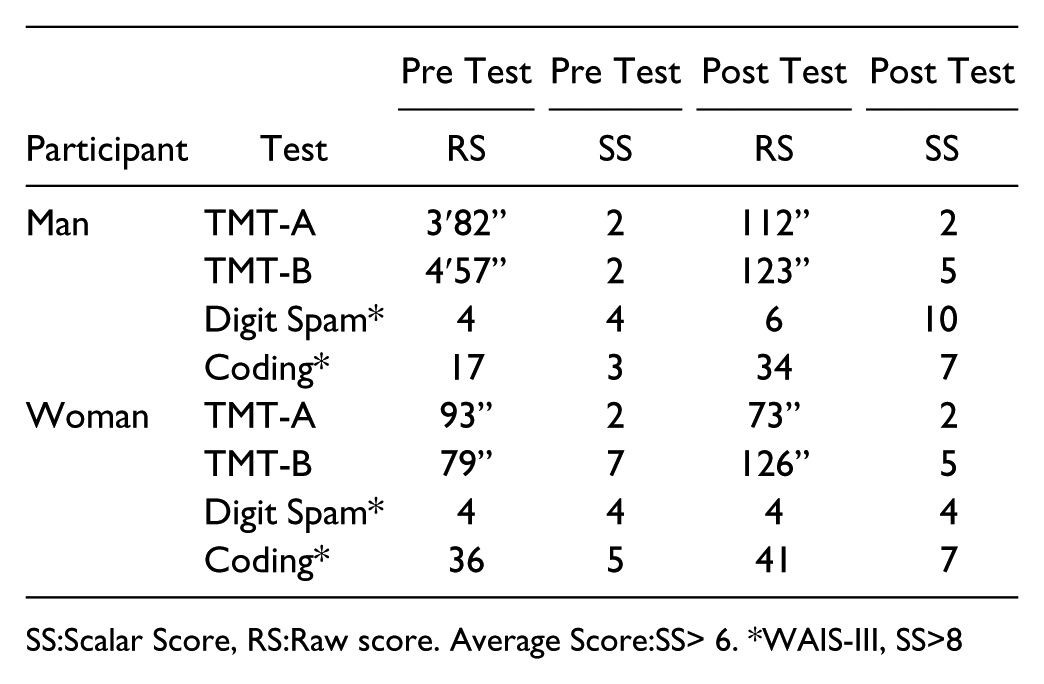
Objectives: To evaluate and describe the attentional mechanisms neuropsychological functioning in two adults diagnosed with Morquio A Syndrome and Hurler Syndrome before and after a cognitive stimulation program.
Methods: Descriptive and cross-sectional case reports with a pre and post test. A 35 years old man with Hurler Syndrome and a 30 years old woman with Morquio A Syndrome who live in Bogotá-Colombia, with scholar skills and a career as a system engineer both of them. The neuropsychological tests used was based on previous studies to assess neuropsychological domains such auditory, visual attention and processing speed, taking the native language of the participants (Spanish) and scales for Hispanic and Colombian population. The cognitive stimulation program was based on the principles of neuropsychological rehabilitation (enhanced learning) whereby the adults work together with a student group to improve attentional mechanisms during 2 weeks, 3 times per week with 12 sessions. Activities were adapted to them, thinking on the physical and cognitive manifestation of the syndromes (Mucopolysaccharidosis). After the program the adults was assessed again with the same neuropsychological tests.
Results:
SS:Scalar Score, RS:Raw score. Average Score:SS> 6. *WAIS-III, SS>8
Conclusions: The cognitive stimulation program was effective in some attentional areas; these results suggest that there are possible improving attentional skills in Hurler and Morquio A patients by different tasks based on the principles of neuropsychological rehabilitation. Even so, it is necessary to extend population and interventions programs.
Rehabilitation Challenges in an Elderly Cauda Equina Syndrome with Concurrent Normal Pressure Hydrocephalus a Case Report
Dr. Wan Najwa Wan Mohd Zohdi1, Dr. Nur Nadia Abd Rahim2, Dr. Nadia Mohd Mustafah3
1Universiti Teknologi MARA, Sungai Buloh, Selangor, Malaysia
2Universiti Teknologi MARA, Sungai Buloh, Selangor, Malaysia
3Universiti Teknologi MARA, Sungai Buloh, Selangor, Malaysia
Objectives: Introduction: Cauda equina syndrome is a known diagnosis that entails neurogenic bladder, neurogenic bowel with or without lower limb neurological impairment. In medically stable and otherwise healthy patients, the functional outcome after optimum rehabilitation are mostly successful with subsequent ability to return to community and premorbid job.
Methods: Case Report: We present a case of a 70 year-old lady with underlying diabetes mellitus type 2 and cauda equina syndrome secondary to degenerative L4/L5 anterolisthesis who had undergone surgical correction with posterior instrumentation. At one month post-surgery, she developed delirium secondary to urinary tract infection which was treated with antibiotic. Post treatment, she only showed partial improvement in cognitive function, with residual disorientation to place and person, poor focus and recall as well as inconsistent comprehension. Training for bed mobility, basic activities of daily living including bowel and bladder care were not attainable. Magnetic resonance imaging of the brain and whole spine revealed communicating hydrocephalus with no obvious cause. With presence of dementia features, she was diagnosed with normal pressure hydrocephalus (NPH) since abnormal gait and urinary incontinence as part of the classic signs of NPH could not be assessed due to the masking cauda equina syndrome. Subsequent assessment predicted poor response to shunt surgery, hence it was not performed.
Conclusions: The challenges to organize rehabilitation in elderly cauda equina syndrome is not only affected by the age factor but also due to the concurrent presence of brain organic condition that affects cognition and motivation significantly. This has resulted in unfavorable rehabilitation potential and outcome. Multidisciplinary approach, caregiver training, social support and serial counselling to the family are the key elements in rehabilitation phase to avoid depression and further deterioration in both the patient and caregiver.
To Test if Separation Anxiety(SA) is Absent/Attenuated in Autistic Children
Dr. Nisreen Bohra1, Dr. Vrajesh Udani2
1PD Hinduja National Hospital & MRC, Mumbai, Maharashtra, India
2PD Hinduja National Hospital & MRC, Mumbai, Maharashtra, India
Objectives: Autism is a common neurodevelopmental disorder with mean age of diagnosis around four years.Routine screening enables early diagnosis and interventionleading to significant improvement.Modified Checklist for Autism in Toddlers (MCHAT), most commonly used screening testin toddlers is very subjective and relies completely on the parent’s observations. One of the authors (V.P.U) observed SA, a normal developmental milestone is absent/attenuated in autistic children.Hence this study was carried out to confirm this observation.
Methods: This was a prospective study in a paediatric neurology clinic.30 toddlers between 1-3 years suspected with autism were recruited as cases and 30 age matched normally developing toddlers as controls after informed consent.Exclusion criteria were motor disability, secondary causes of autism eg epilepsy, syndromes etc.
We devised separation anxiety score (SAS) modified from Ainsworth’s attachment scale. Higher scores(>/= 3) indicate absent/attenuated SA. Cases classified as “At risk” on MCHAT were subjected to SAS.Cases were observed on Childhood Autism Rating Scale/Autism Diagnostic Interview-Revised to confirm autism.Controls who passed the MCHAT were subjected to SAS.
Results: 28/30 cases vis-à-vis 2/30 controls had absent attenuated SA which was statistically significant.The median SAS was 4 for the cases and 2 for the controls. Specificity, sensitivity, Positive and negative predictive value was 93 % each for SAS. Socio demographic factors like gender, birth order, joint/nuclear family, parents education and socio-economic status did not show a significant difference.
Conclusions: The evaluation of SA using SAS is a helpful clinical sign in the early diagnosis of autism in toddlers, and can be easily performed in a busy OPD.
Usefulness of a Brief Cognitive Tool to Predict Progression of Amnestic Mild Cognitive Impairment to Alzheimer’s Disease
Ms. Jwala Narayanan1, Dr. Ratnavalli Ellajosyula2, Ms. Nidhi Dev3
1Manipal Hospitals, Bangalore, Karnataka, India
2Manipal Hospitals, Bangalore, Karnataka, India
3Manipal Hospitals, Bangalore, Karnataka, India
Objectives: To evaluate the usefulness of a brief cognitive tool in predicting progression of patients with amnestic mild cognitive impairment (aMCI) to Alzheimer’s disease (AD). Currently various biomarkers are used to predict conversion of amnestic MCI to Alzheimer’s disease (AD). We investigated the usefulness of a brief cognitive tool as an alternative to predict the conversion.
Methods: Standard neuropsychological tests and the brief cognitive tool, the Addenbrooke’s Cognitive Examination (ACE-III) were administered to a group of aMCI patients (n=55) and healthy controls (n=83). The diagnosis of MCI, aMCI and AD were based on standard criteria and patients were followed over 3 years. Performance at baseline was retrospectively analysed to differentiate aMCI patients who had either progressed (pMCI) to develop AD or remained stable (npMCI). One-way ANOVA and ROC curves were used to analyze data.
Results: The ACE-III total score (p= 0.025), ACE-III memory sub-component (p=0.00) and the visual association test [VAT] (p=0.00) significantly differentiated between the pMCI and npMCI groups. While the verbal and visual memory tests significantly differentiated the healthy controls from other groups, they were not able to differentiate between the pMCI and npMCI.
Conclusions: Our finding suggests that simple bedside cognitive tool like the ACE can be useful in the clinic to identify patients with aMCI who progress to develop AD. This is non-invasive, quick, cost-effective and easily available as compared to biomarkers. Early recognition is the key to therapy and carries prognostic implications.
Is the abstract presented earlier?: Yes
Demography of Cases Referred for Psychological Support and Counselling in a Developing Country a Descriptive Study
Dr. Intan Sabrina Mohamad1, Mr. Hairol Kamal Ab Rahman2, Mrs. Wan Norizan Wan Othman3, Mrs. Norhazniza Mohammad4, Ms. Nur Adhelia Alisa Abdullah5, Ms. Noor Hazwani Zulkifli6, Mrs. Farawahida Ahmad7, Mrs. Zaliah Jusoh8, Ms. Kong Lai Tee9
1Hospital Rehabilitasi Cheras, Kuala Lumpur, Federal Territory, Malaysia
2Hospital Rehabilitasi Cheras, Kuala Lumpur, Federal Territory, Malaysia
3Hospital Rehabilitasi Cheras, Kuala Lumpur, Federal Territory, Malaysia
4Hospital Rehabilitasi Cheras, Kuala Lumpur, Federal Territory, Malaysia
5Hospital Rehabilitasi Cheras, Kuala Lumpur, Federal Territory, Malaysia
6Hospital Rehabilitasi Cheras, Kuala Lumpur, Federal Territory, Malaysia
7Hospital Rehabilitasi Cheras, Kuala Lumpur, Federal Territory, Malaysia
8Hospital Rehabilitasi Cheras, Kuala Lumpur, Federal Territory, Malaysia
9Hospital Rehabilitasi Cheras, Kuala Lumpur, Federal Territory, Malaysia
Objectives: To determine the demographic features of the cases referred for psychological support and counselling in the Ministry of Health, Malaysia.
Methods: A retrospective descriptive study was performed using data obtained from an online-based Case Management System (KesMS) by the Psychological Counselling Unit, Ministry of Health (MoH), Malaysia from January 2013 to October 2017. All cases referred for counselling are entered daily by counsellors from the MoH based on their diagnoses or reason for the referral. Data and graphs were analysed using Excel Version 2010.
Results: A total of 54,943 cases were referred from 14 states nationwide. Two third of the cases comprised Malay Muslim females. The most common causes were depression (10-15%), stress (5-6%), adjustment disorder and unmarried pregnancies (2-5%). Married and single persons were 14 times more likely to seek psychological support compared with divorcees or widows. Muslims were eight times more likely to seek psychological support compared with those from other faiths. The majority of clients were government servants (32.3%), unemployed (30%) and students (8.8%).
Conclusions: Malay Muslim females were more likely to seek psychological support and counselling compared with other ethnic and religious groups. Depression is the most common cause, followed by stress and adjustment disorders. Resources should be allocated for these target groups. Further studies need to be conducted to manage life stressors.
Investigating Causes
Dr. Sumeet Gurunath Sarang1, Dr. Dhruv Suryakant Mehta2
1MGM School of Physiotherapy, Mumbai, Maharashtra, India
2MGM School of Physiotherapy, Mumbai, Maharashtra, India
Objectives: To study in the Indian Context, owing to tremendous constraints of accessibility and architectural barriers how cerebral palsy patients have coped up with their ambulation adult life.
Methods: A questionnaire concerning demographic facts, active participation, environmental factors and structural impairments were investigated. Data was collected through an interview in the area of Mumbai. Thirty five patients answered the questionnaire out of which eight patients were female and twenty seven patients were male, with standard deviation of 5. Mean Age was twenty five years with range of 17 to 40 years. 24 patients were Spastic Diplegic, 5 patients were spastic quadriplegia, 4 patients were spastic triplegia, 1 patient was spastic hemiplegia and mixed cerebral palsy was one.
Results: 54% of the patients reported they were walking independently and 46% patients were using assistive devices, 82% of patients reported having balance problem. 57% patients reported that their strength has improved 31% reported strength has remained same; 9% patients reported they are deteriorating. 82% patients reported they were able to walk on uneven surface, and 97% were able to climb stairs and walk in crowded places but only 34% of patients were able to climb stairs independently and 28% of patients needed railing for support while rest 28% of patients needed assistive device or others assistance
Conclusions: Considering results of the patients, majority of the patients were able to maintain their GMFCS level, It has been seen that few patients, (n=5) who were GMFCS 3 have now become GMFCS 4, (that is now functioning one level lower on mobility). Because of lack of phyiotherapy, caregivers assistance and illness. Special care should be taken for the patient of GMFCS 3 and 4 that as these group of patients (n=5) are going down in their level of GMFCS.
Magnitude of Co-Morbidities in Pediatric Epilepsy: Need for Neuropsychological Rehabilitation
Ms. Somini Haresh Mehta1, Ms. Somini Haresh Mehta2, Dr. Urvashi Rupin Shah3, Ms. Mayuri Kalika4, Ms. Tanvi Dingankar5, Ms. Yuvika Kamdar6, Dr. Sangeeta Ravat7
1K.E.M Hospital, Mumbai, Maharashtra, India
2K.E.M Hospital, Mumbai, Maharashtra, India
3K.E.M Hospital, Mumbai, Maharashtra, India
4K.E.M Hospital, Mumbai, Maharashtra, India
5K.E.M Hospital, Mumbai, Maharashtra, India
6K.E.M. Hospital, Mumbai, Maharashtra, India
7K.E.M. Hospital, Mumbai, Maharashtra, India
Objectives: Chronic epilepsy is associated with significant co-morbidities that are often more disabling than the seizure itself. Children with epilepsy often present with neuropsychiatric issues and cognitive impairments (memory, speed of processing and attention deficits) despite having normal global intellectual functioning. These impairments affect academic performance and overall quality of life yet remain unacknowledged and unaddressed. There is a growing need for early identification to plan effective interventions.
The objective of the study was to examine the prevalence and spectrum of cognitive, mood, behavioural issues and academic difficulties in a group of children with chronic epilepsy in a tertiary hospital epilepsy clinic.
Methods: Retrospective data analysis of 30 children with focal epilepsy with normal intelligence underwent standardised, domain specific neuropsychological evaluation. Cognitive impairments as per the test score percentiles; mood, behaviour rating as per scales and parent report and academic difficulties as per the grades and teacher report were analysed.
Results: The mean duration of seizures was 5.64 years and the mean general intelligence (IQ) was 93.6. 80% had mood and behaviour difficulties (irritability, anger outbursts, oppositional defiant behaviour). 43% had academic challenges and 40% presented with cognitive complaints. On objective testing impairments were seen in executive functioning (73%), attention (>50%), and speed of processing (50%).
Conclusions: Early identification, monitoring, and appropriate intervention and rehabilitation for neuropsychological problems are required. Long-term risk of academic underperformance exists despite normal global intelligence. This study underscores an urgent need for psycho-education and awareness programmes for parents, teachers and health professionals to plan effective neuropsychological rehabilitation.
Effect of Medical Welfare Cooperation of “Vocational Rehabilitation Planning Sheet for People with Cognitive Disorders After Acquired Brain Injury”
Dr. Tamami Aida1, Dr. Takashi Yamada2
1Mejiro University, Saitama Shi, Saitama Pref., Japan
2Mejiro University, Saitama Shi, Saitama Pref., Japan
Objectives: In Japan, social supports in daily living and social activities for clients with Cognitive disorders (CDs) after acquired brain injuries (ABI) were started in 2011. However, VR services were not effectively used by clients with CDs. The purpose of this study is to verify the effect of promoting transition to Vocational rehabilitation (VR), according to professionals’ use of the brochure for consulting clients at hospitals.
Methods: We printed an 8-page brochure in B5 size about the processes for clients and professionals which we found by a qualitative analysis from 12 professionals’ interviews.
Objects are professionals for CDs at seven hospitals who are members of support councils for CDs in Itabashi, Tokyo. We surveyed before and one month after the use of the brochure by the placement method. We constructed a questionnaire that has 20 items, 7 items about the Capacity of VR, 7 items about the Knowledge of VR, 6 items about the Experience of VR. We gave a presentation about the brochure at hospitals that the used it to consult about VR with clients who need RTW or reemployment. We used Mann-Whitney U for comparing before and after using the brochure. This study has been approved by the research ethics committee of Mejiro University (Permission number: 14-016).
Results: The survey period was August to September, 2016. Of 78 people, 68% were Occupational Therapists, 21% were Speech Therapists. 69 people answered again after a month. 30% used the brochure, 70% didn’t have clients who must use the brochure. After using the brochure, knowledges about VR transfer items were significantly increased. There weren’t significantly decreased items. Also, offering VR wasn’t significantly different.
Conclusions: The brochure was effective for improvement of VR knowledge.
Neuropsychology Practice in India: Online Survey Based Study
Ms. Farzana Mulla1, Dr. Aparna Dutt2, Prof. Jonathan Evans3
1Jehangir Hospital, Pune, Maharashtra, India
2Duttnagar Mental Health Hospital, Kolkatta, Maharashtra, India
3University of Glasgow, Glasgow, Scotland, United Kingdom
Objectives: Neuropsychology is a relatively young profession in India. To date, no study has examined aspects of neuropsychological practice in India. Hence, the purpose of this online study was to identify characteristics of individuals working in the area of neuropsychology with a view to understanding their educational background, current work situation, referral pattern, existing professional and organizational support and future needs.
Methods: An online survey was administered to participants via an email link. Participants who completed the online survey were self-identified psychologists who carried out neuropsychology related work. 36 participants from geographically different parts of India completed the survey. The data was analyzed into frequencies.
Results: Findings from the online survey suggested that those practicing in neuropsychology are predominantly females, have limited training in neuropsychology in India, formally through placement during MPhil in clinical psychology or informally through guidance from senior neuropsychologists and/or through attending workshops. Majority are relatively new to the field with less than 5 years of experience and mainly practicing in the private sector. Those practicing in government or teaching hospitals constitute a minor segment. Referral numbers are currently low, and these are primarily for neuropsychological assessment rather than rehabilitation. The findings further identified a huge gap, namely a significant need for formal courses or training in neuropsychology and neuropsychological rehabilitation in India.
Conclusions: Despite some limitations, the online survey was the first evidence based exploration of the environment and needs of those practicing in neuropsychology. It provides an early insight into current working conditions, identifying future needs and direction.
Validation and Proof of Concept Pilot Study on Cognitive Retraining Strategies in Early Dementia
Ms. Nandini Mohanan1, Ms. Mrudula Mathew2, Ms. Indupriya B3, Ms. Manju Mohan4, Ms. Elshal Bava5, Ms. PG Rajesh Pillai6, Dr. Ramshekhar Menon7
1Sree Chitra Tirunal Institute For Medical Sciences And Technology, Trivandrum, Kerala, India-695011
2Sree Chitra Tirunal Institute for Medical Sciences, Trivandrum, Kerala, India
3Sree Chitra Tirunal Institute for Medical Sciences, Trivandrum, Kerala, India
4Sree Chitra Tirunal Institute for Medical Sciences, Trivandrum, Kerala, India
5Sree Chitra Tirunal Institute for Medical Sciences, Trivandrum, Kerala, India
6Sree Chitra Tirunal Institute for Medical Sciences, Trivandrum, Kerala, India
7Sree Chitra Tirunal Institute for Medical Sciences, Trivandrum, Kerala, India
Objectives: Literature from India on cognitive retraining (CR) in mild cognitive impairment (MCI) and early dementia is sparse. To develop and validate a CR strategy and to examine its utility in rehabilitation of subjects with MCI and Early Dementia.
Methods: A CR strategy was devised across multiple domains (attention, orientation, memory, executive function, language, visuospatial). Inter-examiner reliability was calculated and estimated at 0.80. Subjects were evaluated using the Malayalam versions of Addenbrooke’s cognitive examination (ACE), and standard neuropsychological tests to identify the maximal affected domain. Individualised domain-deficit specific cognitive retraining was given with weekly one hour therapy sessions. Clinical dementia rating scale (CDR) and instrumental activities of daily living (IADL) assessments were done on a monthly basis to assess the efficacy of the therapy in addition to follow-up neuropsychology tests at 6 months and 1 year.
Results: Out of a total of 60 early dementia with CDR is less than 2, 13 patients consented for CR therapy (amnestic MCI-6, multi-domain MCI-1; early AD-5; limbic encephalitis-1), based on the scores in neuropsychological test evaluation. CDR ranged from 0.5- 4 (calculated as sum of boxes); ACE ranged from 65-89; age at evaluationranged from 60- 85. Post retraining CDR change ranged from 0.5- 1 in more than 50% of the cohort, with stability and no further decline in the remaining participants. Subjects with amnestic MCI and the single subject with multi-domain MCI were more likely to demonstrate improvement on neuropsychological functions of attention, memory, executive functioning, planning and visuo-spatial skills.
Conclusions: This is a unique validation and proof of concept case series from India on the efficacy of CR in MCI and early dementia.
A New Computer-Based Saccade Training Program for Patients with Hemianopia Preliminary Results
Prof. René M Müri1, Ms. Nadia Schweizer2, Prof. Klemens Gutbrod3, Prof. Tobias Nef4
1University Hospital Inselspital, Bern, BE, Switzerland
2University of Bern, Bern, BE, Switzerland
3University Hospital Inselspital, Bern, Be, Switzerland
4University of Bern, Bern, BE, Switzerland
Objectives: Stroke is the most common cause for hemianopia followed by traumatic brain injury and brain tumors. Though early recovery is expected in around half of cases within the first 3 months after injury, patients with persistent hemianopia are often disabled in activities of daily living. Rehabilitative compensatory approaches generally target eye movement training which are needed to improve explorative behavior in the hemianopic visual field. We developed and evaluated a computer based training tool for patients with homonymous hemianopia that is portable, inexpensive, and easy to deploy.
Methods: The computer based training program consists of two client interfaces, “Therapist” and “Patient” with an automatic difficulty adjustment. The task is a visual search task, presented on pictures or videos during 20-30 seconds. A server based user management system is used to store and synchronize data between both interfaces. The complete application was evaluated with four patients. Participants were instructed to press either the right or left arrow key depending on the stimuli presented on the background. Upon improvement, the background changed from images to videos. Patient’s feedback was collected via the System Usability Score and the Saccadic Eye Training questionnaire.
Results: The average usability score was 96.875. Patients who claimed to work more often with computers, and have better computer knowledge show higher SUS scores. As the difficulty increases, the completion time is longer. The reaction time in the impaired quadrants is slower compared to the other quadrants.
Conclusions: An advantage of the new training program is the automatic adjustment of the difficulty level in par with the performance and individual capacity of the patient, which will insure patient motivation and participation. The new training program will insure patient compliance and improve quality of life of patients suffering from visual field deficits.
Case Study Highlighting the Effectiveness of Intensive Neuropsychological Rehabilitation 13 Years Since Brain Injury
Ms. Shraddha Shah1, Ms. Pooja Gala2, Ms. Vaishnavi Trivedi3
1Synapsium Brain Health Clinic, Mumbai, Maharashtra, India
2Synapsium Brain Health Clinic, Mumbai, Maharashtra, India
3Synapsium Brain Health Clinic, Mumbai, Maharashtra, India
Objectives: To highlight the relevance of rehabilitation interventions even after several years of initial injury. In the present case, neuropsychological rehabilitation began 13 years after injury and therefore, presented with various challenges including, significant amnesia and depression.
Methods: NS, a 47-year old gentleman was a high-functioning businessman prior to contracting TB Meningitis and suffering secondary hydrocephalus in 2002. He suffered several medical complications and remained in intensive care for 8 months.
Cognitively, his profile revealed improvement from 2003 to 2013. However, he remained in a state of posttraumatic amnesia and displayed significant global impairment.
Behaviourally, NS had been severely depressed and frequently threatened suicide. A neuropsychiatrist was treating him with pharmacotherapy but the symptoms persisted.
He was anxious about his right-sided hemiparesis and walking disability. He was severely apathetic, gave up easily and was not cooperative during therapy. His mood and behaviour led to significant strain on the family relations over the years.
In terms of ADLs and IADLs, NS remained dependent on the family and was largely homebound.
In July 2015, an intensive rehabilitation plan was strategized. The daily plan included 4 hours of cognitive therapy and an additional 1-hour of physical therapy from Monday to Friday spanning over 4 months.
The neuropsychological rehabilitation included cognitive stimulation, emotion-oriented psychotherapy, positive psychology interventions, social participation and mindfulness meditation.
Results: After completion of the intervention, there was an improvement in mood as well as social and occupational participation leading to better Quality of Life (QOL). He also displayed an improvement in several cognitive domains. The gains remained even 5 months after discontinuing rehabilitation.
Conclusions: This case highlights the importance of beginning rehabilitative interventions despite several years since injury. Customising rehabilitation sessions according to the patient’s injury profile and functioning can lead to improvement in spite of significant global cognitive impairment.
Is the abstract presented earlier?: Yes
Designing New Technologies to Assist Memory in Patients with Neurological Disorders
Dr. Laurie Ann Miller1, Prof. Elise van den Hoven2, Ms. Natasha Boyle3, Ms. Nicola Charlesworth4, Mr. Matthew Simpson5
1Royal Prince Alfred Hospital, University of Sydney, Sydney, New South Wales, Australia
2University of Technology, Sydney, New South Wales, Australia
3University of Technology, Sydney, New South Wales, Australia
4University of Technology, Sydney, New South Wales, Australia
5University of Technology, Sydney, New South Wales, Australia
Objectives: Memory deficits associated with stroke, mild cognitive impairment or early dementia can include problems with wayfinding, remembering to do things and recalling people’s names. A team of neuropsychologists and designers sought to create appropriate implements to compensate for these deficits.
Methods: Male and female outpatients (and their partners), aged 38 to 69, who had recently been assessed in the hospital’s neuropsychology unit were selected because they were still living at home, but had a memory disorder causing mild-moderate functional impairment. Design students interviewed the subjects to clarify their everyday memory problems and then built three prototype devices. The devices were designed to maximise patient independence, improve quality of life, be easy to use and have visual/tactile appeal.
Results: Design 1 was a compass-like object that provided a simple arrow to indicate the direction home from any location within a specified area. The device would vibrate when the patient wandered outside a designated “safe” zone and also provide reminders of the nature of the outing. Design 2 was a prospective memory (PM) watch-like device. This provided the opportunity to record specific tasks to be carried out at particular times. (These tasks were programmed to reset each night when the PM-watch was re-charged.) Tasks could be recorded and listened to with a press of one of 12 buttons circling the watch face. Design 3 was a tablet-based system for recording names, faces and factual information about individuals in the patients’ network. Specific social settings (e.g., work, sport or relative) could be created to store individuals in relationship-groups. An errorless learning component provided the opportunity to practice the information.
Conclusions: There are a number of exciting possible devices that could assist memory in people with neurological impairment. Testing their effectiveness in everyday life is the logical next step.
Diagnosis and Management of Anti-NMDA Receptor Limbic Encephalitis in a Young Filipino Woman: A Case Report
Dr. Ma Nessa Buniel Gelvosa1, Dr. Mark Edmon Figuerra Tan2, Dr. Patrick Wendel Salonga Zamora3
1Veterans Memorial Medical Center, Quezon City, Metro Manila, Philippines
2Veterans Memorial Medical Center, Quezon City, Metro Manila, Philippines
3Veterans Memorial Medical Center, Quezon City, Metro Manila, Philippines
Objectives: Autoimmune limbic encephalitis is a rare and complex disorder affecting the limbic system. Neurologic manifestations include confusion, seizures, short-term memory loss, and psychiatric disturbances. This report aims to present the case of a 27 year-old female diagnosed with anti-NMDA receptor autoimmune encephalitis confirmed with CSF antibody testing, including the medical and physiatric management done.
Methods: This case report describes a 27 year-old female computer engineer diagnosed with autoimmune encephalitis, referred by her neurologist for rehabilitation. Patient initially experienced short-term memory loss, anorexia, anhedonia, insomnia and mood changes and was treated as a case of depression. However, patient later developed blepharospasms of both eyes and left sided involuntary movements. Cranial CT scan, MRI and EEG were unremarkable. Treatment with IVIg was immediately started while awaiting CSF analysis sent abroad which eventually revealed anti-NMDA receptor antibodies. Tumor work-up was negative. Rehabilitation interventions included speech therapy which addressed problems in cognition, articulation and voice production, as well as occupational therapy which focused on improvement of social and leisure participation.
Results: Involuntary movements decreased with IVIg treatment, however blepahospasm persisted. Improvements have been achieved in communication, social participation and overall function.
Conclusions: Autoimmune encephalitis requires a multidisciplinary approach and involvement of various specialists. Since behavioral and psychiatric symptoms are common presentations of this disease affecting patient’s function, communication and social interaction, physiatric interventions are essential in the management of autoimmune encephalitis.
Efficacy of Cognitive Corrective Therapy in Patients with Mild Cognitive Impaiment
Mrs. Nan Haiou1, Dr. Ma Wen Juan2, Dr. Liu Shuai3, Mrs. Wang Yan4, Mrs. Lu Yanli5
1The Affiliated Hospital of Inner Mongolia Medical University, Huhhot, Inner Mongolia, China
2The Affiliated Hospital of Inner Mongolia Medical University, Huhhot, Inner Mongolia, China
3The Affiliated Hospital of Inner Mongolia Medical University, Huhhot, Inner Mongolia, China
4The Affiliated Hospital of Inner Mongolia Medical University, Huhhot, Inner Mongolia, China
5The Affiliated Hospital of Inner Mongolia Medical University, Huhhot, Inner Mongolia, China
Objective: To evaluate the efficacy and safety of Computerized Cognitive Remediation Therapy (CCRT) in patients with mild cognitive impairment.
Methods: In April 2017 August 2017, a total of 124 subjects were enrolled according to sample size calculation by the statistical superiority design in three rehabilitation centers, and assigned randomly to the experimental group (n = 61) and the control group (n = 63). The CCRT were performed in experimental group in addition to regular treatments including exercise therapy and occupational therapy for four weeks. The control group received only regular treatments without CCRT. After the treatment, Mini-mental State Examination (MMSE) and its latitude scores, Montreal Cognitive Assessment (MoCA), Clinical Dementia Rating (CDR) and Global Deterioration Scale (GDS) were used to evaluate the symptom improvement, and MMSE score changes as the primary outcome. The vital signs and adverse events were applied as safety outcomes.
Results: After treatment, the change of MMSE in experimental group (3.37 ± 1.55) was significantly higher than that in control group (1.26 ± 1.34), and statistically significant superiority test (P <0.05). Among them, the experimental group in MMSE changes in all dimensions compared with the control group were statistically significant (both P <0.05). The evaluation indexes such as CDR score, MoCA score and GDS score improved in both experimental group and control group, but also the improvement in experimental group was more obvious than that in control group (P <0.05). There were no adverse events associated with the trial in both groups throughout the trial.
Conclusions: Computerized Cognitive Correction Therapy System software can effectively treat patients with mild cognitive impairment and is easy and safe to be operatedoperate.
Keywords: Cognitive Remediation Therapy; Mild cognitive impairment
Impact of Electroacupuncture Combined with Behavior Training on Event Related Potential P300 in Rats with Mid/ Advanced Cerebral Infarction
Dr. Qian Yu1, Dr. Hong Xiao Li2, Mr. Wei Jiang3, Dr. Mei Ya Li4, Mrs. Jing Fu5, Mr. Lin Huang6
1The Sichuan Academy of Medical Sciences & Sichuan Provincial People’s Hospital, Chengdu, Sichuan, China
2The Affiliated Hospital of Southwest Medical University, Luzhou, Sichuan, China
3The Affiliated Hospital of Southwest Medical University, Luzhou, Sichuan, China
4The Sichuan Academy of Medical Sciences & Sichuan Provincial People’s Hospital, Chengdu, Sichuan, China
5The Sichuan Academy of Medical Sciences & Sichuan Provincial People’s Hospital, Chengdu, Sichuan, China
6The Sichuan Academy of Medical Sciences & Sichuan Provincial People’s Hospital, Chengdu, Sichuan, China
Objectives: To investigate the impact of electroacupuncture combined with behavioral training (EA-BT) on the learning-memory ability and event related potential P300 in rats with mid/ advanced cerebral infarction (M/ACI).
Methods: The rat M/ACI models were grouped according to different treatment methods and recorded the P300 and Y-maze learning ability using the cranial crest electrode implantation technique.
Results: There existed significant differences in P300 among different intervention groups before and after intervention (P<0.01); there was no significant difference in Group Model between the data before and 4 days after surgery (P> 0.05), but the amplitude exhibited the trend of increase, the latency period exhibited the trend of decrease, and there existed significant differences between Group Model and different intervention groups (P <0.01). There existed significant difference between Group EA-BT and Group EA (P <0.01); Compared with Group BT, the differences were statistically significant (P <0.05). There were significant differences between Group BT and Group EA (P <0.05). The changing trend of Y-maze learning ability was consistent with that of P300 in all the groups before and after intervention.
Conclusions: EA or BT can affect P300 in rats with M/ACI, and the combination of these two methods can significantly improve the learning-memory ability.
Impact of Electroacupuncture Combined with Behavioral Training on Learning Memory and In Vivo LTP in Rats with Advanced Cerebral Infarction
Dr. Qian Yu1, Dr. Qian Yu2, Dr. Hong Xiao Li3, Dr. Mei Ya Li4, Dr. Dong Xiao Duan5, Mr. Wei Jiang6, Dr. Hu Ke Hui 7
1The Sichuan Academy of Medical Sciences & Sichuan Provincial People’s Hospital, Chengdu, Sichuan, China
2The Sichuan Academy of Medical Sciences & Sichuan Provincial People’s Hospital, Chengdu, Sichuan, China
3The Affiliated Hospital of Southwest Medical University, Luzhou, Sichuan, China
4The Sichuan Academy of Medical Sciences & Sichuan Provincial People’s Hospital, Chengdu, Sichuan, China
5The Affiliated Hospital of Southwest Medical University, Luzhou, Sichuan, China
6The Affiliated Hospital of Southwest Medical University, Luzhou, Sichuan, China
7Suining Central Hospital, Suining, Sichuan, China
Objectives: To investigate the impact of electroacupuncture combined with behavioral training on the learning memory and in vivo long-term potentiation (LTP) in rats with advanced cerebral infarction (ACI).
Methods: The ACI rats were randomly divided into several groups for comparing the synaptic effects and behavioral acquisition in the hippocampal CA3 region after related treatments.
Results: The formation speeds of LTP of the synaptic effects in the hippocampal CA3 region in various interference groups were significantly faster than the model group (P<0.05). The formation speeds of LTP in Group EA-BT was significantly faster than those in Group BT and EA (P<0.05). The formation speeds of LTP in BT groups were faster than those in EA groups (P<0.05). The results of related Y-maze learning were consistent with those of LTP.
Conclusions: EA-BT can accelerate the formation of LTP in the hippocampal CA3 region of the rats with middle/advanced cerebral infarction (M/ACI). The combination of these two methods can accelerate the formation speed of LTP and improve the learning efficiencies.
Impact of Heartfulness Meditation on Transmission of Brain Waves
Dr. Snehal Deshpande1, Dr. Mansi Bhartiya2, Dr. Kamlesh Patel3, Dr. Ali Irani4, Mrs. Sukhada Bhatte-Paralkar5
1SNEH Rehabilitation, Education and Research Centre, Mumbai, Maharashtra, India
2Divatia School of Science, SVKM’s NMIMS, Mumbai, Maharashtra, India
3Sri Ramchandra Mission, Mumbai, Maharashtra, India
4Nanavati Hospital, Mumbai, Maharashtra, India
5College of Home Science, Nirmala Niketan, University of Mumbai, Mumbai, Maharashtra, India
Objectives: Heartfulness is a simple meditation practice that relaxes the mind from stress. The study objective was to evaluate the effect of the heartfulness transmission on the wave pattern in the brain.
Methods: A group of 33 practicing and non-practicing individuals were randomly allotted to the study. The preceptors or experienced practitioners assisted the new practitioners to achieve this journey of transmission. They explained the meditation to the participants who had provided written consent to participate in the study. A brief medical history was taken for each participant, and blood pressure and heart rate were monitored before the procedure. Electrodes were applied on the forehead of each participant before beginning the meditation, and the EEG recording was monitored.
Results: After a session of Heartfulness Meditation, a delta wave pattern was observed in all the subjects with a prominent delta wave pattern, in both the hemispheres, seen in 70% subjects (24/33). About 71% of subjects with a prominent delta wave were females. The average time taken to spot the delta wave in the subjects in either hemisphere was 60 seconds or less. Approximately 17 (71%) experienced practitioners (5-36 years practice) had prominent delta wave pattern and about 10 (63%) novice practitioners (with 1-5 years practice) also showed a prominent delta wave pattern. Delta and Theta wave patterns were prominently seen in working professionals (78%) and students (100%) while home makers (46%) showed prominent Beta waves. One’s profession might have a bearing on the utility of the brain, and hence, specific wave patterns are associated with different professionals.
Conclusions: The delta wave pattern in the brain is synonymous with deep sleep, and hence may show physiological benefits in working individuals experiencing stress. The effect of Heartfulness meditation on combating stress in different situations needs further evaluation.
L- Carnosine in Pediatric Cognitive Disorders
Dr. Moses P Moorthy1, Prof. Srinivasan A.V2, Prof. Bhanu K3, Dr. Mugundan K4, Prof. Sivakumar S5
1The Tamilnadu D.R. MGR Medical University, Chennai, Tamilnadu, India
2The Tamilnadu D.R. MGR Medical University, Chennai, Tamilnadu, India
3The Tamilnadu D.R. MGR Medical University, Chennai, Tamilnadu, India
4The Tamilnadu D.R. MGR Medical University, Chennai, Tamilnadu, India
5The Tamilnadu D.R. MGR Medical University, Chennai, Tamilnadu, India
Objectives: This is a double blind placebo controlled study done in pediatric neurology division of Govt Mohan Kumaramangalam Medical College, Salem from January 2016 to December 2016.
Children presented with cognition disorders due to autistic spectrum disorders (ASD), attention deficit hyperactivity disorder (ADHD), cerebral palsy (CP), epilepsy and dyslexia were included.
Methods: L-Carnosine is given as liquid form in the dose of 30-50 mg/kg from 3-12 years of children presented with ASD, ADHD, CP, Epilepsy, and Dyslexia for 10 weeks.
Placebo children were treated with multivitamin supplement for period of 10 weeks, monitored and compared with children on L-Carnosine therapy.
Results: In this study 300 children were studied. L-Carnosine therapy (n=150), placebo therapy (n=150) male children (70%) mean age (7.1years). Female children (30%) mean age (8years) children on placebo therapy didn’t so statistically significant changes in cognitive outcome. Children on L-Carnosine therapy showed statistically significant improvement in cognition in ASD (p<0.00001), ADHD (p<0.00001), CP (p<0.0001), Epilepsy (P<0.00001) and Dyslexia (P<0.00001). Language domains, social communications, memory, behavior, learning ability showed statistically significant improvement. Gross motor ability also showed improvement and treatment of associated co-illness with anti-psychotics, CNS Psycho stimulants gives good clinical outcome.
Conclusions: L-Carnosine acts as neuroprotector; therapy shows statistically significant improvement in cognitive function in pediatric cognitive disorders when compared with placebo therapy.
Marital Quality After Acquired Brain Injury: An Indian Perspective
Ms. Shraddha Shah1, Ms. Rowena Sait2, Mr. Archisman Dash3, Ms. Karishma Jethmalani4
1Synapsium Brain Health Clinic, Mumbai, Maharashtra, India
2Synapsium Brain Health Clinic, Mumbai, Maharashtra, India
3Synapsium Brain Health Clinic, Mumbai, Maharashtra, India
4Synapsium Brain Health Clinic, Mumbai, Maharashtra, India
Objectives: There is a dearth of information about marital quality after Acquired Brain Injury (ABI) in the Indian context. However, Western studies have revealed significant findings about increased marital instability and caregiver burden, and poorer marital quality and life satisfaction after ABI. Given the importance of the family as part of holistic rehabilitation, it is vital that clinicians become aware of these issues and are equipped to address them.
This study is designed to investigate the relationship between various demographics, ABI sequelae, life satisfaction and financial burden with marital quality. Unlike previous studies, both spouse and survivor perspectives will be investigated.
Methods: Couples will be contacted at least 4 months after injury through Neurology Departments of 2 hospitals. Demographic, marital, family data will be obtained from the couple while injury information will be sourced from medical records. Five standardized questionnaires: European Brain Injury Questionnaire (EBIQ), Community Integration Questionnaire-Revised, Satisfaction with Life Scale, Financial Impact Scale and the Dyadic Adjustment Scale-Revised will be administered. Etiologies include non-traumatic cerebrovascular accidents, traumatic and hypoxic injuries. Data from a projected sample size of 30 couples will be analysed using regression and correlational models to evaluate associations between the aforementioned factors and marital quality.
Results: The researchers hypothesize that there will be poorer congruence between patient- and spouse-report EBIQ scores as an effect of severity of injury. Poor marital quality will be correlated with greater financial burden, poor community integration and more severe injury outcomes. Marital length will be associated with poorer marital quality when present with poor community integration. Neurobehavioural sequelae are expected to correlate with poorer marital quality.
Actual results will be discussed after data collection.
Conclusions: The findings will address the need for family psychoeducational models about extent and consequences of injury and its impact on marital relationships.
Meta-Cognition in Children with Attention Deficit Hyperactivity Disorder an Occupational Therapy Cohort Study
Dr. Aishwarya Swaminathan Iyer 1, Dr. Hemant Nandgaonkar2
1Lokmanya Tilak Medical College and Hospital, Sion, Mumbai, Maharashtra, India
2Seth GSMC and KEMH, Mumbai, Maharashtra, India
Objectives: ADHD is also considered as a disorder of executive functions. (Russell Barkley PhD). The objective of this study is to understand the role of occupational therapy in managing meta cognitive functions in children with ADHD
Methods: 10 children with ADHD in the age range 6-10 years participated in the study. They were assessed on Sensory Profile Caregiver Questionnaire (SPCQ). Individualized goals were set for each child using Goal Attainment Scaling (GAS). Occupational therapy program targeting meta cognitive skills/executive functions was provided for 12 weeks. Alert Program © was also used as an adjunct.
Results: 1) SPCQ shows significant improvement as per paired t test (P < 0.05) in sub sections post intervention. The percentage changes being Sensory Processing (4.5%), Modulation (9.2%), Behavior & Emotional Responses (7.9%). SPCQ ADHD worksheet also showed changes post intervention. 2) As per GAS 20% less than expected outcomes, 54% expected outcomes and 22% more than expected outcomes achieved post intervention.
Conclusions: This study, gave an insight into the role of the comprehensive occupational therapy program in facilitating better meta cognitive/executive functions in children with ADHD which was seen in the form of decreased inattention/distractibility, improved on-task behaviors and improved social behaviors at home and school.
Is the abstract presented earlier?: Yes
Narrative Medicine and Patients with Alzheimer’s Disease: The TimeSlips Experience
Dr. Matilde Leonardi1, Dr. Erika Guastafierro2, Dr. Chiara Scaratti3, Dr. Venusia Covelli4
1Neurological Institute C. Besta IRCCS Foundation, Milan, Italy
2Neurological Institute C. Besta IRCCS Foundation, Milan, Italy
3Neurological Institute C. Besta IRCCS Foundation, Milan, Italy
4e-Campus University, Novedrate, Italy, Novedrate, Novedrate, Italy
Objectives: Alzheimer’s Disease has required more attention in the geriatric care system and the researchers have devised innovative paths based on the Narrative Medicine approach. Narrative Medicine is considered a methodology of clinical-care intervention turn to acquire the different perspectives of the story of illness of the patients. The use of Narrative Medicine could develop a tailored treatments of the patients focused on their representations, perceptions, experiences and preferences. The stories of elderly patients and their caregivers allow the interdisciplinary teams to focus on the individual as a complex system.
Methods: The TimeSlips method originates in the frame of Narrative Medicine approach, in which patients of a health care home are invited to tell their story of illness. This technique consists in the presentation of a concrete or abstract stimulus, like a visual stimulation, from which start to telling a story. During a TimeSlips session there are professionals, appropriately formed, that take on the role of facilitator who proposes open questions, related to the stimulus, to elicit the stories.
Results: The elderly people, regardless of health condition, could be able to feel active in the care procedure without feeling uncomfortable in remembering past events. The TimeSlips program permits to the patients to express themselves as the person they are at the moment by using creativity and enriching the personal and group life experiences.
Conclusions: The special space created by TimeSlips could offer to the patients an interval of time in which they could communicate their feelings, about their current state of mind, to allow a care process based on their needs and expectations. The possibility to access to the personal experience of illness could influence positively the diagnostic, therapeutic and rehabilitative decisions.
Shark Attack Memory: A ‘Brain-Training’ App with the Hippocampus in Mind
Dr. Narinder Kapur1
1University College London, London, London, United Kingdom
Objectives: ‘Brain-training’ has become topical in recent years. While there remains uncertainty as to the rehabilitation potential of brain-training, such games can nevertheless represent a source of enjoyment and satisfaction for individuals who have suffered a brain injury. Our objective was to produce a brain-training game that was both engaging and also based around memory research findings and clinical measures of memory.
Methods: We therefore designed a game which aimed to tap the hippocampal network, had some parallels in memory test measures, and had intrinsic motivational value. We used the Unity software platform to instantiate the basic infrastructure for the game.
Results: The game asks the player to save the lives of swimmers by caging sharks. 1-10 swimmers are in a lagoon. Sharks move towards the swimmers and encircle them. The sharks and swimmers temporarily disappear. Swimmers can be saved by the player placing a cage at the shark locations. There are three types of cages Silver cages that appear immediately, Gold cages that take 5s to be formed, and Sapphire (purple) cages that take 10s to be formed. After performance at one level, players can then go on to the next level with a greater number of swimmers.
Conclusions: It is feasible to draw up a brain-training app that in part derives from memory research and from memory assessment. There is scope to try this game out with young and older healthy participants, those with brain injury, and those in the early stages of dementia. There is scope to see how closely performance on the test correlates with clinical tests of memory, and to gather neuroimaging correlates of performance on the game. Critically, it would be important to see whether regular practice on the game improves performance on related and unrelated cognitive tasks, and whether it benefits everyday adjustment.
Telemedicine to Set New Frontiers in Neurorehabilitation: A Cognitive Home-Based Solution
Dr. Federica Alemanno1, Dr. Elise Houdayer2, Dr. Daniele Emedoli3, Dr. Matteo Locatelli4, Dr. Sandro Iannaconne5
1IRCCS San Raffaele Hospital and Vita-Salute San Raffaele University, Milan, MI, Italy
2IRCCS San Raffaele Hospital and Vita-Salute San Raffaele University, Milan, MI, Italy
3IRCCS San Raffaele Hospital and Vita-Salute San Raffaele University, Milan, MI, Italy
4IRCCS San Raffaele Hospital and Vita-Salute San Raffaele University, Milan, MI, Italy
5IRCCS San Raffaele Hospital and Vita-Salute San Raffaele University, Milan, MI, Italy
Objectives: After patient’s recovery, different aspects can limit rehabilitative activities, thus worsening the chances for the patient’s optimal recovery. These limitations are essentially organizational and cost-related and often result in patients’ relapse or worsening of clinical conditions. Telerehabilitation has been developed as an Information and Communication Technology-based solution for home-based rehabilitation with remote supervision from medical staff. We aimed at providing cognitive telerehabilitation to patients with cognitive disorders after hospital discharge in order to prevent clinical worsening.
Methods: 8 patients with cognitive disorders due to acquired brain injuries were included. Patients were provided with a tablet equipped with the VRRS system (Khymeia, Italy) on which a personalized cognitive rehabilitative training was programmed. Patients were asked to perform training at home every day. Moreover, a hospital-based remote supervision was provided, consisting of online interactive exercises with a neuropsychologist (3 times a week) and an offline evaluation of patients’ performance at exercises. Patients were evaluated at hospital discharge and at 3, 6 and 12 months after for follow-up (FU). Evaluations at baseline and FU included: cognitive functions, quality of life (QoL), mood and anxiety. Mood and anxiety of caregivers were also evaluated. A satisfaction questionnaire was submitted at follow-ups. For each patient, evaluations were compared before and after telerehabilitation.
Results: In our preliminary results, 4 patients underwent FU at 6 months and 4 patients underwent FU at 3 months. At hospital discharge, all patients presented with deficits of memory, attention, visuospatial and logical abilities. At FU, patients presented with improvements in the 4 domains, with greater achievements regarding memory and attention. QoL and satisfaction questionnaires showed significant improvements both in patients and caregivers.
Conclusions: Our data showed that telerehabilitation allows medical staff to provide high-quality training and support to patients and caregivers, thus offering an efficient solution to prevent cognitive functions worsening after hospital discharge.
The Relationships Between Mediation Levels in Cognitive Assessments and Mediation Levels in Cognitive-Functional Assessments
Dr. Rachel Kizony1, Prof. Noomi Katz2, Dr. Asnat Bar Haim Erez3, Ms. Rasha Mansour4
1University of Haifa, Haifa, Israel
2Ono Academic College, Kiryat Ono, Israel
3Ono Academic College, Kiryat Ono, Israel
4Bait Balev, Nesher, Israel
Objectives: To examine the relationships between the mediation levels required in the Dynamic Lowenstein Occupational Therapy Cognitive Assessment- Geriatric Version (DLOTCA-G) and the mediation levels required in the Kettle Test and Bill Payment from the Executive Function Performance Test (EFPT) both are cognitive-functional assessments.
Methods: Thirty people with a mean age of 76.6 (SD=7.4) years, receiving in-patient rehabilitation after an acquired brain injury (ABI), participated. Each participant was administered with the DLOTCA-G that assesses visual and spatial perception, praxis, visuo-motor integration and thinking operations, with the Kettle Test that assesses the ability to organize and problem solving during preparation of hot drinks and with the Bill Payment. Order of assessments was randomized between participants. Spearman correlations were used for analysis.
Results: Significant correlations were found between the mediation score of the thinking operations and the Kettle test (rs=.46,p<.05) and Bill Payment (rs=.61,p<.05). Correlations were found between the visuo-motor integration and praxis and Bill Payment (rs=.48, rs=.41 p<.05, respectively). The percent of participants that received same level of mediation (high, low or none) in the DLOTCA-G and the Kettle test ranged between 48.2% for praxis and 20.6% for visual perception, and in DLOTCA_G and Bill payment ranged between 62.9% for praxis and 38.5% for visual perception.
Conclusions: The findings of the current study showed that different levels of mediation were required for different types of cognitive tasks. In order to plan an optimal intervention it is recommended to use dynamic cognitive tests together with dynamic cognitive-functional tests during the evaluation process of people with ABI. This will lead to a more comprehensive understanding of the cognitive abilities and learning potential of the individual.
Using an Eclectic Neuropsychotherapeutic Approach to Improve Quality of Life in a Patient with Mild Cognitive Impairment
Ms. Shraddha Shah1, Ms. Rowena Sait2
1Synapsium Brain Health Clinic, Mumbai, Maharashtra, India
2Synapsium Brain Health Clinic, Mumbai, Maharashtra, India
Objectives: To highlight the benefits of psychotherapeutic techniques, cognitive stimulation and social participation in a case of Mild Cognitive Impairment (MCI) to improve the individual’s Quality of Life (QOL).
Methods: PK, a 74-year old, high-school educated housewife presented with memory difficulties, fatigue, severe anhedonia and social withdrawal. Further investigations revealed anxiety related to health concerns and household responsibilities. The neuropsychological assessment revealed borderline performance across several cognitive domains including orientation, attention, verbal fluency, memory and other executive functions.
A rehabilitation plan with 15 sessions spanning 8 weeks was created. Each therapeutic technique was chosen to target mood and specific behaviours to help improve the overall QOL.
The techniques used were a combination of reminiscence therapy, journal therapy, positive psychology interventions, behavioural activation and social participation. The family members were psychoeducated about her condition to help them cope. Three sessions included the husband’s participation in order to improve their interpersonal relationship.
Results: A pre- and post-intervention measure was administered using the Neuropsychiatric Inventory (NPI), the Geriatric Depression Scale (GDS) and the WHO QOL-Old Scale (WHOQOL-OLD). Scores on the NPI suggest reduction in caregiver distress and domain-specific improvement. Scores on the GDS increased from 22/30 to 5/30 indicating significant improvement in mood. Scores on the WHOQOL-OLD improved from 60 to 79 indicating better QOL across domains. PK reports an improvement in mood, relationships, participation and subjective QOL.
Conclusions: This case highlights the importance of using an eclectic approach when dealing with behavioural and psychological consequences of neurodegeneration in the elderly.
Dysarthrophonia in Assossiation with Voice Analysis: A Case Study
Dr. Nikhila K. Govathi1, Dr. Arun Garg2
1Medanta –The Medicity Hospital, Gurgaon, Haryana, India
2Medanta –The Medicity Hospital, Gurgaon, Haryana, India
Objectives: Stroke is the second leading cause of death worldwide and the brain damage caused by it can affect communication in several aspects. Voice analysis in dysarthria is challenging because of the complexity of the disorder and its effects on the speech production system. In this study we are presenting a 56 years old male who was visited to Medanta Hospital with history of hypertension and chief complaint of Right upper limb weakness and slurred speech to the Emergency and later Clinically and Radio logically Diagnosed as LT MCA Infarct. Later, on the day 3 the patient has underwent Speech and Language Evaluation and Diagnosed with Spastic Dysarthria based on Frenched Dysarthria Assessment scale and later a detail Voice Analysis was done with using PRAAT software and analysed voice features. Voice analysis basically deals with decomposition of voice signal into voice parameters for processing the resulted features in desirable application. The features that are extracted in this paper are: frequency, pitch, voice intensity, formant, speech rate and pulse functions like Jitter (local), Jitter (local, absolute), Jitter (rap), Jitter (ppq5), Jitter (ddp), Shimmer (local), Shimmer (local, dB), Shimmer (apq3), Shimmer (apq5), Shimmer (apq11), Shimmer (dda) and Harmonic coefficients. Over all, we conclude with the voice parameters in spastic dysarthria which reveals interesting data on the voice quality with features which helps the clinician for better management. However, large placebo study is required.
Is the abstract presented earlier?: Yes
What is Stroke? Why I Can’t Swallow!! What are the Risk Factors of Swallowing?
Dr. Nikhila K. Govathi1, Dr. Arun Garg2
1Medanta –The Medicity Hospital, Gurgaon, Haryana, India
2Medanta –The Medicity Hospital, Gurgaon, Haryana, India
Objectives: Dysphagia is a difficulty in swallowing, which can be caused by many pathologies including stroke. It results from lesions in central or peripheral nervous system as well as from diseases of muscle and disorders of neuromuscular junction.
Aim: Aim of the study is to elicit the Common Risk factors of Dysphagia in stroke.
Methods: Among 110 Stroke patients over a period of 2 months (Sept & Oct-2016) there were 72 patients with Dysphagia were selected. Data collection was started during and after swallow recover phase with few dysphagia questionaries’ and also by considering few components of swallowing during dysphagia Evaluation by swallow therapist. Data entry was done in excel sheet with 24 different components of risk factors of stroke & dysphagia for statistical analysis.
Results: However, Dysphagia is most common factor after stroke. Among 100% of stroke patients there are 65.45% dysphagia patients present .In which there are 12(16.66) females and 60(83.3%) males. Later all this patients were divided into three groups based on hemispheric lateralization for data analysis, i.e. Group-1 -38(52.7%) Left Hemisphere, Group-2- 22(30.5%) Right Hemisphere and Group-3- Both RT/LT Hemisphere were 11( 15.2% ) .There was no statically significant difference was noted based on hemispheric lateralization but, Overall there was a statistically significant difference was noted with few common Risk factors with p- value (<0.001).
Conclusions: However, a large population study is required in order to rule out most common risk factor of dysphagia and also to correlate with stroke risk factors.
Simple Self-Paced Yoga-Asana Learning and Home Practice Improves QOL and Cognitive Functioning In Parkinson’s Disease: A Pilot Study
Dr. Rahul V Kulkarni1, Dr. Sanjay Phadke2
1Deenanath Mangeshkar Hospital and Research Center, Pune, Maharashtra, India
2Deenanath Mangeshkar Hospital and Research Center, Pune, Maharashtra, India
Objectives: Logistic difficulties make it hard for patients with PD to participate in regimented rehab programs that require frequent attendance. Our objective was to assess the feasibility and effectiveness of a simple 12-week self-paced Yoga-asana learning and predominantly home practice program on Quality of Life and other neuropsychiatric aspects in a group of Stage 2-3 PD patients.
Methods: Twelve patients (8 males 4 females, average age 65.6 years, illness duration 5 years or more) participated, and tested at baseline and after 12 weeks using the UPDRS, Non-Motor Symptoms Assessment Scale for PD, WHOQOL-BREF, Cognitive-Failures-Questionnaire, Pittsburg Sleep Quality Index, CBM Yoga Questionnaire and HRV-CART (Cardiac Autonomic Reflex Test). Ongoing medication was continued.
Participants attended learning sessions in the hospital about once a week (average 10 sessions). They learnt 4 standing asana/half-asana with or without support, 2 inverted ones with support, and Shavasana in a self-paced manner and correctness of practice checked individually. Participants practiced almost daily at home and were instructed to adjust the duration of practice to stay within their ‘comfort zone’. They were encouraged to apply the learning to daily activities like sitting, standing and walking. Relatives were also encouraged to attend sessions to supervise home practice.
Results: The practice was tolerated well. Significant improvement observed in aspects of QOL that included mood, cognitive functioning and sleep. Improvement in balance and reduction in cognitive failures were other important findings.
Conclusions: The results suggest that simple self-paced Yoga-asana learning and home practice is feasible and improves QOL and cognitive functioning in stage 2-3 PD patients. It is particularly encouraging considering cost-effectiveness of intervention and family participation, and makes a case to undertake proper RCT.
Reliability, Validity and Sensitivity of the Outcome Measures Provided by a Robotic Device for Hand Rehabilitation
Dr. Marco Germanotta1, Dr. Irene Aprile2, Dr. Arianna Cruciani3, Dr. Cristiano Pecchioli4, Dr. Simona Loreti5, Dr. Luca Padua6
1Don Carlo Gnocchi Onlus Foundation, Milan, Italy
2Don Carlo Gnocchi Onlus Foundation, Milan, Italy
3Don Carlo Gnocchi Onlus Foundation, Milan, Italy
4Don Carlo Gnocchi Onlus Foundation, Milan, Italy
5Sant’Andrea Hospital, La Sapienza University of Ro, Rome, Italy
6Università Cattolica del Sacro Cuore Don Carlo G, Rome/Milan, Italy
Objectives: Hand deficits following neurological diseases limit Activities of daily living and quality of life. To evaluate the effects of a rehabilitation intervention, sensitive, reliable and clinically meaningful assessment tools are necessary. Amadeo is a robot designed for hand rehabilitation that provides several outcome measures, but their psychometric properties have not yet been evaluated. We aimed to evaluate the psychometric properties of the metrics provided by Amadeo.
Methods: Thirty subacute stroke patients (age: 70±11 years; mean latency:70±49 days) and 10 age-matched healthy subjects were evaluated with Amadeo. Robotic evaluation provided flexion and extension force, tonus and spasticity (at three different velocities). Participants were evaluated twice to evaluate reliability. Clinical evaluation included: BMRC, ARAT, Frenchay Arm Test (FAT) and MAS. Reliability was measured with the ICC2,k.Validity was evaluated analyzing the correlation between the robotic metrics and the clinical scales. Sensitivity was evaluated comparing data of the two groups.
Conclusions: Force-related metrics provided by Amadeo showed an excellent reliability, validity and sensitivity, demonstrating their potential to be used in clinical practice. Tonus metric is reliable and correlated with the MAS, but showed a poor sensitivity. Finally, spasticity metrics showed a poor to fair reliability, and a very low sensitivity. However, it is noteworthy that hypertonus and spasticity usually occur in the chronic phase of stroke, therefore, studies on chronic patients should be performed.
Are Functional and Anatomical Biomarkers of the Motor System Independent to Explain Upper Limb Motor Impairment After Stroke?
Mrs. Claire Kemlin1, Mr. Eric Moulton2, Dr. Jean-Charles Lamy3, Dr. Charlotte Rosso4
1Institut Du Cerveau Et De La Moelle (ICM), Paris, France, France
2Institut Du Cerveau Et De La Moelle (ICM), Paris, France, France
3Institut Du Cerveau Et De La Moelle (ICM), Paris, France, France
4Institut Du Cerveau Et De La Moelle (ICM), Paris, France, France
Objectives: Accumulated evidence supports motor evoked potential (MEP) status and neuroimaging measures of corticospinal tract (CST) integrity as useful biomarkers for predicting upper-limb motor recovery. Our aim was to determine which biomarkers among electrophysiological and MRI variables can accurately explain the motor impairment in subacute/chronic stroke patients.
Methods: Forty patients (> 1 month) were assessed by the Fugl-Meyer score, grip force and the Jebsen Taylor test. A motor composite score was calculated with principal component analysis for each patient. In both hemispheres were collected resting motor threshold (rMT), MEP amplitude and latency and fractional anisotropy of the CST (FA_CST). Laterality index was computed within the primary motor cortex during a motor fMRI paradigm. Lesion volume, age and time since stroke were also collected. Analysis was done in patients with MEP in the affected hemisphere (MEP+) with ratio values. First, each variable was tested in simple regression with linear or nonlinear effects. Only significant variables (p<0.1) were included in the multiple regression analysis.
Results: In the 34 MEP+ patients, the multiple regression model retained the rMT, the FA_CST and the time since stroke as predictors. FA_CST and time had non-linear effects. This model explained 57 % of the motor composite score’s variance.
Conclusions: In MEP+ patients, electrophysiological and anatomical measures of CST are independent explanatory factors of the upper limb impairment. In contrast to MEP amplitude and latency, the rMT, which is a complex measure of corticospinal excitability and influenced by the corticocortical interactions, was retained. Other studies are required to determine the unexplained variance of the motor impairment.
Calpain-Mediated Beta-Secretase (BACE1) Up-Regulation Induce Neurodegeneration in the Postischemic Basal Ganglia
Dr. Hanbai Liang1, Prof. Yoshio Minabe2, Prof. Tetsumori Yamashima3
1Kanazawa University Graduate School of Medicine, Kanazawa, Ishikawa, Japan
2Kanazawa University Graduate School of Medicine, Kanazawa, Ishikawa, Japan
3Kanazawa University Graduate School of Medicine, Kanazawa, Ishikawa, Japan
Objectives: Brain hypoperfusion may be related to the development of Alzheimer’s disease (AD), which was shown in some neocortex studies. However, the basal ganglia have received less attention. In this study, we focused on the implication of brain ischemia to elucidate the pathogenesis of sporadic AD, using the monkey experimental paradigm.
Methods: The subjects were 12 four- to six-year-old Japanese monkeys (Macaca fuscata) with a body weight of 5–8 kg. The experimental animals were divided into the control and ischemic groups. Under general anesthesia, transient global ischemia was performed by clipping both the innominate and left subclavian arteries for 20 min. Thereafter, the blood circulation of the brain was resumed, whereas the control monkeys underwent a sham operation. Monkeys were sacrificed on different time points after the ischemic insult — days 3, 5, 7 for the immunofluorescence miscoscopic analysis and Western blotting.
Results: As hallmarks of neurodegeneration induced by cerebral hypoperfusion, microtubule-associated protein 2 (MAP2) and glial fibrillary acidic protein (GFAP) were studied by immunohistochemistry. This showed that immunoreactivity of MAP2 was decreased while that of GFAP was increased in the basal ganglia and thalamus. Cerebral ischemia/reperfusion also induced amyloid precursor protein (APP) processing due to μ-calpain activation, which was represented by both up-regulations of β-Site APP-cleaving enzyme 1 (BACE1) and C-terminal fragment of 99 amino acid (β-CTF) protein levels. Moreover, decreases of the cAMP response element-binding protein (CREB) and brain-derived neurotrophic factor (BDNF) were seen in postischemic basal ganglia and thalamus.
Conclusions: These results suggest that brain ischemia plays an important role in the development of neurodegeneration in the basal ganglia. The μ-calpain-induced overexpression of BACE1 in the postischemic basal ganglia of monkeys, may suggest implication of brain ischemia for the development of AD.
Correlation Between Neuronal Regeneration and Expression Levels of Macrophage Migration Inhibitory Factor (MIF) After Ischemic Stroke in the Rat Brain
Prof. Daeyul Kim1, Mrs. Chaeri Park2
1University of Ulsan Collegae Medicine, Asan Medica, Seoul, Korea
2Asan Medical Center, Seoul, Korea
Objectives: To investigate the neuroprotective effects of exercise on brain damage and the expression levels of the macrophage migration inhibitory factor (MIF) and brain-derived neurotrophic factor (BDNF) in treadmill-exercise-induced neuronal regeneration in the ischemic penumbra of rat brains, and whether MIF changes correlated with reduced brain injury induced by treadmill exercise, in rats after cerebral ischemia.
Methods: Rats were assigned to one of four groups: ischemia and exercise group, ischemia and sedentary group, sham-surgery and exercise group, sham-surgery and sedentary group. Rats in the ischemia groups underwent middle cerebral artery occlusion (MCAO) for 60 minutes (n=40). Rats ran on a treadmill for 30 min once a day at a speed of 20m/min for 5days a week. Magnetic resonance imaging (MRI) and diffusion weighted image (DTI) were then acquired 24h, 7days after reperfusion. The total infarction volume was measured in T2WI at 24h, 7days after reperfusion. Behavioral test was evaluated in a 24h, 7days post-infarction. Rats were killed at 7 days after the operation. Then histological study was performed to observe the change of expression level of MIF and BDNF by immunohistochemistry (IHC).
Results: To compare a behavioral ability between exercised and sedentary rat group, MCAO models with 7day after surgery showed a statistical significant difference between the treadmill exercise and sedentary state. BDNF and MIF expression level between exercised and sedentary groups in penumbra region, in the MCAO models showed a statistical significant difference between the treadmill exercise and sedentary state. Correlation between immunohistochemistry (IHC) and relative fractional anisotropy (rFA), were showed a statistical significant.
Conclusions: Treadmill exercise helped a regeneration of the corticospinal tract and ameliorated motor function. Moreover, treadmill exercise induced increasing expression levels of MIF and BDNF after ischemic stroke. These imply that MIF might be an important role for the neuronal regeneration.
Cross-Cultural Adaptation and Validation of Life Satisfaction Checklist-11 Among Persons with Stroke in China at 3 Years After Discharge
Dr. Rongrong Wang1, Prof. Birgitta Langhammer2, Prof. Xiaolei Hu3, Prof. Tong Zhang4
1Oslo and Akershus University College, Oslo, Norway
2Oslo and Akershus University College, Oslo, Norway
3Umeå University, Umeå, Umeå, Sweden
4China Rehabilitation Research Center, Beijing, China
Objectives: The aim of this study was to develop a cross-cultural adaptation and to evaluate the validity and reliability of a Chinese version of LiSat-11.
Methods: LiSat-11 was translated to Chinese according to standardized procedures. A cross-sectional descriptive study was conducted to examine the reliability and validity of the translated version among persons with stroke at approximately 3 years after discharge. Participants completed LiSat-11, and the Chinese versions of 36-Item Short-Form Health Survey (SF-36), National Institutes of Health Stroke Scale (NIHSS), modified Rankin Scale (mRS), Barthel Index (BI), Hospital Anxiety and Depression Scale-Anxiety/Depression (HADS-A/D). To examine the test-retest reliability, thirty of these participants completed LiSat-11 again after 2 weeks.
Results: Sixty stroke survivors (mean age 49.3 years old) were recruited in this study. The Chinese version of LiSat-11 had good internal consistency with Cronbach’s alphas 0.82, and the correlated item-total correlation ranged from 0.45 to 0.62 except for the item “Self-care” 0.17. Ceiling effects were observed in 5 of the 11 domains, i.e. Social contacts, Self-care, Family life, Partner relationship, and Psychological health. Floor effect was only found in the item Sexual life. In concurrent validity test, LiSat-11 had moderate to high correlations with SF-36 with Spearman’s correlation coefficient ranging from 0.44 to 0.73 (p < 0.01). LiSat-11 had high correlations with HADS-A/D (-0.63/-0.67, p < 0.01) in convergent validity test, and low correlations with NIHSS, BI and mRS (-0.25, 0.17, and -0.26, respectively) in divergent validity test.
Conclusions: The current study verified that the translated Chinese version of the Life Satisfaction Checklist-11 is a reliable and valid tool for measuring the life satisfaction of persons with stroke at about 3 years after discharge.
Is the abstract presented earlier?: Yes
Effects of Different HBO Treatment Courses on Perihematomal Edema and Expression of Aquaporin-4 and Superoxide Dismutase in Rats with ICH
Prof. Li Hong Ling 1, Dr. Jin Wang2
1The Second Hospital of Hebei Medical University, Shi jiazhuang, Hebei Province, China
2The Second Hospital of Hebei Medical University, Shi Jiazhuang, Heibei, China
Objectives: To explore the effects of different courses of hyperbaric oxygen treatment (HBOT) on the expression of water channel protein (AQP4) and superoxide dismutase (SOD) in the rat brain after intracerebral hemorrhage (ICH).
Methods: A tolal of 52 male SD rats were randomly divided into 3 groups, normal group (12 rats), ICH group (20 rats) and HBOT group (20 rats,1, 2, 3, 4 week group). HBOT group after ICH were given to the rats in 6h after the model was successfully prepared, the pressure was 2.0ATA, stabilized oxygen time HBO 60min treatment, 1 times /d, 7 times a week. Normal group and ICH group were not given any intervention, all rats were kept in normal environment. The rats in each group were treated with brain tissue to retain part of brain tissue after the end of treatment period. The water content of brain tissue was measured by dry wet weight method, the expression of AQP4 was detected by immunohistochemical staining and determination of SOD by xanthine oxidase activity.
Results: 1. the water content and Expression of AQP4of the brain tissuein ICH group and HBOT group was higher than the normal group in 1-3 weeks(P<05); the HBOT group was lower than the ICH group(P<0.05). Expression of AQP4 in the forth week was lower than the first week and third week (P<0.05); 2. Expression of SOD in brain tissues is same to AQP4,but expression of SOD is high.
Conclusions: HBOT can reduce the water content of the brain tissue around hematoma, down regulate the expression of AQP4 and up regulate the expression of SOD, and continuous HBOT for 4 weeks is better than other treatments.
Endogenous Mobilization of Hematopoietic Stem Cells- a Sub-Acute Stroke Intervention to Improve Functional Outcome
Dr. Sanakr Prasad Gorthi1, Dr. Vikram Asturkar2, Dr. Sitla Prasad Pathak3
1Kasturba Medical College Hospital, Manipal, India, India
2Army Hospital Research and Referral, Delhi, India
3Army Hospital Research and Referral, Delhi, India
Objectives: Subcutaneous injection of GCSF is associated with neuroprotection, reduction in infarct volume, anti-inflammation, angiogenesis and mobilization of bone marrow derived hematopoietic stem cells to the periphery and improvement in neurological outcome. It is hypothesized that intervention with GCSF in AIS with in five days of onset is associated with significant improvement in infarct volume and functional outcome at three months.
Methods: Sixty AIS patients were randomly assigned to either conventional best medical management or to GCSF 15ug/kg/day for five days from 5th post stroke day with best medical management in an open label fashion. Subjects were assessed with NIHSS, Barthel index, modified Rankin scale and Stroke volume on DWI of MRI brain with Analyze soft ware 11.0 at entry and after three months. Appropriate statistical methods were used to analyse categorical and continuous variables.
Results: The mean NIHSS, mRS, Barthel index scores and stroke volume after 90 days in control and intervention group are: 9.87 and 9.07(p=0.30), 3.10 and 2.80 (p=0.10), 45 and 46.83 (p=0.53) and 35.71 and 37.35(p=0.79). A Non-significant (p>0.05) Mean difference of NIHSS, mRS, Barthel index and stroke volume from three months to base line in control and intervention groups is observed: -2. 60 and -2.97, -0.60 and 0.83, 15.67 and 13.50 and -2.83 and -2.8. GCSF therapy is well tolerated.
Conclusions: The safety, feasibility and tolerability of GCSF administration in GCSF is established. There is no statistically significant difference between various study parameters however a trend towards positive outcome in NIHSS and Barthel index was observed in intervention group which needs to be studied further with a large sample size.
Enriched Environment Promotes Motor and Social Function After Stroke in Mice
Dr. Whenhua Chen1
1Shanghai General Hospital, Shanghai, China
Objectives: The purpose of this study was to investigate the effect of enriched environment on the recovery of motor and social function after experimental stroke in mice. Earlier investigations have shown that enriched environment can enhance neuroplasticity and facilitate motor recovery after stroke in mice, but the recovery of social function has not been reported.
Methods: In this study, mice of experimental group were put into an enriched environment on the first day after permanent middle cerebral artery occlusion, while control group were fed in the standard environment. The motor and social function were evaluated by smart cage, rotarod test and modified neurological severity score on day 7, 14, 21, 28 after stroke.
Results: We found that the motor performance of enriched environment treated group was significantly better than control group since the 14th day after stroke. In addition, mice of experimental group seemed to be more interested in strange mice than those of control group in social behavior test, but it was not a significant difference. Although experimental group did not stay long enough in the area of strange mice, the occupancy time in the middle of the experimental device were significantly longer than that in the control group at day 14, 21, and 28, and the velocity at day14 was significantly higher than that in the control group, suggesting that the faster recovery of motor function of enriched environment treated group after day 14 may cover up the recovery of social function.
Conclusions: We conclude that an enriched environment could promote motor and social function recovery after stroke in mice, and the progress of motor function is more obvious.
Hypoxic Encephalopathy: Is It That Bad!
Dr. Navita Purohit1, Dr. Abhishek Srivastav2
1Kokilaben Dhirubhai Ambani Hospital & Medical Research Centre, Mumbai, Maharashtra, India
2Kokilaben Dhirubhai Ambani Hospital & Medical Research Centre, Mumbai, Maharashtra, India
Hypoxic ischemic brain injury is a diagnostic term that encompasses a complex constellation of pathophysiological and molecular injuries to the brain induced by hypoxia, ischemia, cytotoxicity, or combinations of these conditions. The typical causes being cardiac arrest, respiratory arrest, near drowning, near hanging and other forms of incomplete suffocation, which exposes the entire brain to potentially injurious reductions of oxygen (hypoxia) and or blood supply (ischemia).
We are presenting a case study of 69 years old male who is suffering from hypoxic ischemic encephalopathy post cardiac arrest. The presentation focuses on how timely and goal oriented neurorehabilitation has improved the prognosis who continues to improve neurologically and functionally, whilst defying all odds against him put by age and extent of brain damage.
Malignant MCA Infarction: A Catastrophe For Stroke Rehabilitation
Dr. Abhishek Srivastav1, Dr. Navita Purohit2
1Kokilaben Dhirubhai Ambani Hospital & Medical Research Centre, Mumbai, Maharashtra, India
2Kokilaben Dhirubhai Ambani Hospital & Medical Research Centre, Mumbai, Maharashtra, India
Stroke rehabilitation relies on virtually all members of rehabilitation team and demands multifaceted approach. As no two strokes are same and every patient reacts differently to treatments compelling the rehabilitation team to work on a problem based approach and customize the rehab program according to the need. Also involvement of language function significantly worsens the outcome and makes rehabilitation more challenging. The presentation describes our experience with a 37 yr old male suffering from global MCA territory infarct with hemiplegia, global aphasia, right hemi neglect, with a long term follow up of 1 ½ yrs. The case backs up the concept of early stage and multifaceted problem oriented rehab efforts can not only increase the life expectancy of a person but also his quality of life.
Study of Clinical Spectrum, Prevalence, Localization and Etiological Implications of Post Stroke Movement Disorders in Adults
Dr. Neeraj Elango1, Dr. Bhanu Kesavamurthy2, Dr. Chandramouli Venkatraman3, Dr. Sarala Govindharajan4
1Madras Medical College, Chennai, Tamilnadu, India
2Madras Medical College, Chennai, Tamilnadu, India
3Madras Medical College, Chennai, Tamilnadu, India
4Madras Medical College, Chennai, Tamilnadu, India
Objectives: To determine the clinical phenomenology, prevalence, localization and etiologic implications, and treatment options for movement disorders occurring after stroke in adult patients.
Methods: We reviewed consecutive patients with involuntary abnormal movements (IAMs) following a stroke who were included in the Government General Hospital Stroke Registry and they were followed up for at least one year after the onset of the IAM.
Results: Of 500 patients with stroke 62 developed movement disorders up to one year after the stroke. Patients with chorea were older and the patients with dystonia were younger than the patients with other IAMs. In patients with isolated vascular lesions without IAMs, surface lesions prevailed but patients with deep vascular lesions showed a higher probability of developing abnormal movements. One year after onset of the IAMs, 12 patients (19.3%) completely improved their abnormal movements, 42 patients (67.7%) partially improved, four did not improve (6.4%), and two patients with chorea died. In the nested case–control analysis, the patients with IAMs displayed a higher frequency of deep lesions.
Conclusions: The recognition of movement disorders in the setting of stroke can be important in localizing the lesions and in suggesting an underlying etiology. Most strokes associated with movement disorders involve small vessel branches of the middle or posterior cerebral arteries since these supply the basal ganglia, the usual pathological site. The movement disorders can appear acutely at the time of the stroke or they can have a delayed onset. The most commonly observed disorders are hemiballism-hemichorea and dystonia, but other hyperkinetic and hypokinetic disorders can occur as well. The movement disorders may need to be a target for therapy since they can contribute to disability. Involuntary movements tend to persist despite the functional recovery of motor deficit.
Visual Verticality Perception After Stroke: A Systematic Review of Methodological Approaches and Suggestions for Standardization
Prof. Dominic Pérennou1, Dr. Celine Piscicelli2
1Grenoble-Alpes University Hospital & University, Grenoble, X, France
2Grenoble-Alpes University Hospital & University, Grenoble, X, France
Objectives: Visual vertical (VV) are being increasingly used for routine clinical assessment of spatial cognition, to investigate otolithic vestibular function and identify altered verticality perception as a possible cause of postural disorders after stroke. Our objective was to synthesize knowledge of assessment methods for testing VV after stroke.
Methods: This systematic review, following the PRISMA statement, involved a search for articles in MEDLINE via PubMED published up to November 2015 by using the search terms “visual vertical”, “verticality perception” and “stroke”. We included 61 studies (1,982 patients), only case (n=3) or group studies (n=58), on VV perception after hemisphere (n=43), brainstem (n=18) or cerebellar strokes (n=8). Two authors independently assessed data on patients’ and VV assessment characteristics, outcome measures, ranges of normality and psychometric properties.
Results: VV assessment procedures varied widely in paradigm, stimulus, patient’s posture, number of trials and outcome measures. However, some emerging guidelines recommend assessing VV in absolute darkness, with an even number of trials, from 6 to 10, with the body maintained upright. Under these conditions, normal VV orientation can be considered from -2.5° to 2.5° and is highly reliable for use in clinical practice and research. A difference ≥ 2° between repeated measures for a given patient can be interpreted as a real change in VV perception.
Conclusions: This first review of VV assessment methods after stroke showed a great heterogeneity of procedures, settings and parameters, among which only some are eligible for standardization to limit measurement errors and better interpret the results.
Work- Family Conflict Among Health Care Workers in a Rehabilitation Center
Dr. Styliani Konstantinos Papakosta1, Dr. Athanasios Konstantinos Tsivgoulis2, Dr. Dimos Ilias Galliopoulos3, Dr. Christos Pavlos Liaskos4, Dr. Sotorios Vasilieios Valais5, Ms. Maria Athanasios Mpakali6, Mr. Dimosthenis Kleanthis Anastasiou7, Dr. Georgia Ilias Skoufi8
1Arogi, Larisa, Greece
2Arogi, Larisa, Greece
3Arogi, Larisa, Greece
4Arogi, Larisa, Greece
5Arogi, Larisa, Greece
6Arogi, Larisa, Greece
7Arogi, Larisa, Greece
8Arogi, Larisa, Greece
Objectives: Family-to-work conflict (family engagement negatively impacting work) and work-to-family conflict (work negatively impacting family) are associated with negative outcomes for individual employees, their families and company.
The aim was to investigate the WHO-5 and the WAFCS scale test among health care workers (HCW) in a Rehabilitation Center.
Methods: 91 HCW (20 males, 71 females, age 32,2 ± 6,9 y) were surveyed WHO-5 (Well-Being Index-5) and WAFCS (Work-Family Conflict Scale). Each test is a short measure assessing (5-item and 10-item respectively). WAFCS can divide in two subgroups, FWC (Family-to-work-conflict, 5-items) and WFC (work-to-family-conflict, 5-items), Workers rate their level of agreement with each item on a 7-point scale. Items are summed to total score.
Results: Workers had higher score for WFC (mean 16.9 ± 7.2) and WHO-5 score 13.9 ± 5.2 than FWC score (9.7 ± 5.6). Females had worse FWC score than males (19.42 ± 4.2 vs 12.1 ± 3.5, p=0.023). No association was found between sex and WFC/ WHO-5 score. Married workers had higher WFC score than singles (12.2 ± 4.4 vs 7.1 ± 3.4, p=0.21). No statistical status were noticed according to the occupation status of these workers. Statistically significant bivariate correlation was found between WFS/ FWS score (r=0.388, p=0.02), and a reverse correlation between FWC/ WHO-5 score (r=-0.454, p=0.00) but not between WFC/ WHO-5 score (r=-0.059, p=0,581).
Conclusions: The family-to-work conflict had a significant indirect affect to their behavior at work and especially in female workers. On the contrary, single workers had worse work-to family conflict. Awareness of these factors may help health providers to prevent or offset the development of this condition.
Cerebral Cortex Activation Pattern of Unilateral and Bilateral Wrist Extension Task Using 3T fMRI
Ms. Neha Singh1, Mr. Dixit Sharma2, Ms. Megha Saini3, Prof. Sneh Anand4, Dr. Nand Kumar5, Prof. M.V. Padma Srivastava6, Prof. S Senthil Kumaran7, Dr. Amit Mehndiratta8
1Indian Institute of Technology, Delhi, India
2Indian Institute of Technology, Delhi, India
3Indian Institute of Technology, Delhi, India
4Indian Institute of Technology, Delhi, India
5All India Institute of Medical Sciences, Delhi, India
6All India Institute of Medical Sciences, Delhi, India
7All India Institute of Medical Sciences, Delhi, India
8Indian Institute of Technology, Delhi, India
Objectives: While assessing post-stroke recovery of upper-limb, motor control of wrist-extension plays very crucial role. Literature suggests that extra neural activity during bilateral movement might result in neuroplasticity, this encouraged us to study neural activity pattern in bilateral coordination and differences in activation pattern of unilateral and bilateral movement of this complex task.
Methods: Neural activation for unilateral versus bilateral movement of repetitive wrist-extension task were compared; Left hand only (LH), Right hand only (RH) and both hand simultaneously (BH).fMRI dataset for 6 healthy right-handed volunteers were acquired using 3T MR scanner. The tasks consisted of three sessions-LH, RH and BH of 6 minutes each of 40 seconds active and rest state each (4-cycles). General Linear Model was employed to evaluate inter-subject contrast-maps and for second level analysis one sample t-test (p<0.01 FDR-corrected, k=15) was performed to evaluate activation during each task. Paired t-test (p<0.001,k=15) was used for between-task comparison.
Results: Figure-1 and Table-1 shows peak activation in respective contralateral precentral gyrus during all three individual tasks (LH, RH & BH). LH shows activation in contralateral precentral-gyrus, ipsi and contralateral supplementary motor area and ipsilateral cerebellar motor areas along with a few subcortical activations. RH shows comparatively less activation only in contralateral precentral-gyrus and supplementary motor area. BH shows bilateral activation in precentral-gyrus, supplementary motor areas. Table-2 shows between task comparisons.
Activation observed in cerebral regions during BH, LH and RH task. p<0.01 (FDR corrected), minimum 15 voxels per cluster
Conclusions: Peak activation was observed in precentral-gyrus during all three individual tasks. Bilateral activation was considerably higher than unilateral activations.
Cognitive Communicative Deficits in Adults with Stroke
Mrs. Jaya Venkatagiri1, Mrs. Johnsi Rani R2, Mr. Monish Vasudevan3
1Madras Medical College and Rajiv Gandhi Government General Hospital, Chennai, Tamilnadu, India
2Madras Medical College and Rajiv Gandhi Government General Hospital, Chennai, Tamilnadu, India
3Madras Medical College and Rajiv Gandhi Government General Hospital, Chennai, Tamilnadu, India
Objectives: • To compare the cognitive communicative performance of, Stroke subjects with normal subjects
• Across stroke subgroups of cortical lesion and subcortical lesion.
Methods: A total of 30 subjects participated in this study. Among 30 subjects, 15 subjects with stroke (experimental group) and 15 subjects with age matched normal adults (control group) were taken. Among experimental group, 7 had cortical and 8 had subcortical lesion. All the subjects included were right handed. Both adult males and females were included (10 males and 5 females). Subjects were tested in the post stroke period from 1 month to 16 months. Equal number of controls were matched for age, gender, handedness and educational level. All the subjects had history devoid of any psychological, cognitive, peripheral, sensory deficit. Scale of Cognitive and Communicative Ability for Neurorehabilitation (SCCAN) which includes oral expression, orientation, memory, speech comprehension, reading comprehension, writing, attention and problem solving was used for assessing cognitive communicative abilities of all subjects.
Results: The stroke subject as a group performed significantly poorer compared to that of normal aging adults. The performance of subjects with stroke was more impaired on memory scale compared to other scales of SCCAN. The performance of subjects within the stroke showed that subjects with subcortical stroke performed poorer when compared to the subjects with cortical stroke
Conclusions: This study highlights on nature of cognitive communicative deficits in stroke individuals. It also gives information regarding which type of lesion (Cortical or Subcortical) would probably lead to more problems in cognitive functioning of stroke patients. The findings of this study will help in setting appropriate rehabilitative strategies in individuals with stroke.
Correlation of Muscle Activity During Trunk Training Activities with Trunk Performance in Stroke Survivors
Ms. Arunima Biswas1, Dr. Manikandan N2, Dr. Senthil Kumaran D3
1Emp 62/603 Sector 4 Thakur Village Kandivali East, Mumbai, Maharashtra, India-400101
2School Of Allied Health Sciences, Manipal University, Udupi, Karnataka, India
3School Of Allied Health Sciences, Manipal University, Udupi, Karnataka, India
Objectives: To determine correlation between trunk muscle activation during pelvic bridging and sit-to-stand transition with trunk performance using surface electromyography and Trunk Impairment Scale in stroke survivors
Methods: Study approval was obtained from the Institutional Ethics Committee prior to onset of study. Trial was registered under clinical trial registry of India. Twenty-eight stroke survivors with less than six month post onset duration, able to sit for 30 seconds and perform sit to stand transition will be included in the study. Participants with pre-existing conditions which limit transition movements and severe spasticity will be excluded from the study. Selected participants will be explained about the study and a written informed consent will be obtained. Demographic characteristics of all the participants will be recorded followed by assessment of trunk muscle activation and performance. Surface electromyography (EMG) will be used to assess trunk muscle activity during pelvic bridging and sit-to-stand transition from rectus abdominis, external oblique, internal oblique and erector spinae muscles. Trunk Impairment Scale (TIS) will be used to measure the trunk performance under static, dynamic and coordination components. The order of measurement will be randomly chosen using lottery method for each participant. Post measurement, trunk muscle activation values obtained from surface EMG will be correlated with the trunk performance measured by TIS using Spearman’s rank correlation coefficient. Level of significance will be set at p<0.05 for the analysis.
Results: Awaited
Conclusions: Awaited
Development of a Screening Tool for Visuo-Perceptual Impairments in Indian Stroke Population
Dr. Poonam Ramesh Bajaj1, Ms. Karishma Jetmalani2, Dr. Aashish Contractor3
1Sir H.N. Reliance Foundation Hospital & Research Centre, Mumbai, Maharashtra, India
2Sir H.N. Reliance Foundation Hospital & Research Centre, Mumbai, Maharashtra, India
3Sir H.N. Reliance Foundation Hospital & Research Centre, Mumbai, Maharashtra, India
Objectives: Post-stroke visual impairments affect approximately 65% of the stroke survivors (Rowe et al., 2009). These can have a huge impact on the patients independence and subsequent recovery. Hence, it is crucial that these impairments are screened to allow therapists to plan rehabilitation appropriately and expedite recovery. The main objective of this study is to develop a comprehensive and standardized visual screening tool that can assess several domains of visuo-perceptual abilities which can be administered in less than 30 minutes, and furthermore to develop a test that is aphasia-friendly and requires less motor involvement.
Methods: A pilot study was conducted on seven stroke patients and five healthy controls to determine internal consistency reliability of the test (mean age = 64 years). A normative data was created by comparing the two groups on their test performance and time taken to complete the test. The tool includes 15 items in five sub-domains of visual perception, including; agnosia, visual discrimination, visuo-spatial perception, visual memory, and visual processing speed. An MCQ like option was provided as an alternate method of administering patients with aphasia.
Results: The results revealed no overall significant difference between the two groups (this could be attributed to small sample size). However, a significant visual perceptual difference was seen between the two groups on the clock-drawing task, trail-making task, and total time taken to complete the test (p<.05). Alpha coefficient revealed a high inter-test variability (.63). Similarly, a high intra-group reliability was found for agnosia (.92), discrimination (.89), and visual memory (.98). An alpha coefficient of .30 was found for visuo-spatial perception.
Conclusions: In conclusion, a high internal consistency reliability of this test makes it suitable to use it as a comprehensive screening tool to determine the level of impairment and plan rehabilitation interventions appropriately.
Kinematic Analysis of Physiological Impairment of Dysphagia in Stroke Survivors –A Videofluroscopic Based Study
Ms. Ankita Murlidhar Bhutada1, Ms. Bhavna Singh2, Dr. Navita Purohit3, Dr. Abhishek Srivastava4
1Kokilaben Dhirubhai Ambani Hospital, Mumbai, Maharashtra, India
2Kokilaben Dhirubhai Ambani Hospital, Mumbai, Maharashtra, India
3Kokilaben Dhirubhai Ambani Hospital, Mumbai, Maharashtra, India
4Kokilaben Dhirubhai Ambani Hospital, Mumbai, Maharashtra, India
Introduction: It is well established that Videofluroscopy (VFSS) is a Gold standard tool for assessment and planning therapy based on physiological impairments. However, literature is scarce to identify the underlying impairments based on objective measures utilizing the temporal, and structural kinematic measures of oro-pharyngeal structures.
Objective:
To study the patterns of impairments of dysphagia following stroke in terms of Superior, Anterior hyoid displacement
To study the kinematic impairments of dysphagia following stroke using the Peak Hyoid Measurement
Methods: This is a retrospective study done at Kokilaben Dhirubhai Ambani Hospital during the period of 2016 till March 2017. All the recordings were done on Siemens Fluoroscope with a constant 30frames per seconds. Microbar Barium Sulphate was used. The VFSS was done in adherence to the standardized protocol. Total 15 stroke patients VFSS were studied of stroke including, right, left, cortical, sub cortical. Those with multiple stroke were excluded from the analysis. Each of the samples was analyzed using pixel-based measures using Image J. The c2-c4 was used as internal Scalar (Molfenter and Steele, 2014)
Results: All the data was recorded into SPSS 18 software for analysis. The maximum Peak hyoid position was 119.2, while Superior displacement was47.68, while Anterior displacement was 36.24. These values were less when compared with the normative (Molfenter, Steele 2014) for 5ml bolus.
Conclusions: Thus, results of our investigation indicate differences in superior, vertical and anterior displacement of the hyoid bone between stroke survivors versus normals. These markers would help objectively to quantify impairment as Vertical and anterior displacement of the hyoid bone during pharyngeal swallow is considered to be important for maintaining airway protection and for opening the upper esophageal sphincter.
Lower Extremity SEMG Activity During Functional Mobility in a 22 Years Old with Postpartum Cerebral Venous Thrombosis: A Case Report
Mrs. Jayasree T.J1, Dr. Suruliraj Karthikbabu2
1Manipal University, Bangalore, Karnataka, India
2Manipal University, Bangalore, Karnataka, India
Objectives: Stroke, the sudden onset of brain dysfunction from a vascular cause, is one of the most common causes of long-term disability. Pregnancy and the postpartum period are associated with an increased risk of ischemic stroke and intracerebral haemorrhage. This impairments can generate many deviation in muscle activity and gait kinematics. The purpose of this study was to assess the lower extremity muscle activity during different levels of walking using Surface EMG. And to set a rehabilitation design.
Case description: The patient, 22-year-old primigravida gave birth to a healthy boy by LSCS. Post 1day of delivery she felt sudden onset of headache and lost consciousness. The initial examination showed that she had pre-eclampsia. Emergency left craniotomy was performed and she was unconscious and was in critical care unit for one month.
Outcome measures:
Methods: SEMG ACTIVITY: Mrs SA’s Muscle activity pattern was observed during various range of functional activities such as sit to stand, level walking, ascending stairs, descending stairs, curved walking and walking through the ramp with self-selected speed with the wireless SEMG electrodes placed on the paretic side Vastus medialis, Tibialis Anterior, Semitendinosus and Gastrocnemius Medial head based on the maximal innervation Zone.
Results: The Surface EMG analysis along with observational Gait analysis showed that her Muscle Co-contraction is the mechanism that regulates simultaneous activity of agonist and antagonist muscles crossing the same joint throughout the activity.
Conclusions: In order to improve the selective muscle activity a similar circuit training can be employed instead of conventional training
Neurorehabilitation in Post Partum Hypernatremic Encephalopathy: A Case Report
Dr. Varsha Soni1, Dr. Vinit Banga2, Dr. Atulabh Vajpeyee3
1Pacific Medical College & Hospital, Udaipur, Rajasthan, India
2Pacific Medical College & Hospital, Udaipur, Rajasthan, India
3Pacific Medical College & Hospital, Udaipur, Rajasthan, India
Objectives: A 23 year old, G2P1L1 patient presented with acute onset generalized weakness associated with decreased level of consciousness on her 2nd week of post partum. She was found to be having hypernatremia associated with post partum intracranial hemorrhage, rhabdomyolysis and acute kidney injury. Medical and rehabilitative management was instituted.
This case concerns a rare presentation of the postpartum hypernatremic encephalopathy with cerebral hemorrhage with normal pregnancy, delivery and with no other associated risk factor in past. This case report highlights the importance of early diagnosis and multidisciplinary approach in managing post partum stroke. Early rehabilitative care with home program and follow up is helpful for good functional outcome in young stroke.
Profile of Cognitive Impairment Post Stroke within In Patient Rehabilitation
Dr. Lydia Abdul Latif1, Dr. Mohaneswary Sandrajagran2
1Faculty of Medicine, University Of Malaya, Kuala Lumpur, Malaysia
2Faculty of Medicine, University Of Malaya, Kuala Lumpur, Malaysia
Objectives: Stroke is a leading cause of disability for adults. It often results not only in physical disability, but also in significant cognitive impairment. Current literature suggests that 20–30% of patients will have dementia 3 months post stroke but up to two-thirds of patients will have cognitive impairment. There is limited data in Malaysia on this subject. The objective of this study was to profile cognitive impairment among first time stroke survivors undergoing rehabilitation, factors associated with it and to compare cognitive impairment on admission and upon discharge populations.
Methods: A prospective observational cohort study was conducted on patients who underwent intensive stroke rehabilitation in rehabilitation medicine ward, UMMC from October 2014 till March 2016. Participants were evaluated twice, on admission followed by discharge. Cognitive impairment was measured using the Montreal Cognitive Assessment (MoCA). Subsequently, assessments using Modified Barthel Index (MBI), Patient Health Questionnaire-9 (PHQ-9) were administered to the patients.
Results: A total of 134 patients who fulfil the inclusion criteria were recruited. Over half of the sample (66.4%) were found to have cognitive impairment upon discharge.Mean MOC 23.5(SD +/-4.9) Patients who are functionally independent and have normal mood are significantly associated with normal cognition. There was no significant association between cognitively impaired group with any of the demographic factors and vascular risk factors.
Conclusions: The frequency of post-stroke cognitive impairment in stroke survivors is high, alarming, and leads to significant disability and morbidity. The recognition of cognitive impairment and other outcomes in the acute phase after stroke may offer vital information to the clinician for early cognitive rehabilitation, preventing mortality and significant disability by improved management. This study also highlights that upon discharge from rehabilitation, there is evidence of improvement in cognition.
Swallowing Dysfunction in Patients with Supra Medullary Infarct (SMI) & Lateral Medullary Infarct (LMI): A Comparative Study
Dr. Silky L Arora1, Dr. Deepak Goel2
1Max Super Speciality Hospital, Dehradun, Uttarakhand, India
2Max Super Speciality Hospital, Dehradun, Uttarakhand, India
Objectives: To analyze differential features of swallowing function in patients with SMI & LMI.
To apply rehabilitative strategies for the targeted swallowing dysfunction.
Methods: Study investigated 5 patients of SMI & LMI having persistent severe dysphagia following 6 weeks post stroke. Mann Assessment of Swallowing Ability (MASA), Functional Oral Intake Scale (FOIS) & Flexible Laryngoscopy have been used for clinical evaluation.
Results:
Conclusions: Study findings: During Dysphagia evaluation, oro-motor sensory symptoms should be observed in SMI cases, while pharyngeal phase with laryngeal symptoms should be evaluated in LMI cases. Therefore, intervention strategies for improving oro-motor sensitivity works well in cases with SMI, while hyperadduction exercises compensatory maneovers and stimulation strategies work well in LMI cases. 3 out of five cases recovered completely.
Sympathetic Skin Response May Help with the Diagnosis of Post-Stroke Complex Regional Pain Syndrome
Mr. Justin Li1, Dr. Joseph S.K. Kwan2, Dr. Mandy C.M. Leung3, Dr. Raymond T.F. Cheung4, Prof. S.L. Ho5, Prof. Leonard S.W. Li6
1University of Hong Kong, Pokfulam, Hong Kong
2Queen Mary Hospital and University of Hong Kong, Pokfulam, Hong Kong
3Tung Wah Hospital, Sheung Wan, Hong Kong
4Queen Mary Hospital and University of Hong Kong, Pokfulam, Hong Kong
5Queen Mary Hospital and University of Hong Kong, Pokfulam, Hong Kong
6Tung Wah Hospital and University of Hong Kong, Sheung Wan, Hong Kong
Objectives: To investigate whether absolute amplitude differences in sympathetic skin responses (SSR) and ratio of SSR amplitudes between hemiplegic and normal sides can help with the diagnosis of type I complex regional pain syndrome (CRPS) after stroke.
Methods: We included 51 hemiplegic patients (13 with CRPS, 38 without CRPS) undergoing stroke rehabilitation, and 27 healthy age- and sex-matched controls. CRPS was diagnosed according to the Revised CRPS Clinical Diagnostic Criteria proposed by the International Association for the Study of Pain. SSR were measured in both hands by a standardized method of stimulating the median nerve. We compared the ratio of SSR amplitudes between hemiplegic and normal sides for the hemiplegic patients, and the maximum ratio of SSR amplitudes between left and right hands for the healthy controls, using non-parametric tests for statistical analysis.
Results: Baseline characteristics including age, sex, side of hemiplegia and types of strokes were similar among groups. Mean SSR amplitudes of hemiplegic patients with CRPS vs. hemiplegic patients without CRPS vs. healthy controls were not significantly different. However, the ratio of SSR amplitudes of hemiplegic to normal sides in hemiplegic patients with CRPS was significantly higher than that in hemiplegic patients without CRPS (p<.001), and the maximum ratio of SSR amplitude in healthy controls (p<.001).
Conclusions: The ratio of SSR amplitude between the hemiplegic and normal sides, but not the absolute amplitude differences in SSR, may help with the diagnosis of post-stroke type I CRPS.
The Study of the Stretch Reflex Measurements in the Elbow Spasticity Using Color Augmented Depth Tracking
Dr. Sungmin Cho1, Prof. Won-Seok Kim2, Mrs. Seo Hyon Park3, Prof. Nam-Jong Paik4
1Seoul National University Bundang Hospital, Seongnam-Si, Gyeonggi-Do, Korea
2Seoul National University Bundang Hospital, Seongnam-Si, Gyeonggi-Do, Korea
3Seoul National University Bundang Hospital, Seongnam-Si, Gyeonggi-Do, Korea
4Seoul National University Bundang Hospital, Seongnam-Si, Gyeonggi-Do, Korea
Objectives: Existing methods of measuring spasticity have been reported to have insufficient confidence in test-retest or inter-rater. In general, the way to catch the Angle of Catch (Aoc) depends on the catch feeling. Measuring AoC by goniometer causes errors in repositioning the joint. In this study, we developed a system to measure AoC using Color Augmented Depth Tracking (CA-DT) and investigated the reliability enhancement for the measurement.
Methods: Skeleton tracking by depth cameras is used in many areas of rehabilitation such as virtual rehabilitation and assessment. However, due to the nature of the camera, it is vulnerable to occlusion. The CA-DT method can solve the occlusion problem by fusion of color and depth information, unlike conventional skeleton tracking, which uses depth information only. In this study, we have devised an algorithm to calculate AoC using Sigmoid function fitting in time-series angle data as well as CA-DT application.
Results: Two rater and 19 patients were examined three times each with R1 catch angle and data were recorded by CA-DT system at the time of examination. The reliability of manual (goniometer), CA-DT as goniometer (recording feeling AoC) and CA-DT Automatic was statically analyzed by ICC. ICC for the inter-rater reliability of manual was 0.692, which was fair to good. CA-DT improved it to the ICC of 0.751 and 0.766 based on the CA-DT goniometer and CA-DT auto, respectively, which was near the low bound of the excellent.
Conclusions: The increase in reliability using CA-DT can reduce the error in measuring AoC, the sample size, and the need for the same rater. Although CA-DT is vulnerable to sunlight because of uses infrared pattern, it can be seen that the reliability has increased. Therefore, depth sensing using the stereo camera method can solve these problems and expect a higher reliability increase.
A Prospective Observational Study of Early Seizure in Stroke Patients in a Tertiary Care Center (On Going Study)
Dr. Sanjeev Kumar1, Dr. Ashok Kumar2, Dr. Abhay ranjan3, Dr. Bishwanath Kumar4
1Indira Gandhi Institute of Medical Sciences, Patna, Bihar, India
2Indira Gandhi Institute of Medical Sciences, Patna, Bihar, India
3Indira Gandhi Institute of Medical Sciences, Patna, Bihar, India
4Indira Gandhi Institute of Medical Sciences, Patna, Bihar, India
Background: The most common cause of seizure in elderly is stroke. Incidence of seizure in stroke patient is approximately 10%.
Methods: In this prospective observational study, 226 patients of stroke underwent detailed clinical, laboratory, neuroimaging and electroencephalographic evaluation. Seizure were classified according to onset and semiology.
Results: Out of 216 patients of stroke, we found that ischemic, hemorrhagic and subarachnoid hemorrhage in 122(54%), 67(30%) and 37(16%) patients respectively. Seizure occurred in ischemic, hemorrhagic and subarachnoid hemorrhage were 9(7.38%), 11(16.42%) and 4(10.81%) respectively. Seizure semiology were focal (14;58.33%), focal with secondary generalization (5;20.83%), generalized (4;16.67%) and status epilepticus(1;4.17%). Seizure mainly associated with Cortical stroke (22;91.67%). EEG was abnormal in 13(54.17%) patients. Most of seizure event was occurred within 72 hours of stroke (19;79.17).
Conclusions: Incidence of early Poststroke seizure in our study was 10.62%. seizure was common in hemorrhagic and cortical strokes. Focal seizure was commonest semilogy.
Is the abstract presented earlier?: Yes
Association of Hip Muscular Strength with Weight Bearing Asymmetry, Pelvic Tilt and Gait Speed in Patients Post Stroke
Ms. Vishakha Bhagwandas Darak1, Dr. Suruliraj Karthikbabu2, Ms. Divya Mohan3
1Manipal Hospital, Bangalore, Karnataka, India
2Manipal Hospital, Bangalore, Karnataka, India
3Manipal Hospital, Bangalore, Karnataka, India
Objectives: Hip muscular strength is weaker in ambulatory patients following stroke. The objective of this study was to examine the association of hip muscular strength with weight bearing asymmetry, pelvic tilt and gait speed in patients post stroke.
Methods: Eighty patients with stroke aged 30-75 years (mean age of 57 years and post stroke duration 11 months), independent standing ability and supervised walking capacity over 10 meter distance were assessed for hip muscular strength, weight bearing asymmetry (WBA), pelvic tilt and gait speed using hand held dynamometer, weighing scales and PALM device respectively.
Results: The mean muscular strength (lb.) of hip flexors, extensors, abductors and adductors ranged between 22 and 24.4 pounds. The mean WBA and gait speed were 0.64 percentage and 0.49 meter/second. The degree of pelvis tilt was excessive towards most affected side (3.84) and also in anterior direction (5.72). All the variables were analyzed using the Pearson product moment correlation at an alpha level of 0.01. The hip muscles strength was negatively correlated to WBA, lateral and anterior pelvic tilt (LPT and APT). The corresponding r values are as follows: hip flexor (0.47, 0.31, 0.44); hip extensor (0.45, 0.38, 0.37); hip abductor (0.49, 0.32, 0.38) and hip adductor (0.45, 0.31, 0.23). There was a positive correlation of hip muscles strength with gait speed and the r value for flexor, extensor, abductor and adductor was 0.44, 0.35, 0.40 and 0.34, respectively.
Conclusions: Hip muscle strength is weak in patients after stroke and their poor muscles strength is moderately related to weight bearing asymmetry, altered pelvic alignment and gait speed.
Burden of Post Stroke Seizures in a Tertiary Care Centre in South India
Dr. Shubha Subramanian1, Prof. Chandramouleeswaran Venkatraman2
1Madras Medical College, Chennai, Tamil Nadu, India
2Madras Medical College, Chennai, Tamil Nadu, India
Objectives: Post stroke seizures are defined as those at the beginning of or after stroke in a patient without prior onset of seizures.They are classified into early (< 2 weeks) and late onset(>2 weeks) seizures based on differences in their presumed pathophysiology.Semiology could be focal seizures,focal with secondary generalization and generalized tonic clonic seizures. Aim is to study the clinical and etiological profile of 100 consecutive patients presenting with post stroke seizures to a tertiary care centre in south India.
Methods: 100 consecutive patients with post stroke seizures presenting to the Department of Neurology, Madras Medical College were studied.All patients were evaluated with a detailed structured format.
Results: Mean age of all patients was 50.4 years. Out of 100 patients males were 71 and females were 29 in number. Thrombotic stroke as a cause was 83, Hemorrhagic stroke was 12, embolic stroke was 5 in number.Patients had 65 % early onset and 35 % late onset seizure. Out of 88 patients with infarct 67 had cortical, 21 had subcortical infarcts. In 12 patients with hemorrhage 8 had >30 ml hematoma volume and 4 had <30 ml. 7 patients had focal seizures, 57 had focal with secondary generalization and 34 had generalized tonic clonic seizures and 2 had atypical semiology.
Conclusions: In my study patients with thrombotic stroke presented with post stroke seizures more commonly compared to stroke of hemorrhagic or embolic origin. Of them cortical infarcts were common compared to subcortical and larger infarcts commoner than subcortical. Focal seizures with secondary generalization followed by generalized tonic clonic seizures were found to be the commonest semiology. In Intra cerebral hemorrhage early onset seizures was higher than late onset. Larger hematoma volume was more common with post stroke seizures. Recurrence was associated with late onset seizures.
Investigating the Prevalence of Upper-Limb Spasticity and Associated Problems Among Nursing Home Residents with Stroke
Dr. Steven G Faux 1, Dr. Yuriko Watanabe2, Ms. Angela Vratsistas-Curto 3, Ms. Valerie Bramah4, Dr. Christine T Shiner5
1St Vincent’s Hospital Sydney, Darlinghurst, Sydney, NSW, Australia
2St Vincent’s Hospital Sydney, Darlinghurst, Sydney, NSW, Australia
3St Vincent’s Hospital Sydney, Darlinghurst, Sydney, NSW, Australia
4St Vincent’s Hospital Sydney, Darlinghurst, Sydney, NSW, Australia
5St Vincent’s Hospital Sydney, Darlinghurst, Sydney, NSW, Australia
Objectives: Post-stroke upper limb spasticity can result in harmful effects including pain, deformity and decreased passive and active function, which may complicate everyday activities such as showering, dressing, grooming and feeding. Consequently, some stroke survivors require high-level residential care in nursing homes. Community-dwelling stroke patients with spasticity can have assessment and treatment through a hospital-based spasticity clinic. However, nursing home residents have limited access to such services. There have been very few studies of stroke patients with spasticity who reside in nursing homes. This study aimed to quantify the prevalence of post-stroke upper limb spasticity among nursing home residents, and its associated problems.
Methods: In this cross-sectional cohort study, four nursing homes located in metropolitan Sydney were approached, and all residents with stroke were invited to participate in the study. Observational data regarding limb position and skin condition were recorded, in addition to descriptive measurements of passive range-of-motion, spasticity via the Tardieu Scale, pain rating scores and the Arm Activity Measure (ArmA). Nursing staff were asked to complete the passive ArmA to reflect ease/difficulty of care.
Results: A total of 263 nursing home residents were screened across all facilities. 47 residents were identified with a diagnosis of stroke. Assessments have been completed with 13 to date, with 7 displaying some degree of upper-limb spasticity. A number of challenges related to conducting research in residential care facilities were identified, largely concerned with consent, ethical and governance considerations. These are being explored as assessments and detailed analyses continue.
Conclusions: In a metropolitan sample, we identified 17.8% of nursing home residents with a diagnosis of stroke, of whom a considerable proportion (54%) demonstrated upper limb spasticity which contributed to discomfort and/or complicated care needs. Our preliminary data suggest that an outreach spasticity management service for nursing home residents may be warranted.
Ischemic Stroke and Migraine in a Tertiary Care Centre
Dr. Venkatesan Manickam1, Prof. Chandramouleeswaran Venkatraman2, Dr. Ramakrishnan Venugopal3
1Institute of Neurology, Madras Medical College, Chennai, Tamilnadu, India
2Institute of Neurology, Madras Medical College, Chennai, Tamilnadu, India
3Institute of Neurology, Madras Medical College, Chennai, Tamilnadu, India
Objectives: To determine the characteristics of Acute Ischemic Stroke (AIS) in patients with active migraine in a tertiary care centre.
Methods: The authors studied the features of patients admitted to a tertiary care centre with active migraine. The diagnosis of previous episodes of migraine was made on answers to International Headache Society questionnaire. Those with migraine were divided into two age groups, one below 50 years and ther above 50 tears and compared by uni and multivariate analysis with age matched control subjects with AIS but without migraine.
Results: Of 102 patients with acute AIS, 10 patients had active migraine(10%). 8 of them were younger than 50 and 2 were above 50 years. Women proportion was higher in both groups of patients with migraine. In younger patients, PFO and posterior circulation stroke was charecteristic. In older group, hypertension and smoking were common. In younger group, 1 patient developed AIS during active migraine.
Conclusions: Migraine is common in young patients with AIS and infrequent in older patients.Patients with AIS and migraine are mainly women with stroke features that are age dependent.
Profile of Acute Stroke Patients in Relation with Duration of Stay in an Urban Tertiary Health Care Centre & Recovery Pattern
Dr. Kushal Agrawal1, Dr. Nirmal Surya2, Dr. Hitav Pankaj Someshwar3
1Bombay Hospital and Research Institute, Mumbai, Maharashtra, India
2Bombay Hospital and Research Institute, Mumbai, Maharashtra, India
3Surya Neuro Center, Mumbai, Maharashtra, India
Objectives: To determine demographic factors, co-morbid conditions, clinical, biochemical and imaging variables that affect the severity, recovery and length of hospital stay.
Methods: A prospective observational study was done at an urban tertiary care hospital in Mumbai. Patients with acute stroke admitted in the stroke unit were included in this study. Data of all stroke inpatients related to demographic variables (age, gender, eating habits), co-morbid conditions, risk factors, radiological imagining, haematological blood test and various complications during hospital course was collected from patients’ medical records. The location, extent and type of injury were noted from the MRI brain reports of the stroke patients. The patients were then assessed using the stroke rating scales (National Institute of Health Stroke Scale & Functional independence measure scale). The data was statistically analysed and then presented.
Results: The study took place for 6 months from Jan 2017- May 2017.
Serum homocystein level raised, vitamin B12 deficiency, vitamin D3 deficiency in 26, 21 and 18 patients respectively. Out of the 40 patients, 7 had intra cranial bleed, 25 had MCA territory infarct, 3 had watershed infarct and 5 had PCA territory infarct. 11 patients had developed complications like pressure sores, seizure and infections.
22 patients required ICU stay, the average ICU stay was 7.09 days and hospital stay was 12.9 days.
Conclusions: The present study is of 40 subjects predominantly male average age of 62.1 years (34years to 83years), the patients with one risk factor were 12 (30%), two risk factors were 9(22.5%) & three risk factors were 10(25%). The average stay in hospital was 12.9 days and ICU stay was 7.09 days (minimum-3 days, maximum-55 days). Thus, the stay in the hospital depends upon the severity, number of risk factors and complications present.
Risk Factors, Etiology, and Outcome of Stroke in Young Adults in a Tertiary Care Centre
Dr. Aqila Babu Abdullah1, Dr. Sarala G2, Dr. Ramakirshnan V3, Dr. Krishnamoorthy K4
1Madras Medical College, Chennai, Tamilnadu, India
2Madras Medical College, Chennai, Tamilnadu, India
3Madras Medical College, Chennai, Tamilnadu, India
4Madras Medical College, Chennai, Tamilnadu, India
Objectives: Stroke in people under 45 years of age is less frequent than in older populations but has a major impact on the quality of life for the individual and society. We aimed to evaluate risk factors, etiology, and morbidity at the time of discharge in patients with stroke under the age of 45 years.
Methods: It is a hospital-based prospective observational study of 250 stroke patients.Patients with Acute CVA aged 15–45 over 2 years were included in the study. Risk factors like smoking, alcohol, hyper coagulable states, heart diseases, dyslipidemia, DM, hypertension, Vasculitis and AV malformations were analysed and documented. Morbidity was assessed with MRS scoring during admission and at discharge. Comparisons were done between groups stratified by gender and age.
Results: Although male patients predominate in our study, females outnumber males significantly at ages under 30. Smoking (65%) and dyslipidemia (39%) were the most frequent risk factors. Small vessel disease was identified as cause of stroke in 25%, whereas cardioembolism caused 13.4% of all strokes. No definite etiology was found in 33.6%, whereas other causes of stroke, including dissection (6.7%), were documented. At the time of discharge, the disability scoring (MRS) was done.Most patients (90% of survivors) were independent.
Conclusions: There are gender- and age-related differences regarding risk factors and causes of stroke in young patients. With appropriate treatment and intense rehabilitation,survival and outcome is generally favorable in this study group.
Sexual Dysfunction After Stroke a Review Paper
Dr. Intan Sabrina Mohamad1
1Hospital Rehabilitasi Cheras, Kuala Lumpur, Federal Territory, Malaysia
Objectives: Stroke is the third largest cause of disability in Malaysia and worldwide. Stroke survivors may experience changes in their motor, sensory, cognitive and psychosocial function, which affect intimacy and sexual activities. Sexual concerns are prevalent stressors for stroke survivors and their partners, which affect their quality of life (QoL). The aim of this study is to describe the common features of sexual dysfunction (SD) after stroke.
Methods: A descriptive study was conducted using the PUBMED search based on the following keywords: intimacy, sexuality, stroke and sexual function. Out of 26,650 papers identified, only 21 papers fulfilled at least two of the following keywords: sexual desire, sexual arousal, orgasmic and/or ejaculatory dysfunction, sexual dissatisfaction, dyspareunia and stroke. Of the 21, only eight articles were reviewed for SD after stroke.
Results: Of the eight studies reviewed, the most common SD after stroke are reduced sexual desire, libido, arousal, frequency of sexual activity and sexual dissatisfaction (orgasmic dysfunction)l in both genders; erectile and ejaculatory dysfunction in men and lack of lubrication in women. The causes of SD after stroke are physical (organic), psychosocial and medical factors. The use of beta-blockers and antidepressants were reported to be associated with SD in men. Psychosocial issues related to SD in stroke patients are general attitude towards sexuality, fear of impotence, inability to discuss sexuality, unwillingness to participate in sexual activity and the degree of functional disability. Stroke survivors and their partners reported that healthcare providers should offer information on intimacy and sexuality after stroke using available resources.
Conclusions: The most common sexual dysfunction after stroke are reduced sexual desire and arousal, erectile and ejaculatory dysfunction, lack of lubrication, orgasmic dysfunction, sexual dissatisfaction. Psychosexual counselling should be offered to stroke survivors and their partners in the inpatient and outpatient settings.
Understanding Demographic Variables of Stroke Patients Admitted to an Indian Post Hospital Inpatient Rehabilitation Center: To Pave the Way Forward
Dr. Vijay Janagama1, Dr. Phani Sagi2, Ms. Sunanda Kandiraju3, Ms. Sheila Addanki4
1Suvitas Holistic Health Care Pvt Ltd, Hyderabad, Telangana, India
2Suvitas Holistic Health Care Pvt Ltd, Hyderabad, Telangana, India
3Suvitas Holistic Health Care Pvt Ltd, Hyderabad, Telangana, India
4Suvitas Holistic Health Care Pvt Ltd, Hyderabad, Telangana, India
Objectives: To explore demographic data of stroke patients admitted to an Indian post hospital inpatient rehabilitation unit.
Methods: We have collected data from patients admitted to our inpatient rehabilitation center in Hyderabad, India. Total 95 patients admitted between the period of July 2015 to March 2017 (20 Months) underwent comprehensive inpatient stroke rehabilitation program.
Results: The mean age of patients was 58.4 (SD= 13.55). Out of the total patients, 69.5% were male and 0.5% were females. 32.6% patients were retired while 37.9% were employed and 2.1% were still tudents. 88.4% were directly admitted from hospital where as 11.6% came from home. 83.2% of the total patients had associated comorbid conditions. The comorbidities included hypertension (67.4%), type 2 diabetes mellitus (49.5%), coronary artery disease (3.2), hyperlipidemia (1.1), trial fibrillation (2.1), alcohol consumption (6.3%), tobacco use (8.4%), and others (14.7%). atients were also found to have post- stroke emotional disturbances (30.5%) and behavioral disturbances (14.7%). On admission, more than half (55.8%) of patients needed artificial feeding and while 29.5% had tracheostomy and a significant number of patients (74.7%) had urinary catheter. Also 62.1% of patients required speech therapy due to speech impairment.
Conclusions: With increasing incidence of stroke patients in India, there is a need for understanding demographic variables and establish quality rehabilitation care units with multidisciplinary team to address the complex needs of stroke patients.
Effects of Post-Stroke Fatigue on Functional Outcome During Stroke Rehabilitation Program
Prof. Yoon Ghil Park1, Prof. Jinyoung Park2, Dr. Soeun Pyo Pyo3, Dr. Joowon Lee4
1Gangnam Severance Hospital, Yonsei University Coll, Gangnam-gu, Seoul, Korea
2Gangnam Severance Hospital, Rehabilitation Instit, Gangnam-gu, Seoul, Korea
3Gangnam Severance Hospital, Gangnam-gu, Seoul, Korea
4Gangnam Severance Hospital, Yonsei University Coll, Gangnam-gu, Seoul, Korea
Objectives: We sought to investigate the effects of post-stroke fatigue (PSF) on the rehabilitation outcomes in stroke patients with functional improvements in activities of daily living (ADL), cognition, and ambulatory function
Methods: The Fatigue Severity Scale (FSS) score was measured in 33 patients who scored at least 20 points or more on the Korean Mini-Mental State Examination (K-MMSE) and who were admitted to stroke rehabilitaion unit. Based on the FSS score, the patients were divided into two groups; fatigue (FSS ≥ 3.7) and control group (FSS < 3.7). Rehabilitation outcomes were assessed by several scales; Korean version of the Modified Barthel Index (K-MBI), Functional Independence Measure (FIM), K-MMSE, Clinical Dementia Rating (CDR), Global Deterioration Scale (GDS), and Functional Ambulation Category (FAC). These were evaluated at the time of admission and after 3 weeks of inpatient rehabilitation programs.
Results: The fatigue group showed significantly less improvement on th MBI score than controls, especially on bladder management, toilet, stair climbing, dressing and ambulatory subscales. There was no direct correlation between cognitive function and PSF.
Conclusions: PSF can negatively affect functional improvement, especially in the aspect of ADL. Some subscales of MBI should be carefully evaluated for concomitant PSF
Is the abstract presented earlier?: Yes
Stroke in Pregnancy: Rehabilitation Challenges and Experience from Malaysia
Dr. Nor Faridah Ahmad Roslan1, Dr. Nor Azira Ismail2, Prof. Lydia Abdul Latif3
1Universiti Teknologi Mara, Sungai Buloh, Selangor, Malaysia
2Hospital Sungai Buloh, Sungai Buloh, Selangor, Malaysia
3University Of Malaya, Kuala Lumpur, Wilayah Persekutuan, Malaysia
Objectives: Estimated incidence of stroke in pregnant women varies from 4.3 to 210 per 100,000 deliveries. Risk of having stroke in pregnant women higher during peripartum and post partum stages. Most literitures reported acute management dillema in handling stroke during pregnancy, as the main purpose is to save both lives. Stroke rehabilitation for pregnant women encompassess unique challenges at different points of pregnancy phases. We highlighted the challenges and suggestion from our experiences managing the cases.
Methods: We described 2 cases presented during antepartum and post partum period separately, reffered and admitted for further stroke rehabilitation.
Results: Case 1. A 38-year old women at 26 weeks of gestation with right middle cerebral infarct due to thromboembolism, presented with left hemiparesis, hemisensory impairment, dysathria and facial asymmetry. Case 2. A 37-year old women, presented with sudden onset of left sided body weakness and eclampsia at 36 week of gestation. She was referred post emergency caeserean section, for rehabilitation following haemorrhagic stroke as the complication of her eclampsia.
Conclusions: The management of stroke rehabilitation requires multidisciplinary approach with different aims targetted at different phase of pregnancy. Proper guideline is important to establish ownership of care and role of each discipline involved at different stages in the management of stroke in pregnancy.
A Paradigm Shift in Rehabilitation Robotics: Moving Toward More Functional Outcomes After Stroke?
Prof. Susan Eleanor Fasoli1
1MGH Institute of Health Professions, Boston, MA, United States
Objectives: Despite a growing number of studies investigating the effects of robot-assisted therapy for the paretic upper limb after stroke, systematic reviews suggest that this treatment is not superior to standard rehabilitation and does not generalize well to paretic arm use during daily functional activities. We propose that an integrated treatment approach based on principles of exercise dependent neuroplasticity and motor learning may produce improved outcomes. The presentation objectives are:
To highlight strengths and limitations of previous trials regarding motor learning theory and patient-centered treatment goals.
Discuss results of a hybrid pilot intervention that combined highly repetitive robotic training with a goal-specific, task-oriented home program focused on active problem solving and transfer of acquired skills.
Methods: Participants were randomly assigned to receive 18 one-hour sessions of robot-assisted therapy, with or without task-oriented training, in conjunction with an individualized home program. Principles of motor learning and experience dependent neuroplasticity guided the selection of training activities. Quantitative outcomes spanning ICF domains were administered pre/post intervention and at 1-month follow-up.
Results: Moderate to large post intervention gains were evident for the clinical scales in both treatment groups. Preliminary results indicate that participants with greater distal function at baseline were more likely to follow through with home program activities for the paretic arm and hand and reported greater ability to independently apply problem solving strategies to optimize upper limb functioning.
Conclusions: Previous robotic therapy studies have not well-addressed the transfer of robot-trained movements to functional use of the paretic limb during daily activities. These results indicate the need for an integrated approach that utilizes theory-based motor learning strategies and explicit skills training to optimize robotic therapy outcomes and enhance carryover of upper limb function in the home and community.
A Six-Year Follow-Up of Working Capacity After a Subarachnoid Haemorrhage
Dr. Emma Westerlind1, Dr. Hanna C Persson2, Prof. Katharina S Sunnerhagen3
1Institute of Neuroscience and Physiology, Gothenburg, Västra Götaland, Sweden
2Institute of Neuroscience and Physiology, Gothenburg, Västra Götaland, Sweden
3Institute of Neuroscience and Physiology, Gothenburg, Västra Götaland, Sweden
Objectives: Subarachnoid haemorrhage (SAH) can lead to long-term disabilities. It is a major health issue for the affected person and can affect work capacity. The aim was to investigate working capacity after SAH with a long-term perspective, using national sick leave records. Additionally, factors associated with working capacity were analysed.
Methods: A consecutive total sample of 38 participants with first ever SAH who were of working age were included in this retrospective study. Working capacity of the 30 participants not on early retirement prior to the SAH, was analysed up to 6 years post SAH. Deemed to have working capacity were counted as no longer being registered on sick leave or early retirement in the Social Insurance Agency and not being age retired or deceased.
Results: Regain of working capacity continued until 2.5 years post SAH and 73 % of the participants were deemed to have working capacity at end of follow up. Functional independence at discharge from hospital, and higher responsiveness at admittance were the main factors associated with an earlier regain of working capacity.
Conclusions: Nearly three quarters of the persons were deemed to have working capacity within 2.5 years post SAH with a non self-reported outcome. This information can help individualize the rehabilitation for the affected person.
Adherence to Home-Based Exercises in Community Dwelling Stroke Survivors a Pilot Cross-Sectional Study
Ms. Amreen Mahmood1, Dr. John Solomon2, Dr. N Manikandan3
1Manipal University, Manipal, Karnataka, India
2Manipal University, Manipal, Karnataka, India
3Manipal University, Manipal, Karnataka, India
Objectives: Stroke leads to disabilities that need to be managed for a long term. Home-based exercise programs are most suitable for these patients. For the success of such programs, patient’s adherence to exercises is essential. Benefits of adherence to long-term exercises in stroke survivors cannot be ignored as it improves the function and health-related quality of life. But the level of adherence to home-based exercises among stroke survivors in India has not been studied. Therefore, the objective of the study is to understand the degree of adherence to home-based exercises among community-dwelling stroke survivors
Methods: A questionnaire to assess the adherence to home-based exercises among stroke survivors was developed and content validated by five experts. A pilot cross-sectional study was conducted among 19 stroke survivors who had come for the follow-up to Neurology OPD of a tertiary hospital in coastal Karnataka and those living in the Urban and semi-urban region. The questionnaire had two sections. In section one participant had to describe the prescribed treatment and section two, the participant had to report the details of exercises done at home. Consistency between both the sections was used to determine the adherence rate and the threshold for adherence were set at 70%. Data was analyzed using SPSS 15 software.
Results: 79% of stroke participants were found to be nonadherent to prescribed home program. Subgroup analysis showed no association between the level of adherence and demographic factors like age, gender, occupation or duration of the stroke.
Conclusions: The degree of adherence to home-based exercises is poor among community-dwelling stroke survivors in India. Factors which influence adherence need to be explored to improve adherence to exercises after stroke
Keywords: Adherence, community-dwelling, stroke, home-based exercises, compliance
Application of Mirror Therapy in Rehabilitation of Stroke Hemiplegia in China
Ms. Yang Liu1, Dr. Rui Qi2
1Yueyang hospital of Integrated Traditional Chinese and Western Medicine, Shanghai University of Trad, Shanghai, China
2Yueyang hospital of Integrated Traditional Chinese and Western Medicine, Shanghai University of Trad, Shanghai, China
Objectives: To explore the possibility of Chinese traditional therapy to improve the curative effect of mirror therapy by analyzing the deficiency of mirror therapy, and to promote the integration of traditional Chinese medicine and Western medicine.
Methods: MEDLINE, PUBMED, EMBASE, CNKI, WAN FANG DATA databases were retrieved by computer, and the retrieval time was only in October 31, 2016 since the establishment of the database. The literature included the mechanism of mirror therapy, as well as mirror therapy and traditional Chinese medicine treatment of stroke disorders clinical research. By reviewing the literature, the current shortcomings of mirror therapy are summarized, and the problems that can be solved by traditional Chinese medicine are discussed.
Results: Mirror therapy has a poor effect on relieving spasticity of stroke hemiplegia, and the accuracy of the motion decreased, which affected the effect of mirror training to a certain extent.Acupuncture and Traditional Chinese massage (Tuina)have definite curative effect in improving spasticity and paresthesia of stroke hemiplegia.
Conclusions: Mirror therapy has less effect on relieving spasticity. Acupuncture and Tuina can significantly relieve the spasm and make mirror therapy more effective.Acupuncture can also help relieve pain and other sensory abnormalities.There is a lot of research space in the combination of mirror therapy and traditional Chinese therapy, which is worth exploring actively.
Assessing the Effect of CIMT and rTMS on Cortical Reorganization of Stroke as Shown by Clinical Scales and Neuroimaging Studies
Ms. Neha Kuthiala1, Dr. Padma Mv2, Ms. Sakshi Sharma3, Mr. Rahul Sharma4
1All India Institute of Medical Sciences, New Delhi, India
2All India Institute of Medical Sciences, New Delhi, India
3All India Institute of Medical Sciences, New Delhi, India
4All India Institute of Medical Sciences, New Delhi, India
Objectives:
To evaluate any demonstrable changes in cortical re-organization on functional imaging (fMRI) following either regime.
To correlate the clinical outcomes to the changes on fMRI-BOLD with stroke topography that is infarct volume, pre and post stroke status.
Methods: This study is a randomised controlled trial conducted. This included (N=60) chronic stroke patients from 3 to 18 months of index event with atleast 10° of wrist extension, 10° of thumb abduction, Brunnstorm stage 2-4; NIHSS 4–20. Patients were randomized to CIMT alone (Group A n=30) & rTMS with CIMT (Group B n=30). rTMS (10 Hz, 750 pulses with 110%RMT) was administered for 3 weeks (5days/week). Radiological Assessment of the patients was done with fMRI (BOLD) along with assessment of Fugl Meyer (FM), Barthel Index, and modified Rankin Scales, at baseline, 15th & 90th day.
Results: The mean FMA score at baseline, 15th and 90th day in Group A was 34.53±6.02, 42.47±6.1, 42.50±6.0 in comparison to Group B 34.07±6.4, 51.47±7.7&52.07±7.6 showing the significant improvement (p<.000) in Group B. there was no statistical significant improvement seen in other two scales. The BOLD cluster activation of Group A and Group B was analysed using SPM12 software. The BOLD cluster activation was compared between two groups, there was increase in the number of clusters found in Group B.
Conclusions: Alterations in cortical activations (fMRI-BOLD) was observed after intensive rehabilitation with rTMS in patients with chronic stroke. Activation and functional changes in fMRI and TMS correlated significantly with the degree of clinical improvement in upper extremity function.
Atrial Myxoma’s Embolization and Stroke Causing Aphasia in a Bilingual (Persian And Portuguese) Iranian Girl: A Case Report
Dr. Eliane Cespedes Paes Huard1, Mrs. Ana Luisa Moretto2, Mrs. Juliana Braga Class De Souza3, Dr. Marcus Vinicius Telles4, Dr. Bernardo Jose Alves Ferreira Martins5
1Sarah Network of Rehabilitation Hospitals, Brasilia, DF, Brazil
2Sarah Network of Rehabilitation Hospitals, Brasilia, D, Brazil
3Sarah Network of Rehabilitation Hospitals, Brasilia, DF, Brazil
4Sarah Network of Rehabilitation Hospitals, Brasilia, DF, Brazil
5Sarah Network Of Rehabilitation Hospitals, Brasilia, DF, Brazil
Objectives: To reinforce the importance to think about atrail myxoma as a diferential diagnosis for strokes in children and teenagers. To describe its consequences on a bilingual Iranian girl, and describe her rehabilitation program.
Methods: A case report of a 12 years old Iranian girl, who lived in Brazil for 5 years previously to the stroke, and was bilibgual (persian and portuguese).
Results: On October 2016 the girl suddenly presented seisures, hemiparesis and aphasia. Tests for imunological, infectious and coagulation diseases were normal; echocardiogram showed a 39 X 17 mm tumor in the left atrial cavity, that was surgically excised 20 days after. The patient came to ou Rehabilitation Hospital only 6 months later, and today she presents a mild right side hemiparesis. Her first language was persian, and started learning Portuguese when she was 5 years old. Prior to the stroke, she was fluent for both languages, either for speaking, reading and writing. Now, she has aphasia for both languages, facing more probems with her first language. In portuguese, she presents expression aphasia, with anomias, semanthical, phonemic and morphemic paraphasias, besides paralexias and paragraphias.
Conclusions: It’s very important to consider rare conditions as a cause for a stroke in children and teenagers. Although it’s described a better recovery in bilingual patientes facing aphasia, its fundamental a precoce, detailed and interdisciplinary evaluation, followed by an individualized rehabilitation program.
Cerebral Venous Thrombosis with Polyneuropathy Following Diabetic Ketoacidosis: Case Report
Dr. Sangwon Min1, Prof. Ghichan Kim2
1Kosin University Gospel Hospital, Pusan, Seogu, Korea
2Kosin University Gospel Hospital, Pusan, Seogu, Korea
Objectives: Cerebral venous thrombosis (CVT) is a rare cerebrovascular disease that accounts for 0.5% of strokes, occurring in 5 out of 1 million people. We report a case of CVT following diabetic ketoacidosis (DKA) in a 27-year-old man.
Methods: A 27-year-old man visited the emergency room with general weakness. He was diagnosed with type 2 diabetes mellitus (DM) before 1 year. The results of laboratory tests showed a severe DKA. Sepsis due to mycoplasma infection occurred during treatment for DKA. Mechanical ventilation, extracorporeal membrane oxygenation (ECMO) and continuous renal replacement therapy (CRRT) were performed due to multi-organ failure.
A week after admission he had incompelete tetraplegia and hypoesthesia on both lower limbs. High signal intensity lesions in both high fronto-parietal regions were seen on Magnetic resonance T2 Fluid-attenuated inversion-recovery (FLAIR) images and diffusion weighted images of brain. We diagnosed him as cerebral venous thrombosis with hemorrhagic transformation and started intravenous low molecular weight heparin injection.
Results: At 8 weeks after CVT onset, he was stabilized and transferred to rehabilitation department. The incomplete tetraplegia was improved and hypoesthesia was limited to right calf and foot. The result of electrophysiologic study showed peripheral polyneuropathy (PPN) and Lt. facial nerve palsy (Table 2).
At 18 weeks after CVT onset, he discharged. Incomplete tetraplegia was improved. He was able to walk about 30 meters with Rt. ankle foot orthosis and walker.
At 28 weeks after CVT onset, the result of electrophysiologic studies showed improved Bell’s palsy but no interval changes on PPN.
Conclusion: Rapid diagnosis and treatment have great impact on the prognosis of CVT. So we must consider taking evaluations about cerebral venous system if the patients with uncontrolled DM have neurological deficits, seizures, and loss of consciousness.
Challenges in the Design of Randomized Controlled Trials Using New Technologies for Motor Rehabilitation in Early Stroke
Prof. Andrea Serino1, Ms. Gaelle Minger2, Dr. Friedemann Müller3, Dr. Jesse Dawson4, Dr. Valeria Caso5, Dr. Valentina Arnao6, Dr. Cyntia Duc7, Dr. Karen Kerman8
1Lausanne, Vaud, Switzerland-1006
2MindMaze SA, Lausanne, Vaud, Switzerland
3Schön Klinik Bad Aibling, Bad Aibling, Bavaria, Germany
4University of Glasgow, Glasgow, Glasgow, United Kingdom
5University of Perugia, Perugia, Umbria, Italy
6University of Perugia, Perugia, Umbria, Italy
7MindMaze SA, Lausanne, Vaud, Switzerland
8MindMaze SA, Lausanne, Vaud, Switzerland
Objectives: Each year, over 5 million stroke survivors worldwide experience motor disorders, limiting their independence in activities of daily living. Technology-mediated rehabilitation (e.g. robotics, virtual reality) can provide intensive, task-specific and motivated training to promote neuroplasticity during early stages of recovery. We aim to describe the challenges around designing an RCT for such rehabilitation tools, which is required to demonstrate the benefit of such interventions.
Methods: We reviewed challenges encountered in developing a multicenter, multinational RCT in acute/subacute post-stroke for virtual reality based motor rehabilitation system (ClinicalTrials.gov: NCT02688413).
Results:
Spontaneous recovery within first 5 to 10 weeks post-stroke is a strong confounding factor, which leads to uncontrolled recovery patterns, limiting within-subjects crossover designs.
Variability in stroke lesion location and size, and associated disorders, creates a heterogeneous population requiring a large sample size.
Stroke often results in cognitive disabilities and intense fatigue, limiting trial compliance.
Standard rehabilitation differs between countries and rehabilitation units, adding variance to the measured variables.
Lack of adequately sensitive, widely accepted objective measures to quantify motor function.
Conclusions: First, when designing an RCT for early post-stroke motor rehabilitation technologies, the enrollment criteria should aim to recruit a homogeneous target population, without being too restrictive for recruitment time limit. Second, the selection of a control treatment shall represent a fair comparator, ideally the standard practice. Third, objective assessments should be added to the standardized scales complementing the outcomes measures. Finally, we highlight a need for uniformed consensus document for technology based RCTs for stroke.
Clinical Analysis of Three Kinds of Exercise Therapy on Knee Control in Hemiplegic Patients
Dr. Zhong Hua Yang1, Dr. Hao Yun Wu2, Dr. Ming Yi Shen3, Dr. Jun Qing Yang4, Dr. Li Ma Cao5
1Hunan province Changsha Wanjiali Road, No. 61, Changsha, Hunan, China-Wanjiali R
2Xiangya Boai Rehabilitation Hospital, Changsha, Hunan, China
3Xiangya Boai Rehabilitation Hospital, Changsha, Hunan, China
4Xiangya Boai Rehabilitation Hospital, Changsha, Hunan, China
5Xiangya Boai Rehabilitation Hospital, Changsha, Hunan, China
Objectives: To study clinical observation of three kinds of exercise therapy on the recovery of knee function in patients with hemiplegia.
Methods: Our hospital 60 cases of patients with hemiplegia were randomly divided into A, B, C three group of 20 cases. Group A was treated by Bobath of the concept of knee joint motion control training; B group was added on hemiplegic lower limb flexor and extensor isokinetic muscle training based on A group; group C was added lower limb rehabilitation robot traning based on A and B group.Before and after treatment, isokinetic muscle strength test was used to analyze the peak torque of knee joint, Fugl-Meyer motor function score, functional independence score and Berg balance function score analysis of the function of patients.
Results: After 6 weeks of treatment, the peak torque of knee joint, Fugl-Meyer motor function score, functional independence score and Berg balance function score in both groups were significantly increased, the difference was statistically significant (P < 0.01),the indexes in B group were significantly improved than those those in group A(P < 0.05).and the indexes in C group were better than those those in group B (P < 0.05).
Conclusions: Effectively strengthening the knee control training of hemiplegic patients can improve the recovery of lower limb function obviously.
Compare Effect of PNF for Trunk Versus Weight Shift Therapy on Trunk Control and Dynamic Balance in Chronic Hemiplegia
Dr. Ravi Jyotikant Shah1, Dr. Abhipsa Pandya2
1The Sarvajanik College of Physiotherapy, Surat, Gujarat, India
2The Sarvajanik College of Physiotherapy, Surat, Gujarat, India
Objectives: To evaluate the effects of PNF technique for trunk versus weight shift therapy on the trunk control and dynamic balance in chronic hemiplegic stroke patient
Methods: 20 patients with chronic hemiplegic stroke were randomly divided into Group 1 (n=10) received PNF technique for Trunk and conventional exercise and Group 2 (n=10) received Weight shift therapy and conventional exercise for 4 days per week for 4 weeks. Trunk Impairment Scale, Berg Balance Scale and Functional Reach Test were applied at 1st day of 1 week, after completion of 2 weeks and after completion of 4 weeks.
Results: There was significant improvement within both Groups at the end of 4th week for all 3 outcomes measures; Trunk Impairment Scale (0.00), for Berg Balance Scale (0.047) and for Functional Reach Test (0.00) in both the Groups. There was significant difference between both groups for all 3 outcomes measures; Trunk Impairment Scale (0.01), for Berg Balance Scale (0.00) and for Functional Reach Test (0.01) in both the Groups at the end of 4th week.
Conclusions: PNF technique for trunk was more effective to improve trunk control and dynamic balance in hemiplegic chronic stroke patients than compare to weight shift therapy
Comparison of Mirror Therapy and Action Observation Therapy on Lower Limb Function, Ambulation, Functional Independence, Participation in Early Subacute Stroke
Dr. Esha Raman1, Dr. Ramakrishnan Kalpathy Sundaram2
1D.Y.Patil University, Mumbai, Maharashtra, India
2D.Y.Patil University, Mumbai, Maharashtra, India
Objectives: The aim of this study is to compare the effect of Mirror Box Therapy and Action Observation Therapy as an adjunct on lower limb function, ambulation, functional independence and participation in society in early sub-acute stroke patients
Methods: 26 stroke patients were randomly assigned to either group for a 6 weeks protocol of ankle plantarflexion-dorsiflexion, inversion-eversion and rotation movements of contalateral side for the mirror box therapy group and watching videos of these movements for the action observation therapy group for 30 minutes (1/day, 3/week). They were then compared for effect on lower limb function, ambulation, functional independence and participation in society using Fugyl Meyer-Lower extremity, Functional Ambulation Categories, Functional Independence Measure and London Handicap Scale respectively
Results:
Baseline values are similar except for ‘Reflex activity’ component of Fugyl Meyer –Lower Extremity.
Action Observation Therapy showed a significant difference on all the outcomes measures except for ‘Reflex activity’ component of Fugyl Meyer- Lower Extremity
Mirror Box Therapy showed a significant difference on all except for individual components and total scores of Functional Independence Measure-Cognition and ‘Awareness of your surroundings’ and ‘Affording the things you need’ components in London Handicap Scale
Both the therapies significantly differ on
Fugyl Meyer Motor-Lower Extremity (p value-0.031) (Cohens d-0.91-large effect size) (Bayes factor-2.28)
Functional Ambulation Categories (p value-0.059) (Cohens d-0.77-moderate effect size) (Bayes factor-1.46)
Conclusions: Mirror Box Therapy is better than Action Observation Therapy on lower limb function and ambulation but not on functional independence and participation in society in early sub-acute stroke patients
Determination of Intramuscular Motor Endpoint for the Effective Administration of Botulinum Toxin in the Treatment of Spasticity
Dr. Alexandr Kovalenko1, Dr. Victor Misikov2, Mr. Konstantin Sinelnikov3
1Medical Military Academy, St. Petersburg, St. Petersburg, Russian Federation
2Moscow Regional Research Clinical Institute, Moscow, Moscow, Russian Federation
3Pain Management Clinic, St. Petersburg, St. Petersburg, Russian Federation
Objectives: Neuromuscular transmission is carried out by the axon terminals to the restricted parts of the muscle the Intramuscular motor endpoint (IME), also called Neuromuscular junctions (NMJ). Ultrasound (US) navigation is the optimal way of introducing botulinum toxin (BT) in the treatment of spasticity. But targeting in MT can make injections more effective. IME is found by means of electromyography (EMG). The study was conducted to create a map of motor points of a person’s muscles.
Methods: 40 healthy people were examined. Using EMG carried out a full examination of muscles of upper limbs and shoulder girdle. 2 groups of patients with post-stroke spasticity of the upper limb was introduced 1000 U abobotulinumtoxinA. Group 1 (6 patients) with US control. Group 2 (6 patients) with US and EMG control of the injection in the IME. Checkpoint study 2,4 weeks, MAS and Tardue was used.
Results: IME was localized anatomically. The location of the motor points is identical and does not depend on gender and age of the dominant limb. Results were confirmed in all 40 observations and decorated in the form of tables and maps of locations of IME.
A more rapid effect and greater reduction of spasticity was observed in group 2 with the use of targeted injection BT in the IME. Confirmed by MAS and Tardue.
Conclusions: This study showed motor points of the muscles of the upper limb and shoulder girdle and allow us to create a surface map with location of IME (NMJ). This data might improve the clinical efficacy and the feasibility of motor point targeting, when injecting botulinum neurotoxin in spasticity.
Development & Validation of New Age Goniometer
Dr. Hitav Pankaj Someshwar1, Dr. Ashish John Prabhakar2
1Dr. Vithalrao Vikhe Patil Foundation’s College Of Pharmacy, Ahmednagar, Maharashtra, India
2Dr. Vithalrao Vikhe Patil Foundation’s College Of Pharmacy, Ahmednagar, Maharashtra, India
Objectives:
To prepare a new age goniometer which overcomes the limitations of the universal goniometer.
TO validate the newly developed new age goniometer for measuring the range of motion.
Methods: 60 subjects in the age group of 20 -40 yrs with no h/o surgery or injury to joints were taken. 30 were measured for elbow R.O.M and rest for knee R.O.M. The joint range of motion was first measured by universal goniometer followed by new age goniometer, readings were recorded accordingly for both A.R.O.M and P.R.O.M.
Results: After recording the readings, the mean of the R.O.M measured by universal goniometer and the new age goniometer was statistically analyzed by t test using openepi, version 2, open source calculator–t_testmean), where the p value for the elbow ranges are 0.9235 and that for the knee are 0.9940
Conclusions: According to the statistical analysis,it is concluded that the ranges taken from universal goniometer and new age goniometer has no significant difference. Also it was found that the limitations of the universal goniometer were not seen in the new age goniometer.
Is the abstract presented earlier?: Yes
Development and Validation of Physical Activity Measurement Scale in Stroke Survivors
Dr. Apoorva M S1, Dr. Manikandan N2, Dr. John Solomon M3
11987, 8th main, E block, 2nd stage, Rajajinagar, Bangalore, Karnataka, India-560010
2Manipal University, Manipal, Karnataka, India
3Manipal University, Manipal, Karnataka, India
Objectives: Physical Activity (PA) in stroke survivors is considered important to reduce morbidity as well as to prevent the secondary stroke. Measurement of PA is important to identify the level of activity and prescribe exercises with adequate intensity in stroke survivors. The objective of this study was to develop a scale that measure PA in stroke survivors and content validate the same. To establish the criterion validation of the scale.
Methods: Study was divided into 3phases: Phase 1 included generation of items through literature search; direct interviews with patients who had more than 3months of post stroke duration and expert interviews. Phase 2 included scale drafting and content validation by the seven experts who were involved in stroke rehabilitation and PA activity promotion in stroke survivors. Percentage level of agreement was fixed at 70% to accept or delete the item from scale. Comments from the experts were also used to modify the items as per the requirement. Phase 3 included pilot testing and criterion validation of the scale by doing a pilot study from 14 participants.
Results: 281 items related to PA in stroke survivors were identified from phase 1. Screening of these items, removal of duplicates resulted in 46 items. Content validation resulted in further reduction of 9 items and modifications of items to make it comprehensible and improve clarity. Final scale consisted of 37 items ranging from self-care to therapy. Criterion validation was done usuing Spearman’s Rank Correlation Coefficient which revealed a good validation with r=0.070, p=0.015.
Conclusions: A new Physical Activity Measurement scale for stroke survivors has been developed and validated. Evaluation of other psychometric properties are warranted before its clinical application.
Development of a Hierarchy of Tasks for Functional Training of Stroke Survivors
Mr. Vasanthan Rajagopalan1, Dr. Manikandan Natarajan2, Mr. Subramanian Dharmaraj3, Mr. Akash Shah4, Dr. John Solomon5
1Manipal University, Manipal, Karnataka, India
2Manipal University, Manipal, Karnataka, India
3Manipal University, Manipal, Karanataka, India
4Manipal University, Manipal, Karnataka, India
5Manipal University, Manipal, Karnataka, India
Objectives: Evidence based guidelines recommend functional training for upper extremity after stroke. However, a review of published intervention protocols reveal selection of training activities either based on patient preferences or random set of task necessary for activity or training affected joint movements necessary for functional activity. Hence, our objective was to identify a list of hierarchical task components based on movement capabilities for functional activity training of stroke patients.
Methods:
Step I: We enlisted functional activities from ICF activity domains. We then identified component tasks for each of these functional activities and arranged them in the chronological sequence as performed during daily life. This comprehensive task list was validated by 40 normal individuals.
Step II: We videotaped sub-acute and chronic stroke survivors with a range of movement capabilities for performance on 8 common ADL function in a standardized home environment from the above developed list to verify the task components that prevented successful completion of the functional activity. An observational coding scheme was developed to categorize reasons for failure to complete the tasks.
Step III: We identified Rasch-analyzed functional activity scales through review of literature and ordered the identified functional activities and task components of the list into a hierarchy using the item difficulty scores of the activity and task components from these scales.
Results: Each functional activity was composed of 40-120 essential tasks. Video analysis of the tasks revealed differences in movement pattern across patients that prevented completion of these tasks. A number of essential task components could not be arranged in a hierarchy as the available scales did not incorporate these components.
Conclusions: In addition to patient preferences, identifying and progressing tasks to train for a functional activity can be performed using the above developed list during delivery of functional task oriented interventions.
Development of a Physical Activity Program for Stroke Survivors Living in Community
Dr. John M Solomon1, Mr. Vasanthan Rajagopalan2, Ms. Apoorva M Sankar3, Dr. Senthil D Kumaran4, Dr. Manikandan Natarajan5
1School of Allied Health Sciences, Manipal University, Manipal, Karnataka, India
2School of Allied Health Sciences, Manipal University, Manipal, Karnataka, India
3School of Allied Health Sciences, Manipal University, Manipal, Karnataka, India
4School of Allied Health Sciences, Manipal University, Manipal, Karnataka, India
5School of Allied Health Sciences, Manipal University, Manipal, Karnataka, India
Objectives: Physical activity for stroke survivors is of paramount importance as it can directly influence their quality of life and social participation. There is no available model to improve physical activity in stroke.
The objective of the study was to develop a comprehensive physical activity program or stroke survivors living in community.
Methods: We did a thorough literature review for available models for promoting physical activity in stroke survivors. We used secondary analysis of data of two projects which involved development of stroke-related physical activity items and the factors influencing the physical activity behavior in stroke survivors. Both involved face to face interviews of stroke survivors living in community. We also interviewed seven experts in the field of physical activity and stroke rehabilitation to identify the professionals who could be involved in the model and the method of implication
Results: We identified 12 studies and analyzed it. We also identified 38 different types of activities that was applicable for stroke survivors to participate in physical activity. The qualitative interview identified the factors influencing their physical activity behavior. Experts agreed upon involvement of 9 professionals in implementing the IPE program for physical activity promotion which was further modified for a community-based program.
Conclusions: A structured, comprehensive physical activity promotion program for Stroke survivors was developed.
Different Perspectives of Rehabilitation Protocol for Dominant and Non-Dominant Stroke ?
Ms. Neha Singh1, Mr. Dixit Sharma2, Ms. Megha Saini3, Prof. Sneh Anand4, Dr. Nand Kumar5, Prof. M.V. Padma Srivastava6, Prof. S Senthil Kumaran7, Dr. Amit Mehndiratta8
1Indian Institute of Technology, Delhi, India
2Indian Institute of Technology, Delhi, India
3Indian Institute of Technology, Delhi, India
4Indian Institute of Technology, Delhi, India
5All India Institute of Medical Sciences, Delhi, India
6All India Institute of Medical Sciences, Delhi, India
7All India Institute of Medical Sciences, Delhi, India
8Indian Institute of Technology, Delhi, India
Objectives: Despite several factors affecting recovery after stroke, few studies have indicated towards relation between hand dominance and recovery potential. Also, wrist being a complex joint and involving bigger muscles, is not much studied in stroke but in literature attaining wrist extension is considered as an recovery marker after stroke for improvement of activities of daily living.
Methods: Twelve right-handed patients with stroke–6 Left Middle Cerebral Artery (LMCA) and 6 Right Middle Cerebral Artery (RMCA) (<2 years chronic) were enrolled after written consent. Patients performed self-paced wrist-extension repeatedly for 6 minutes each during block-design paradigm of 40 seconds rest and active for right-hand only (RH) and left-hand only (LH). Data was acquired using 3TMR scanner with 31 slices, TR=2000ms, TE=30ms, voxel-size:1.8x1.8x5 mm. General Linear Model (GLM) was used on preprocessed and smoothed images. Constructed contrast-maps were used for group-analysis. One sample t-test was performed to evaluate activation for each task with voxel-level threshold with p <0.001 (uncorrected).
Results: Greater ipsilesional activation-pattern (M1=244, S1=88) was observed in LMCA with activation in motor, sensory area and cerebellum as compared to no ipsilesional activation-pattern (M1=0, S1=0) in RMCA stroke (table-1). Activation of ipsileison cortex might indicate better recovery reorganizational-pattern in stroke as compared to activation of contralateral cortex favoring dominant stroke recovery as compared to non-do minant stroke.
Conclusions: Results showed to have different effects of dominant and non-dominant stroke, further encouraging studies with specific rehabilitation protocol according to dominant and non-dominant hand affected.
Direct Modulation of Functional Connectivity Through Neurofeedback in Chronic Stroke Patients
Dr. Anaïs Mottaz1, Ms. Tiffany Corbet2, Ms. Naz Doganci3, Ms. Cécile Magnin4, Dr. Pierre Nicolo5, Dr. Armin Schnider6, Dr. Adrian G. Guggisberg7
1University Hospitals Geneva, Geneva, GE, Switzerland
2University Hospitals Geneva, Geneva, GE, Switzerland
3University Hospitals Geneva, Geneva, GE, Switzerland
4University Hospitals Geneva, Geneva, GE, Switzerland
5University Hospitals Geneva, Geneva, GE, Switzerland
6University Hospitals Geneva, Geneva, GE, Switzerland
7University Hospitals Geneva, Geneva, GE, Switzerland
Objectives: Synchronization of neural activity as measured with functional connectivity (FC) is increasingly used to study the neural basis of brain disease and to develop new treatment targets. However, we lack solid evidence for a causal role of FC in disease and therapy. Here, we manipulated FC of the primary motor cortex in human stroke patients through neurofeedback technology, and tested whether this had a behavioral effect on motor performance.
Methods: Ten patients with chronic stroke voluntarily enhanced the global FC of their ipsilesional hand motor area (as measured with the graph theoretical measure weighted node degree, i.e., the sum of FC with the entire cortex) during eight neurofeedback sessions over one month. In a control condition, they underwent the same number of sessions training the node degree of their contralesional dorsolateral prefrontal cortex in a crossover design. FC was reconstructed in real-time from high-density EEG recordings as the imaginary component of coherency in the alpha frequency band, and visually presented to the patients using a vertical cursor. Upper extremity motor performance was assessed before and after the month of training with the Fugl-Meyer Assessment.
Results: Patients selectively increased the node degree in each target area during the respective neurofeedback sessions. During the month of motor coherence training, a significant increase in motor performance was observed, which was significantly greater than during training of the control area. The improvement in motor performance correlated significantly with the increase in FC of the targeted motor area during neurofeedback.
Conclusions: Hence, a direct modulation of FC was specifically associated with proportional behavioral improvements. This opens the way to new approaches for neurorehabilitation, complementing therapies that target activation of circumscribed brain areas.
Does Low Frequency rTMS Play a Role in Elucidating Clinical and Neurophysiological Parameters in Motor Recovery of Acute Ischemic Stroke ?
Ms. Hina Sharma1, Dr. Rohit Bhatia2, Dr. Nand Kumar3, Dr. Rajeshwari Moganty4, Dr. Vasantha Padma Srivastava5, Dr. Vishnu Sreenivas6
1All India Institute of Medical Sciences, New Delhi, Delhi, India
2All India Institute of Medical Sciences, New Delhi, Delhi, India
3All India Institute of Medical Sciences, New Delhi, Delhi, India
4All India Institute of Medical Sciences, New Delhi, Delhi, India
5All India Institute of Medical Sciences, New Delhi, Delhi, India
6All India Institute of Medical Sciences, New Delhi, Delhi, India
Objectives: To Study Neurophysiological parameters using low frequency repetitive transcranial magnetic stimulation (rTMS) to the contralesional (M1) cortex with intensive physiotherapy in motor recovery of patients with acute ischemic stroke (AIS)
Methods: In this Double blind, sham controlled study, patients diagnosed with AIS, NIHSS = 2-15 were recruited. Randomization was done at 75±7 days. Total 750 pulses,1Hz rTMS at 110 % RMT with inter train stimulus interval ~ 45 seconds to contralateral primary motor cortex (M1) area for 10 days ( 5 days /week) was administered to the rTMS group. Physical therapy regime was administered after rTMS application in both the groups. National Institute of Health and Stroke Scale, modified Rankin Scale, modified Barthel Index, FMA Upper Extremity assessment and MEP, LP and RMT were comparable at the Baseline and post rTMS intervention.
Results: Hundred patients were randomized into rTMS group ( N =50) and Control group (N =50). Mean age was (54.2±15.65; {M:F = 2:1}). Non parametric Wilcoxon rank sum tests between the groups revealed a trend of significant changes in the delta Motor Threshold (P = 0.08), Latency period (P <0.001), MEP (P = 0.16) and clinical outcome in delta NIHSS, mRS, mBI and FMA Upper Extremity (P<0.001) was seen.
Conclusions: Low frequency rTMS in study group for 10 days along with physiotherapy for 90 days causes changes in the neurophysiological parameters indicating cortical reorganization and thus contributes motor recovery in patients with AIS.
Does Neuromuscular Electrical Stimulation of Scapular Muscles Enhance Upper Extremity Functional Recovery Following Stroke? A Randomized Clinical Trial
Dr. Vijaya K Kumar1, Dr. Abraham Joshua 2, Ms. Meeta Loya3
1Kasturba Medical College, Manipal University, Mangal, Mangalore, Karnataka, India
2Kasturba Medical College, Manipal University, Mangal, Mangalore, Karnataka, India
3Kasturba Medical College, Manipal University, Mangal, Mangalore, Karnataka, India
Objectives: Upper extremity (UE) hemiparesis is a common consequence following the stroke. It can also lead to abnormal biomechanics at shoulder girdle with reduced activation of scapular stabilizing muscles resulting in scapular malalignment, scapula-thoracic muscle imbalance and incoordination with altered scapulo-humeral rhythm which prevents the functional use of their paretic arm. Hence, this study aimed to investigate the efficacy of NMES on scapular muscles to improve paretic UE functional recovery among stroke survivors.
Methods: Twenty acute stroke subjects stroke with hemiparesis, referred for rehabilitation with brunnstorm upper extremity recovery stages ≥2 were participated in the study and randomized into two groups [Group A (n=10)-Eclectic therapy and Group B (n=10) -NMES+ Eclectic therapy]. Both the groups underwent UE eclectic therapy for 30 minutes daily. Group B received additional 30 minutes of NMES via surface electrodes for four scapula-thoracic muscles namely Serratus Anterior, Lattismus Dorsii, Rhomboids, and Upper Trapezius. Stroke Rehabilitation Assessment of Movement (STREAM-UE), Wolf Motor Function Test (WMFT), and Stroke Impact Scale (SIS -16) were examined at baseline and after one week of intervention by an independent blinded observer.
Results: After the intervention, compared to baseline values, there were significant improvements (p=0.01) for all the outcome variables for both the groups. However, between-group comparison had not shown any significant improvement with STREAM-UE (p=0.06), WMFT (p=0.791), SIS-16 (p=0.520) respectively.
Conclusions: Our results indicate that eclectic approach found to improve UE functional recovery may be recommended during acute stroke rehabilitation. However, further studies are necessary to identify the long-term application of NMES on scapular muscles for added beneficial effects for UE recovery.
Keywords: stroke, hemiparesis, upper extremity, neuromuscular electrical stimulation
Early Endoscopic Evacuation of Spontaneous Supratentorial Intracerebral Hematomas Case Selection, Surgical Technique, and Outcome
Dr. Suresh Sankhla1, Dr. Gm Khan2
1Global Hospital, Mumbai, Maharashtra, India
2Global Hospital, Mumbai, Maharashtra, India
Objectives: Surgical management of spontaneous intracerebral hematoma (ICH) is controversial. Conventional procedures, including craniotomy, have been associated with high mortality and poor outcome. Recently, the minimally invasive technique susing endoscopic methods for hematoma evacuation have shown promising results and a trend towards improved outcomes. In this report, the authors present their experience with early evacuation of ICH using this technique and evaluate its feasibility and effectiveness.
Methods: The clinical and radiographic data of 72 patients treated consecutively with endoscopic evacuation of ICH in last 5 years were analyzed retrospectively. The timing of surgery, surgical technique, and outcome at follow-up were described in detail and the pertinent literature was reviewed.
Results: Surgery was performed within the first 24 hours in all patients. The volume of ICH ranged from 30-100 ml and the GCS of the patient on admission varied from 5- 13. The hematoma evacuation and rebleeding rates were 94% and 1.38% respectively. Four (5.5%) patients died postoperatively, and at the follow-up 72% patients demonstrated a good clinical outcome and 23% patients were clinically stable.
Conclusions: Our results suggest that early evacuation of spontaneous ICH using endoscopic technique is safe and effective. Compared with the traditional surgery using craniotomy method, the endoscopic evacuation, in our experience, is associated with higher rates of hematoma evacuation and improved outcomes, and significantly lower rates of rebleeding, mortality, and procedure related morbidity.
Early Shoulder Internal Rotator Chemo Denervation May Significantly Decrease Spasticity of Elbow Flexors and Wrist Flexors Retrospective Cohort Study
Dr. Sanjay Chatterjee1, Dr. Nirmal Surya2, Dr. Pooja Ghosh3
1Apollo Gleneagles Hospitals, Kolkata, West Bengal, India
2Surya Neuro Centre, Mumbai, Maharashtra, India
3Apollo Gleneagles Hospitals, Kolkata, West Bengal, India
Objectives: To assess the efficacy of Early Chemo denervation of the Shoulder Internal Rotator in preventing complex Upper Limb spasticity patterns.
Methods: 33 patients, in the age group 45 -69years, with dominant side Hemiplegia, presenting with Internal Rotation and adduction of the shoulder in supine and sitting at 6 weeks with a Modified Ashworth scale (MAS +1), at the shoulder, with no distal spasticity patterns were counselled for 100U Botulinum A Chemo denervation of the Shoulder Internal Rotator for prevention of appearance of significant Spastic patterns in future. 12 patients, who gave consent were given 100U Botulinum A as follows (Latissimus Dorsi 50 U, Teres Major 25 U and Subscapularis 25U), whereas remaining patients received conventional Physical Therapy regime. Patients with Voluntary Motor Control (VMC) 1 were included in the study. Data for Modified Tardieu Scale were assessed at 10, 14 and 18 weeks by 2 senior Neurotherapists and Interrater and Test-retest reliability of MTS were statiscally (ICC, Spearmans co-efficient and LOA) analysed retrospectively.
Results: Modified Tardieu Scale (MTS) scores revealed improvement in the Elbow Flexors, followed by Modified Ashworth scale of the shoulder, whereas the Wrist Flexors showed the least improvement. 18.18% patients in the Neurolysed group and 28.5% in Conventional Physical therapy group had to be excluded at 14 weeks, due to development of MTS > 2, which was considered to be an end point for the study. The study also revealed that MTS>2 were found in the non neurolysed group for Brachialis and BrachioRadialis muscle, whereas in both the groups wrist flexors (Flexor Carpi Ulnaris, Palmaris Longus, Flexor Digitorum Superficialis) were the commonest muscle requiring re –chemodenervation.
Conclusions: Early Chemodenervation of the Shoulder Internal rotator clearly benefits the Shoulder and Elbow. Subsequent dosage requires less muscle groups and dosage of Botulinum A. A more comprehensive study, ideally RCT needs to be done to substantiate the findings.
Efectiveness of Constraint Induced Language Therapy Combined with Low Frequency Repetitive Transcranial Magnetic Stimulation for Non-Fluent Aphasia
Dr. Qiumin Zhou1, Dr. Ying Shen2, Dr. Sicong Zhang3, Dr. Chunlei Shan4
1First Affiliated Hospital of Nanjing Medical University, Nanjing, China
2First Affiliated Hospital of Nanjing Medical University, Nanjing, China
3Shanghai University of Traditional Chinese Medicine, Shanghai, China
4Shanghai University of Traditional Chinese Medicine, Shanghai, China
Objectives: To observe the effects of constraint induced language therapy (CILT) combined with low frequency repetitive transcranial magnetic stimulation (rTMS) on language functions and communication skills of non-fluent aphasia patients.
Methods: Forty aphasic patients after stroke were randomly assigned to four groups accepting conventional therapy (conventional group), conventional therapy and CILT (CILT group), conventional therapy and 1Hz rTMS (rTMS group) and conventional therapy, CILT and 1Hz rTMS (combined group) for 10 days respectively. Before and after therapy, language functions were evaluated with the first 4 items of Western Aphasia Battery (WAB) and the Communicative Abilities in Daily Living (CADL) test was adopted to assess the patients’ communication skills.
Results: After treatment for 10 days, WAB and CADL scores in combined group increased significantly (P<0.05) which is higher than that in CILT group and conventional group. More progress on WAB scores including spontaneous speech, listening comprehension, naming and CADL scores in combined group were observed comparing to rTMS group (P<0.05). However, no obvious statistical differences was found between combined group and rTMS group on WAB score of repetition. WAB scores and CADL scores obtained in rTMS group were increased more significantly than in conventional group. The progress was achieved significantly only on WAB score in conventional group (P<0.05) rather than on CADL scores.
Conclusions: For non-fluent aphasia, CILT and low frequency rTMS were more effective than conventional therapy. Furthermore, the combination of CILT and low frequency rTMS could obtain more satisfactory effectiveness for non-fluent aphasias.
Efficacy of Meta-Cognitive Strategy Training on Functional Performance and Community Reintegation of Patients with Chronic Acquired Brain Injury
Dr. Kshanaprava Mohakud1, Dr. Surjeet Sahoo2
1S.V. National Institute of Training and Research, Cuttack, Odisha, India
2IMS & SUM Hospital, Bhubaneswar, Odisha, India
Background: Rehabilitation of patients with brain injury is aimed at optimizing functioning at all levels defined by the World Health Organisation (WHO); body function and structures, activities and participation. But participation in community after discharge from the rehabilitation unit is a challenge for the persons with acquired brain injury. Meta-cognitive strategy training may be used to augment inpatient rehabilitation to promote active engagement and subsequent benefit in community reintegration.
Objectives: To examine the efficacy of meta-cognitive strategy training in chronic acquired brain injury patients on functional performance and community reintegration.
Methods: Methods: Design: single group experimental ABA design. Participants 18 participants of chronic acquired brain injury. Outcome measures: LAWTON -IADL for evaluation of functional performance and CIQ for community reintegration. Intervention: Meta-cognitive strategy training, 5 sessions /week for 6 weeks. Evaluation was done pre, post intervention and after three months of follow up.
Results: After receiving Meta-cognitive strategy training the participants demonstrated statistically significant improvement in the functional performance (p=.001).Patients also showed improvement in community reintegration after 6 weeks of intervention (p=.003) and at three months of follow up (p=.001).
Conclusions: Meta-cognitive strategy training is effective and can be incorporated in therapy program for better community reintegration.
Key words: metacognition, strategy training, community reintegration, functional performance, Acquired brain injury.
Effect of Aquatic Therapy on Balance and Gait in Stroke: A Rapid Review
Mr. Pradeepa Nayak1, Ms. Amreen Mahmood2, Dr. Manikandan N3, Mrs. Aditi Pawan Hombali4, Mr. Prashant C. G.5, Dr. John Solomon M6
1SOAHS, Manipal University, Manipal, Karnataka, India
2SOAHS, Manipal University, Manipal, Karnataka, India
3SOAHS, Manipal University, Manipal, Karnataka, India
4Institute of Mental Health, Singapore, Singapore, Singapore
5SDM, Dharwad, Karnataka, India
6SOAHS, Manipal University, Manipal, Karnataka, India
Objectives: Impairments in balance and gait after a stroke leads to significant functional deficits. Aquatic therapy is one of the modes of therapeutic exercises done in water, and it has been widely applied in various neuromotor disorders, including stroke. It is important to know the level of evidence before recommending aquatic therapy for clinical practice. Hence, we aimed to review and summarize the evidence on the effect of aquatic therapy on balance and gait in stroke.
Methods: This review followed the Preferred Reporting for Systematic Reviews and Meta-Analysis (PRISMA) guidelines. The studies were included when patients with stroke received aquatic therapy as the primary intervention and if the comparison group received either standard physical therapy interventions or placebo intervention or any other intervention and if they assessed either balance or gait or both as primary outcome measures. We searched seven databases from inception to 10th July 2017 limiting to studies published in the English language Cochrane Central Register of Controlled Trials (CENTRAL), EMBASE, CINAHL, PubMed, Web of Science, Aqua4balance, and EWAC using the keywords ‘aquatic therapy’, ‘cerebrovascular accident’. We included only randomized controlled trials and quasi-randomized controlled trial. Two reviewers independently screened and assessed the eligibility for inclusion using Rayyan software and assessed risk of bias of each study. Any disagreements were resolved through discussion with the third reviewer. Data-extraction is ongoing using a pre-piloted data-extraction form.
Results: Electronic search identified 800 citations, after excluding duplicates 692 citations were screened for eligibility. After reading the full-texts, 19 RCTs were included. Currently, we are at the stage of data-extraction. Hence the results and conclusion of this review are awaited.
Conclusions: Awaited
Effect of Elite Manual Therapy on Radicular Lower Back Pain: A Pilot Study
Dr. Naveen Ganer1, Dr. Vijay Kumar2, Dr. Kulandaivalen S3
1Jindal Institute of Medical Sciences, Hisar, Haryana, India
2Jindal Institute of Medical Sciences, Hisar, Haryana, India
3Guru Jambheshwar University of Science & Technology, Hisar, Haryana, India
Objectives: The objectives of this single group design study was to find out the efficacy of Elite Manual Therapy (EMT) on radicular lower back pain (RLBP) patients.
Methods: 8 subjects (5 males and 3 females) suffering from radicular lower back pain were selected from Department of Physiotherapy of Jindal Institute of Medical Sciences, Hisar; who met selection criteria. The subject’s age and duration of symptoms were ranging from 32.75 (SD= 7.40) yrs and 19.81 (SD= 20.13) months respectively. Functional disability, pain intensity and nature of pain were measured using Modified Oswestry Disability Questionnaire (MODQ), Visual Analogue Scale (VAS) and Leeds Assessment of Neurological Signs and Symptoms (LANS) respectively. Difference between variables was measured using related t-test. Significance level was set at p d” 0.05.
Results: On analysis of MODQ, VAS & LANSS after three week there were strong significant changes were there in MODQ (t=4.41), VAS (t=9.57) & LANSS (t=0.015) respectively.
Conclusions: Results of present study indicate that EMT is very helpful in reducing pain as well as disability in patients suffering from RLBP. Also application of EMT in RLBP condition resulted in better outcome in minimum possible time.
Effect of Fatigue Management Protocol in Patients with Stroke a Randomized Controlled Trial
Dr. Manikandan Natarajan1, Mrs. Esha Seth2
1School of Allied Health Sciences, Manipal, Karnataka, India
2School of Allied Health Sciences, Manipal, Karnataka, India
Objectives: Post stroke fatigue has detrimental effects on functional recovery, ADL, quality of life and emotional status of the patient. Fatigue management is generally overlooked in patients with stroke. Suggestions and recommendations exist for fatigue management, however there is no evidence to prove the effect of these interventions on post stroke fatigue.Hence the objective of this study was to determine the effect of fatigue management protocol on fatigue and activities of daily living (ADL) in patients with stroke.
Methods: Twenty stroke survivors who were medically stable, able to sit independently and with fatigue severity score ≥4, were randomly allocated into control and experimental group. Control group participants received standard hospital protocol for stroke management while experimental group participants received fatigue management protocol in addition to the standard exercises. Fatigue management protocol consisted of education about fatigue, its management strategies and specific exercises to improve muscular strength and cardiovascular endurance. Treatment was given for 30 minutes per day with adequate rest periods for 5 days a week for 4 weeks. Baseline and post treatment assessment was done using fatigue severity scale and fatigue assessment scale and Barthel index for all the participants. Statistical analysis was done for the data using non parametric tests.
Results: Fatigue severity scale scores showed statistically significant change in the experimental group over control group (p=0.001) whereas fatigue assessment scale and Barthel index scores improved equally in both groups (p=0.067).
Conclusions: Fatigue management protocol was more effective than standard treatment in alleviating subjective fatigue of stroke patients. However, it is equally effective as standard treatment in improving independence of activities of daily living in stroke patients with fatigue.
Effect of Functional Electrical Stimulation (FES) on Dorsiflexor Muscle Strength and Gait Training with Dorsiflexor Assist in Acute Stroke Patients
Mr. Senthil Kumar1, Dr. K.Nithya Manoj2
11A/1, No-2, Pilaiyar Kovil St, Salem, Salem, Tamilnadu, India-636001
2Neurofoundation Hospital, Salem, Tamilnadu, India
Objectives: To evalute the therapeutic effect of Functional Electrical Stimulation (FES) On Dorsiflexor strength,active ankle range of motion & lower extremity functional recovery in acute stroke patients.
Methods: Prospective interventional study conducted at Neuro rehabilitative department, Neuro foundation hospital, salem.45 patients with unilateral limb weakness following acute stroke were divided into Control &Test groups.The FES (TEST) GROUP (n=24) received 30 minutes of FES to the peroneal nerve & anterior tibial muscle of the ankle dorsiflexors,5 days a week for 2 weeks along with conventional rehabilitation program of gait training with dorsiflexor assist for 30 minutes CRP -THE CONTROL GROUP ( n=21) was treated with conventional rehabilitation program of gait training with dorsiflexor assist for 1 hour per day for 2 weeks main outcome measures are,
1. Manual Muscle Test (MMT) for dorsiflexor assist
2. Goniometry for ankle joint range of motion
3. Fugl-Meyer Assessment (FMA) score for lower extremity functional recovery
Results: After 2 weeks of treatment dorsiflexor muscle strengthh increased significantly by 56.6% & 27.7 % in the FES group & Control group respectively. Voluntary ankle dorsiflexion & lower extremity motor function improved significantly in both the groups. When compared with control group a significant improvement (p<0.05) was measured in all assessment parameters in the FES group
Conclusions: Therapy combining FES & conventional rehabilitation program of gait training with dorsiflexors assist was superior to a conventional rehabilitation program of gait training with dorsiflexor assist alone, in terms of improving dorsiflexor strength, active anle range of motion & lower extremity functional recovery in acute Stroke patients
Effect of Mirror Therapy on Lower Extremity Muscle Activation in Patients with Stroke
Dr. Asha G1, Dr. John Solomon2, Dr. Senthil Kumaran D3
1School of Allied Health Sciences, Manipal University, Manipal, Karnataka, India
2School of Allied Health Sciences, Manipal University, Manipal, Karnataka, India
3School of Allied Health Sciences, Manipal University, Manipal, Karnataka, India
Objectives: To determine the effect of mirror therapy in patients with acute stroke on: the level of muscle activation in ankle dorsiflexor (Tibialis anterior) and knee flexors (Hamstrings) and lower extremity motor function
Methods: Design Assessor blinded randomized controlled trial
Participants: Participants with first episode of unilateral acute stroke with hemiparesis and has the ability to complete full range of motion of lower limb on the normal side were enrolled.
Intervention: Participants in the experimental group underwent conventional rehabilitation and Mirror therapy using mirror box for the lower limb in semireclined and sitting. Participants in the control group underwent sham therapy and conventional rehabilitation. Participants in both the group received intervention for 30 minutes/session, 12 sessions for a period of 2 weeks.
Outcome measures: Muscle activation amplitude in root mean square-rms (Normalized to % maximal voluntary contraction %MVC), Fugl Meyer Assessment-Lower extremity scale (FMA-LE) and Motricity Index- lower extremity scale
Results: 18 participants completed the study. No pre existing differences between the experimental and control group were found on demographic and clinical variables. There was no significant difference observed for dorsiflexors(semireclined:p=0.735, sitting:p=0.513) and hamstring muscle activation (supine:p=0.50, prone:p=0.48), though experimental group showed better activation compared to control group. There was significant difference seen in motricity index score(p=0.014) between group. No significant difference was found in the FMA-LE scores(p=0.38) between group
Conclusions: This study shows that administration of Mirror therapy in addidtion to Conventional rehabilitation did not produce significant recovery in lowerlimb muscle activation and motor function compared to conventional rehabilitation alone in patients with acute stroke
Effect of Neuromuscular Electrical Stimulation (NMES) on Management of Dysphagia in Patients with Stroke
Ms. Nikita Uday Kirane1, Ms. Gayatri Attmaram Hattiangadi2, Ms. Gayatri Attmaram Hattiangadi3
1T.N.M.C. & B.Y.L. Nair Charitable Hospital, Mumbai, Maharashtra, India
2T.N.M.C. & B.Y.L. Nair Charitable Hospital, Mumbai, Maharashtra, India
3T.N.M.C. & B.Y.L. Nair Charitable Hospital, Mumbai, Maharashtra, India
Objectives:
To study efficacy of NMES as an adjunct to traditional therapy (TT) in management of dysphagia.
To study outcome measures of NMES as compared to TT in improving swallow function.
Methods: Quasi-experimental non-concurrent cohort design was used. Participants with oropharyngeal dysphagia were recruited in the study using pre-set inclusion and exclusion criterions.
Study consisted of two groups: experimental and control group. Experimental group (n=10), was subjected to NMES with TT. Existing data from historical study (Jirgale, 2014) was used as control group (n=10), where only TT was given.
Protocol for placement of electrodes and administration of NMES was constructed.
Nair Hospital Bedside Swallowing Assessment (NHBSA) and Nair Hospital Swallow Ability Scale (NHSAS) was administered to measure pre-therapy scores. NHBSA recorded: swallow duration for dry swallow, thin, thick liquid, semi-solid; yielding type of dysphagia. NHSAS recorded severity of dysphagia.
Number of therapy sessions were pre-decided depending upon severity of dysphagia. Current level varied between 12-16 mA. After completion of therapy, NHBSA and NHSAS were repeated to obtain post-therapy scores. Non-parametric statistical analysis of pre-post measures across experimental group; and pre-post measure differences across experimental and control group were done.
Results: All participants were shifted from complete non-oral to complete oral mode in both groups. Significant difference (p<0.01) across all parameters was seen, for within-experimental group comparison. Analysis across experimental and control group revealed significant difference in swallow duration of thin, thick liquid, semi-solid and severity of dysphagia (p<0.05).
Conclusions: Study concludes that there is a strong beneficial effect of NMES as an adjunct to TT, which is over and above the effect of TT, in improvement of swallowing ability.
Effect of Observation with Intent to Imitate Therapy on Upper Limb Muscle Activation In Patients with Stroke- Randomized Controlled Trial
Ms. Dhwani Bhadresh Doshi1, Dr. Senthil Kumaran D2, Dr. Manikandan N3
1School of Allied Health Sciences, Manipal University, Manipal, Karnataka, India
2School of Allied Health Sciences, Manipal, Karnataka, India
3School of Allied Health Sciences, Manipal University, Manipal, Karnataka, India
Objectives: To determine the effect of Observation with intent To Imitate therapy (Action observation +Motor practice) on the activation of upper limb muscles in patients with stroke.
Methods: Eighteen individuals within seven days of first ever stroke, who had an intact cognition, ability to imitate with the non-paretic upper limb and Brunnstrom grade 2 to 4 in the paretic upper limb were randomized into a control (CG; n=9) or an experimental group (EG; n=9). CG participants received sham therapy + motor practice (MP), while EG participants received OTI therapy. OTI therapy was delivered via videos which involved observation with an intention to imitate drinking activity. Sham therapy included observation of random images not related to motor activity. MP included the practice of drinking activity. Both the group participants received the routine physical therapy as well. All participants received treatment for a total of 15 sessions of 30 minutes each, twice a day, over a period of 7 days. Maximal voluntary contraction (% MVC) in six muscles of upper limb via surface electromyography was recorded at baseline and after treatment of 15 sessions. Statistical analysis was done for the data using Mann Whitney U test and Wilcoxon signed ranked test.
Results: The EG revealed a statistically significant improvement in the activation of Biceps Brachii (BB) and Triceps Brachii (TB) muscles (p= .009 and .012 respectively) as compared to sham therapy.
Conclusions: OTI therapy may be effective in improving activation of BB & TB and hence could be advocated as an adjunct to the routine rehabilitation in an acute phase post stroke to enhance the voluntary control in upper limb muscles.
Effect of Robot-Assisted Gait Training on Motor Recovery, Balance and Gait in Stroke
Dr. Palak Thaker Malhotra1, Dr. Abhishek Srivastava2, Dr. Navita Purohit3
1Kokilaben Dhirubhai Ambani Hospital, Mumbai, Maharashtra, India
2Kokilaben Dhirubhai Ambani Hospital, Mumbai, Maharashtra, India
3Kokilaben Dhirubhai Ambani Hospital, Mumbai, Maharashtra, India
Objectives: Stroke survivors have impaired motor control leading to impaired balance and alteration in gait. The objective of the study is to evaluate the effect of robotic-assisted gait training (LokomatTM) on motor recovery, Balance and gait in stroke survival with hemiparesis.
Methods: 40 subjects (M:F =15:25 and average age 52) diagnosed with acute and chronic stroke were assessed for their motor recovery using Fugl-Meyer scale for lower extremity and balance and gait function using Tinetti scale and were assessed pre-post robotic-assisted gait training. They were trained for 12 sessions for the duration of 30 minutes thrice a week for 4 weeks each on robotic-assisted gait training.
Results: Subjects showed significant improvement in scores of Fugl Meyer and Tinetti scale when compared for pre with post scoring (p<0.05).
Conclusions: Findings of study concludes that robotic-assisted gait training enhances the motor recovery and improves the balance and gait function in stroke survivors. Long term randomized controlled trials are required to prove the further efficacy.
Effect of Spatial Versus Temporal Training Using CDP on Balance in Subacute Stroke
Dr. Geeson David Arumbur1
1Kokilaben Dhirubhai Ambani Hospital and Research Centre, Mumbai, Maharashtra, India
Objectives: To assess the effect of spatial versus temporal training using CDP on balance in sub acute stroke.
Methods: Two groups of subacute stroke patients given balance training using either spatial method or temporal method
Results: Both groups showed improvement in balance scores
Conclusions: Both training methods had an effect on improving balance in subacute stroke patients
Effect of Training Eye Hand Coordination Using Emerging User Interface Technologies in Stroke Survivors a Randomized Controlled Study
Mr. Nikhil Choudhary1
1Dr S.N. Medical College, Jodhpur, Rajasthan, India
Objectives: Eye-hand coordination is essential for humans, as many activities in daily life require precise eye and hand functions. Eye gaze supports hand movement planning by marking key positions to which the fingertips or grasped object are subsequently directed. The stroke survivors have poorer eye-hand coordination, in terms of slower movement and reduced accuracy when using their affected hand. The present study examined a novel way to train eye hand coordination in stroke patients by gamification using emerging natural user interface technologies.
Methods: A total of 6 subjects (mean age 59.6 years) with chronic stroke were screened and randomized into experimental and control groups with n=3 each. Control group received 1 hour of standard upper limb physical therapy interventions like strengthening exercises, grasp and release activities, reaching and dexterity tasks once daily, 6 days a week for total 4 weeks. Along with the above interventions experimental group also played a customized eye and hand coordination computer game using Tobii eye x and Leap motion controller for 3 sessions of 2 minutes each. The primary outcome measure was Box and Block test.
Results: There was a significant improvement in hand function scores of the affected hand in the experimental group (F= 4.471, p= 0.025) compared to the control group. Total scores of the hand function test also correlated significantly with hand eye coordination performance scores (r= 0.643)
Conclusions: By using emerging natural user interface technology and gamification techniques eye hand coordination performance and hand function scores can be improved in stroke survivors.
Is the abstract presented earlier?: Yes
Effect of Unstable Surface Sitting on Anterior Tibial Muscle Activity Following Stroke
Dr. Ramachandran Sivakumar1, Ms. Jena Sonali2
1Sri Ramachandra University, Chennai, Tamilnadu, India
2Sri Ramachandra University, Chennai, Tamilnadu, India
Objectives: To compare the effect of stable and unstable sitting conditions on anterior tibial muscle activity in acute stroke individuals and healthy age matched adults.
Methods: 15 First-time acute stroke patients with cerebral hemispheric infarcts and 15 age matched healthy adults participated in the study. Subjects performed static sitting, forward reach and lateral reach in stool and Swiss ball. The anterior tibial activity was recorded in normal and affected lower limb in hemipertic subjects and both lower limbs of healthy subjects. Friedman test was used to test the difference in activity within stool sitting and Swiss ball sitting. Wilcoxon signed-rank and Mann-whitney U were used to test the difference between similar sitting reaching activities from stool and Swiss ball, within and between study groups.
Results: All the study subjects showed increase in anterior tibial activity in Swiss ball sitting compared to stool sitting. Lateral reaching resulted in higher level of anterior tibial activity among the participants. In stroke patients, anterior tibial activity in affected side was lesser than normal side in stable and unstable surface. In healthy adults statistically significant inter limb difference was not noted. The normal side activity in stroke patients was greater than the recording in healthy individuals (p<0.05). The anterior tibial activity in affected side reached values equal to those of healthy adults, while using Swiss ball. Side of hemiplegia did not influence the anterior tibial muscles.
The Swiss ball sitting activities provoke greater anterior tibial activity in paretic lower limb compared to activities in stable base of support in patients with hemiplegia.
Effect of Upper Extremity Chemodenervation and Stretching Casting on Gait Parameters in Spastic Acquired Brain Injury Patients
Mr. Carl Ganzert1, Dr. Paul Winston2, Dr. Rajiv Reebye3
1New Westminster Rehabilitation Group & Hodgson Ort, New Westminster, British Columbia, Canada
2New Westminster Rehabilitation Group & Queen Alexa, Victora, British Columbia, Canada
3New Westminster Rehabilitation Group & UBC, New Westminster, BC, Canada
Objectives: A case series study to prospectively investigate the changes in temporospatial and kinematic gait parameters in Acquired Brain Injury (ABI) patients treated for upper limb flexor pattern spasticity with botulinum toxin A injection and adjunctive therapy involving stretching casting.
This study is intended to guide future research.
Methods: Once selected, patients were treated with ultrasound guided incobotulinum toxin type A injection to the Brachialis (100 units) and Brachioradialis (75 units) muscles. Two weeks following injection, patients received a single upper extremity stretching cast for a duration of seven days.
Outcome measures consisted of: Modified Ashworth Scale (MAS), Tardieu Spasticity Angle (TSA), maximum elbow extension range of motion (ROM), two minute walk test (2MWT), Edinburgh gait score (EGS) and a calculation of step length symmetry. Outcomes were selected to show the changes in functional gait parameters and how they compare to upper extremity spasticity attributes.
Results: Clinically important improvements of up to 9 points were noted in EGS results. Step length symmetry was also shown to normalize by greater than 5% along with improvements in 2MWT results. These improvements corresponded with improvements in TSA results pre and post casting.
Conclusions: Combined intervention of toxin injection and stretching casting to the upper extremity has been shown to affect improvements on functional gait parameters of ABI patients with spasticity.
Effectiveness of an Action-Observation Based Interactive Platform (Mirrorable) in Improving Hand Function in Children Suffering from Unilateral Cerebral Palsy
Dr. Arturo Nuara1, Dr. Pietro Avanzini2, Mrs. Francesca Fedeli3, Dr. Roberto D’Angelo4, Prof. Giacomo Rizzolatti5, Dr. Maddalena Fabbri Destro6
1Consiglio Nazionale delle Ricerche (CNR), Parma, Italy
2Consiglio Nazionale delle Ricerche (CNR), Parma, Italy
3Fight The Stroke, Milan, Italy
4Fight The Stroke, Milan, Italy
5Università di Parma, Parma, Italy
6Consiglio Nazionale delle Ricerche (CNR), Parma, Italy
Objectives: Engaging the Mirror Neuron System and brain networks shared with action execution, Action Observation Therapy (AOT) has been found to be effective in improving hand motor function in children with unilateral-cerebral-palsy (UCP). The aim of this study is to assess the effectiveness of an AOT-based home interactive platform combining video-stories with child-to-child remote sessions (Mirrorable) in improving hand function in children suffering from UCP due to perinatal stroke.
Methods: 14 UCP Children (age 5-10 y.o.) with mild-to-moderate hand impairment underwent 20 Mirrorable sessions structured as follows: first, subjects had to observe and imitate a wizard performing specific dexterity-demanding magic tricks, than, a child-to-child interactive video training session took place. Hand motor function measured with Besta Scale, as well as Modified-Ashworth-Scale (MAS), segmental strength (MRC), mood VAS and Global Impression of change score (GIC), were assessed 1 month before (T-1), at baseline (T0) and at end of treatment (T1).
Results: Subjects showed a T0-T1 improvement in paretic hand global motor function (Besta Scale Mean score 0,53+0,47 vs 0,56+0,45, p=0,015) and a trend to significant T0-T1 improvement in Grasping (Besta Scale Grasp score 0,56+0,34 vs 0,58+0,32, p=0,082) Moreover, a significant improvement of GIC (0,0+0,0 vs 0,50+0,65, p=0,013) between T0 and T1 was observed. No significant changes were found for MAS, MRC and VAS.
Conclusions: Mirrorable showed a potential helpful role in home hand-rehabilitation program for children with UCP, opening the “traditional” AOT approach to novel social-enriched scenarios through child-to-child remote interaction. As putative neuro-biological substrate of AOT, Mirror Neuron System could drive the imitative processes underlying motor learning, leading to functional-motor improvement
Effectiveness of Visual Conflict Exercises in Computerized Dynamic Posturography on Dynamic Balance Post Stroke Pilot Study
Mrs. Chaitali Anup Vaidya1, Ms. Vishakha Darak2
1301, Matrusmruti Chs, Karve Road, Vishnu Nagar, Dombivali (W), Mumbai, Maharashtra, India-421202
2Kokilaben Dhirubhai Ambani Hospital, Mumbai, Maharashtra, India
Objectives: Balance impairment is commonly found in individuals post stroke. Balance training should involve a comprehensive training focusing on challenging the integration of visual, somatosensory and vestibular input. Thus the objective of the study is to evaluate the effectiveness of Computerized Dynamic Posturography (CDP) using visual conflict exercises on dynamic balance in patients post stroke.
Methods: 24 patients with sub-acute to chronic single hemisphere stroke were divided into 2 groups group A (CDP with visual conflict exercises) and group B (CDP without visual conflict exercises). They were accordingly trained thrice a week; for 12 sessions, 25 minutes each. Outcome measures were Berg Balance Scale and Timed Up and Go test. Evaluation was done pre, 6th session, 12th session and 1 week post follow-up.
Results: Both the groups showed improvement in dynamic balance scores. However inter-group analysis showed statistically significant difference in group A.
Conclusions: Balance training using CDP with visual conflict exercises was found to be more effective than control on dynamic balance in stroke.
Effects and Mechanisms of Therapeutic Instrumental Music Performance Combined with Scalp Acupuncture on Upper Limb of Patients with Stroke
Dr. Wenxi Li1, Dr. Meiyi Wei2, Dr. Sicong Zhang3, Dr. Hong Zhang4, Dr. Guohui Zhang5, Dr. Xiayan Xue6
1Shanghai University of Traditional Chinese Medicine, Shanghai, China
2Shanghai University of Traditional Chinese Medicine, Shanghai, China
3Shanghai University of Traditional Chinese Medicine, Shanghai, China
4Shanghai University of Traditional Chinese Medicine, Shanghai, China
5Shanghai University of Traditional Chinese Medicine, Shanghai, China
6Shanghai University of Traditional Chinese Medicine, Shanghai, China
Objectives: Stroke are the most common causes of long-lasting hemiplegia. Up to 85% of the patients have upper limb and fine motor dysfunction during the onset of the disease. Only half of the patients regained their function in varying degrees through standard neuro-rehabilitation approaches. This study aims to establish theoretical basis for Therapeutic Instrumental Music Performance (TIMP) combined with scalp acupuncture in addressing upper limb dysfunction.
Methods: Therapeutic Instrumental Music Performance (TIMP) is one of the recently developed approaches which addresses motor rehabilitation by using musical instruments. It will be combined with single ipsilateral scalp acupuncture applying on the affected side of the patient’s head. By this means, patients will be able to perform therapeutic exercise during scalp acupuncture therapy.
Results: Therapeutic Instrumental Music Performance (TIMP) can help the patients to address appropriate muscle tone, range of motion, muscle strength, coordination, finger dexterity and grasp in the upper extremities. It has been proved by Functional Magnetic Resonance Imaging (fMRI) that TIMP can lead to the plasticity in sensory and motor areas of the brain. Besides that, as a Traditional Chinese Medicine approach, the application of scalp acupuncture on the scalp corresponding to motor cortex areas of the infarct hemisphere shows positive effect of the establishment in collateral circulation.
Conclusions: Combination of multiple rehabilitation approach for patient with stroke have shown to be more effective than application of single approach. The combination of Therapeutic Instrumental Music Performance (TIMP) and scalp acupuncture enhances reorganization of the cortex and the retraining of the functional movement in upper extremities for stroke patients.
Effects of Number of Repetitions of Shaping Practice in Acute Stroke Patients: Preliminary Results of a Randomized Controlled Trial
Mr. Auwal Abdullahi1
1Bayero University Kano, Nigeria
Objectives: The aim of this study is to compare the effects of protocols of hours of shaping practice and the number of repetitions of shaping practice during constraint induced movement therapy (CIMT) on upper limb function, impairment, self-efficacy and real world arm use following stroke.
Methods: Seventy six consecutive stroke patients will be simply randomized into groups A, B, C and D. Group A will receive 3 hours of traditional therapy. Group B will receive modified CIMT which consists of 3 hours of task practice per session per day, group C will receive 300 repetitions of task practice in 3 sessions per day and group D will receive 600 repetition of task practice in 3 sessions per day. Groups B, C, and D will use constraint for 90% of the waking hours. All treatment protocols will be administered 5 times per week for 4 weeks. The outcome of the study will be evaluated using Upper Limb Fugl Meyer, Motor activity log, Wolf motor function test, Upper Limb self-efficacy at 2, 4 weeks post intervention and 3 months follow up. The data collected so far, was analyzed using ANOVA.
Results: So far, 18 participants (A=5, B=4, C=5 and D=4) were 4 weeks post intervention. The result showed that, the groups did not differ significantly in demographics, p greater than 0.05, aside from time since stroke. Similarly, there were significant differences, p less than 0.05 in Upper Limb Fugl Meyer, Motor activity log and Wolf motor function test scores across the time periods in groups B, C and D, but Upper Limb self-efficacy test score only in group D. However, there are no significant differences, p less than 0.05 between groups in all the outcomes.
Conclusions: Number of repetition of tasks practice seems effective.
Effects of Static Progressive Splinting on Shoulder Contracture in Patients with Spastic Hemiparesis: a Pilot Randomized, Single-Blind, Controlled Trial
Dr. Michal Riha1, Dr. Petra Dvorakova2, Dr. Ondrej Bradac3, Ms. Tereza Sadlova4
1Central Military Hospital, Prague, Czech, Czech Republic
2Central Military Hospital, Prague, Czech, Czech Republic
3Central Military Hospital, Prague, Czech, Czech Republic
4Central Military Hospital, Prague, Czech, Czech Republic
Objectives: Patients with spastic hemiparesis after stroke often exhibit limited shoulder movement. The purpose of this study was compared the effectiveness of static progressive shoulder splinting (JAS Joint Active Systems) with traditional neurophysiological approaches (PNF, Vojta).
Methods: In this pilot trial, we enrolled adults at least 1year after stroke. A volunteer sample of 23 patients were randomly assigned to the experimental group A or control group B. Intervention includes 4 weeks of intensive institutional treatment and then 4 weeks of home based self rehabiliation treatment. In addition, the experimental group received static progressive stretching 2 times per day for 30 minutes, 2 times 30sec series of repeated active shoulder flexion and abduction efforts at maximal speed and aerobic activities. The control group recieved 30 minutes of individual physiotherapy, 30 minutes of occupational therapy and robotic rehabilitation. Assessment performed at the start, 4 and 8 week. The primary outcome, measures by blinded assessor, was change of external rotation passive range of motion (PROM). Secondary outcome were changes in spasticity (Tardieu scale) and AROM.
Results: In comparison of the experimental (n=10) with the control subject (n=13) PROM (Graph 1) and AROM (Graph 2) of shoulder external rotation was significantly different in both groups F(2,42)=7.68, p=0.001, F(2,42)=3.51, p=0.039. Post-hoc analysis showed significant difference between input and subsequent measurements (p < 0.001 in both cases) in the experimental group. Control group did not achieve any significant increase PROM and AROM. Spasticity was not significantly affected.
Conclusions: The outcome measures of this study indicate that static progressive splinting may be useful in treating shoulder contracture in pacients with spastic hemiparesis compare to traditional rehabilitation.
Efficacy of Inpatient Rehabilitation by Multidisciplinary Team for Stroke Patients in Improving Functional and Cognitive Outcomes
Dr. Vijay Janagama1, Dr. Phani Sagi2, Ms. Sunanda Kandiraju3, Ms. Sheila Addanki4
1Suvitas Holistic Health Care Pvt Ltd, Hyderabad, Telangana, India
2Suvitas Holistic Health Care Pvt Ltd, Hyderabad, Telangana, India
3Suvitas Holistic Health Care Pvt Ltd, Hyderabad, Telangana, India
4Suvitas Holistic Health Care Pvt Ltd, Hyderabad, Telangana, India
Objectives: Stroke is a major disabling health problem in developing countries like India. In most cases, stroke leaves a challenging impact on individual’s functional and cognitive status to take care of thier daily activities. With increasing burden and complexity of requirements, there is significant need for comprehensive multidisciplinary rehabilitation centers to improve functional and cognitive outcomes and improve independence for daily activities.
Methods: The data was collected through retrospective chart review of neurorehabilitation patients admitted from July 2015 to March 2017 (20 months), at our comprehensive inpatient rehabilitation center. The main outcomes reviewed were Barthel Index, Mini Mental State Examination and Berg balance scale at admission and discharge. We used descriptive and inferential statistics to analyze the data.
Results: A total of 95 patients underwent intensive stroke rehabilitation. A paired t-test was conducted to see the difference on admission and discharge for Barthel Index, Mini Mental Status Examination and Berg balance. There was a significant difference in scores for Barthel Index at admission (M=3.28, SD=3.90) and discharge (M=14.28, SD=3.42), t(95) =29.03, p=0.01; Mini Mental Status Examination at admission (M=19.44, SD=9.5) and discharge (M= 24, SD=8), t(95) =3.84, p=0.01; Berg balance at admission (M= 6.04, SD= 8.18) and discharge (M=31.01, SD= 12.03), t(95) =23.32, p=0.01.
Conclusions: From the above results, it was found that there was a significant improvement in the functional and cognitive status of patients going through intensive inpatient stroke rehabilitation by multidisciplinary team. Multidisciplinary approach ensures holistic approach to alleviate the impact of stroke and improve their functional and cognitive status to enhance indepedence for daily activities.
Efficiency of Physical Therapy on Postural Imbalance After Stroke. Preliminary Results of First Meta-Analysis
Mr. Aurelien Hugues1, Dr. Julie Di Marco2, Dr. Perrine Janiaud3, Dr. Yufeng Xue4, Dr. Jennifer Pires5, Dr. Hooman Khademi6, Dr. Michel Cucherat7, Prof. Isabelle Bonan8, Prof. Francois Gueyffier9, Prof. Gilles Rode10
1Service De Médecine Physique Et Réadaptation, Hôpi, Lyon, Rhone-Alpes Auvergne, France
2Service De Médecine Physique Et Réadaptation, Hôpi, Lyon, Rhone-Alpes Auvergne, France
3Umr 5558 Cnrs Lyon, Université De Lyon 1, France., Lyon, Rhone-Alpes Auvergne, France
4Pôle Information Médicale Évaluation Recherche, Ho, Lyon, Rhone-Alpes Auvergne, France
5Rovisco Pais Rehabilitation Centre, Tocha, Portuga, Tocha, Centre, Portugal
6International Agency Of Research On Cancer, World, Lyon, Rhone-Alpes Auvergne, France
7Umr 5558 Cnrs Lyon, Université De Lyon 1, France., Lyon, Rhone-Alpes Auvergne, France
8Service De Médecine Physique Et De Réadaptation, C, Rennes, Bretagne, France
9Service De Pharmacologie Clinique Et Essais Thérap, Lyon, Rhone-Alpes Auvergne, France
10Service De Médecine Physique Et Réadaptation, Hôpi, Lyon, Rhone-Alpes Auvergne, France
Objectives: Stroke involves postural imbalance (PI) with a negative impact on the level of activity and autonomy. Current physical therapies (PT) have shown a large variety of effects. There are few meta-analyses assessing the evidence of PT for rehabilitation of PI after stroke. The aim is to compare the efficiency of all PT used after a stroke to one another on the reduction in PI in adult patients after stroke.
Methods: A systematic review and meta-analysis were performed according the Cochrane recommendations (Hugues et al., 2017). All randomised controlled trials have been included until august 2015. Outcomes were the Berg Balance Scale (BBS), posturographic parameters in static condition and measures of independence in activities of daily living. The selection has been performed by two independent authors.
Results: 173 RCT from 9337 studies have been included. 20 RCT (588 participants, sample size (mean: 29.4; range: 8 to 61)) have compared PT with no treatment. PT is more effective than no treatment on the BBS (mean difference (MD): 3.61; 95% confidence interval (CI) [0.63; 6.58]) for immediate effects and (MD: 2.29; 95% CI [0.35; 4.23]) for persisting effects; the body weight bearing on lower limbs (standardised mean difference (SMD): 0.29; 95% CI [0.09; 0.49]) for immediate effects; the anteroposterior velocity of centre of pressure with eyes closed (SMD: -0.80; 95% CI [-1.57; -0.03]) for immediate effects; the maximum excursion to the affected side of the limit of stability with eyes opened (SMD: 0.24; 95% CI [0.03; 0.45]) for immediate effects and the Barthel Index (MD: 31.34; 95% CI [21.85; 40.83]) for immediate effects.
Conclusions: These preliminary results show a reduction in the postural asymmetry, a more efficient balance and a better autonomy after PT compared to no treatment.
Electrophysiological Study of Muscle Activity of Lower Limb in Affected and Unaffected Limb While Performing Mirror Movements in Stroke
Ms. Soniya Chhibber1, Prof. Meruna Bose2
1MGM Institute ‘S University Department of Physiotherapy, Navi Mumbai, Maharashtra, India
2MGM Institute ‘S University Department of Physiotherapy, Navi Mumbai, Maharashtra, India
Objectives: To study muscle activity as well as affect of visual feedback in affected and unaffected lower limb while performing movements of unaffected lower limb in subjects with
Methods: 30 stroke subjects participated in study without any deficits in language and attention, presence of peripheral neuropathies, cognitive impairment. Spasticity was assessed by Modified Ashworth Scale(MAS), motor function by Fugl Meyer Lower Extremity Scale and cognitive functioning using Mini-Mental State Examination(MMSE).
Each participant performed 3 trials of 3 different lower limb activities (knee extension, knee extension with dorsi flexion, heel slide).Electromyography surface electrodes were placed on Vastus Medialis and Vastus Lateralis for knee extension, Gastrocnemius and Quadriceps for knee extension with dorsi flexion, Hamstrings and Quadriceps for heel slide for both affected and unaffected limb. sEMG variables i.e. amplitude,peak frequency and median frequency depicting muscle force, muscle activation and muscle fatigability respectively was recorded using Biograph Infinity equipment and its software during all lower limb activities of affected & unaffected lower limb.
Results: Linear regression analysis of recorded sEMG variables of affected lower limb muscle activities during all movements of unaffected lower limb reveals that a considerable amount of muscle activity in affected limb can be predicted significantly by movement of unaffected lower limb (p<0.05). When same activities were performed with mirror, sEMG variables showed higher values, however were statistically not significant
Conclusions: This study concludes that there is considerable amount of activation of muscles on affected lower limb while moving unaffected lower limb in both situations i.e. without and with mirror. All sEMG variables i.e. amplitude, peak frequency and median frequency depicting muscle force, muscle activation and muscle fatigability were influenced by visual feedback using mirror during all lower limb activities.
Embodied Cognition Theory Based Action Verb Processing Treatment for Stroke Patients’ Upper Extremity Function Recovery: Behavioral Effects and Neurophysiological Mechanisms
Dr. Chunlei Shan1, Dr. Sicong Zhang2, Dr. Ruiping Hu3, Dr. Xiangtong Ji4, Dr. Xiaodan Liu5, Dr. Yijie Liu6, Dr. Qian Xu7, Dr. Zhu Chen8, Dr. Ping Wang9, Dr. Jian Chen10
1Shanghai University of Traditional Chinese Medicine, Shanghai, Shanghai, China
2Shanghai University of Traditional Chinese Medicine, Shanghai, Shanghai, China
3Huashan Hospital Affiliated To Fudan University, Shanghai, China
4Huashan Hospital Affiliated To Fudan University, Shanghai, China
5Shanghai University of Traditional Chinese Medicine, Shanghai, China
6Shanghai University of Traditional Chinese Medicine, Shanghai, China
7The Second Rehabilitation Hospital of Shanghai, Shanghai, China
8The Second Rehabilitation Hospital of Shanghai, Shanghai, China
9The Second Rehabilitation Hospital of Shanghai, Shanghai, China
10Dahua Hospital, Xuhui Distinct, Shanghai, China
Objectives: Embodied cognition theory suggests language especially action verb process is implemented by motor cortex. This study was to reveal whether the action verb process could facilitate the plasticity of motor cortex and promote the recovery of upper extremities motor function for stroke patients. Furthermore, transcranial magnetic stimulation motor evoked potentials (TMS-MEP) and functional magnetic resonance imaging (fMRI) were adopted to explore the mechanism underlying this recovery.
Methods: In Experiment 1, eight stroke patients received three weeks’ training procedure in A-B-A sequence, with one protocol for a week each (6 days a week). They implemented Protocol A (read and understand the action verb such as “peel a peanut”) in the first and third weeks and carried out Protocol B (read and understand the abstract verb such as “envy someone”) in the second week. Evaluations of upper extremities function, such as Fugl-Meyer assessment and Simple Test for Evaluating Hand Function, were adopted before and after treatment. In Experiment 2, 18 health subjects received TMS-MEP assessment to show the different effects on pyramidal tracts by action verb or abstract verb process. In Experiment 3, fMRI was adopted to illustrate the cortex activation difference by action or abstract verb process in 2 healthy subjects.
Results: The results of Experiment 1 showed that action verb process training (protocol A) can improve motor function of upper extremities significantly rather than by abstract verb process training (Protocol B). Experiment 2 indicated that action verb process induced higher amplitude and shorter latency of TMS-MEP than abstract verbs process. Experiment 3 showed action verb process resulted in more activations in motor cortex than by abstract verb process.
Conclusions: Action verb process training, as an innovative treatment, could promote the recovery of upper extremities for stroke patients, which may be relevant to facilitated plasticity in motor cortex and pathway.
Evolution of Resting-State Functional Connectivity in Relation to Motor Performance and Recovery After Stroke: A Pilot Study Using fNIRS
Dr. Sungmin Cho1, Dr. ByungHee Park2, Dr. Jihong Park3, Prof. Won-Seok Kim4, Dr. SuYeon Kwon5, Dr. MiKyong Oh6, Prof. Nam-Jong Paik7
1301-1221, Seoul National University, Daehak-dong, Gwanak-gu, Seongnam-si, Gyeonggi-do, Korea, Republic of-151-744
2Seoul National University Bundang Hospital, Seongnam-Si, Gyeonggi-Do, Korea
3Seoul National University Bundang Hospital, Seongnam-Si, Gyeonggi-Do, Korea
4Seoul National University Bundang Hospital, Seongnam-Si, Gyeonggi-Do, Korea
5Seoul National University Bundang Hospital, Seongnam-Si, Gyeonggi-Do, Korea
6Seoul National University Bundang Hospital, Seongnam-Si, Gyeonggi-Do, Korea
7Seoul National University College Of Medicine Seoul National University Bundang Hospital, Seongnam-Si, Gyeonggi-Do, Korea
Objectives: The purpose of this study is to investigate the longitudinal changes in the resting-state functional connectivity in stroke patients.
Methods: 28 first-ever stroke patients and 13 age-matched control subjects were recruited. Participants were classified to mild-impairment (MI) and severe-impairment (SI) group according to the initial Fugl-Meyer Assessment Upper Extremity score (FMA-UE). All subjects were evaluated with fNIRS at around 7 days after stroke (T1) and post-stroke 3 month (T2). A total of 40 channels positioned according to International 10-20 EEG System. 6 regions of interest (ROI) which were dorsolateral prefrontal cortex (DLPFC), supplementary motor area (SMA), premotor area (PMA), primary motor cortex (M1), primary sensory cortex (S1) and parietal association area (PA) were included in the analysis.
Results: The changes in interhemispheric connectivity of PMA was independent predictor of FMS-UE at T2 only in MI group with age, NIHSS, and CST involvement adjusted (p=0.045). In the MI group, the significant changes over time from T1 to T2 were towards the value in normal control group. However, in the SI group, the connectivity between ipsilesional SMA and contra-lesional M1 showed a shift away from normal (. SI group showed higher increase of connectivity in general, and there were more effective connections in SI group at T2 under the 5% threshold in the whole brain correlation analysis for comparison of the brain network between MI and SI groups (. Changes in global efficiency and clustering coefficient was significant only in the MI group.
Conclusions: The evolution of functional connectivity in the resting state of post-stroke brain, which reflects the changes in efficiency of information transfer and the new features of network, differs in MI and SI group.
Is the abstract presented earlier?: Yes
Exploring Stroke Patient’s Expectations for Upper Limb Motor Recovery and Barriers to Achieving Those Expectations
Dr. Katie Meadmore1, Dr. Ann-Marie Hughes2, Dr. Kelly Lundrigan3, Dr. Matthew Spraggs4, Dr. Chris Freeman5, Dr. Neil Grabham6, Prof. Steve Beeby7, Dr. John Tudor8, Dr. Kai Yang9
1University of Southampton, Southampton, Hampshire, United Kingdom
2University of Southampton, Southampton, Hampshire, United Kingdom
3University of Southampton, Southampton, Hampshire, United Kingdom
4University of Southampton, Southampton, Hampshire, United Kingdom
5University of Southampton, Southampton, Hampshire, United Kingdom
6University of Southampton, Southampton, Hampshire, United Kingdom
7University of Southampton, Southampton, Hampshire, United Kingdom
8University of Southampton, Southampton, Hampshire, United Kingdom
9University of Southampton, Southampton, Hampshire, United Kingdom
Objectives: Upper limb (UL) impairment is a common consequence post stroke and affects the ability to undertake many activities of daily living. However, the UL is often neglected in rehabilitation. The data presented here explore the relationship between stroke survivors’ expectations for their own UL recovery and barriers to achieving these expectations.
Methods: Ten people with chronic stroke and 4 partners of people with stroke completed a semi-structured interview at the University of Southampton with an experienced researcher.
Results: Thematic analysis of transcripts revealed two main themes for expectations of recovery that were not being achieved. Amount of motor recovery reflected impairment and activity improvements, and goals for recovery reflected personal goals that participants set themselves in relation to life before and after stroke. Participants knew that recovery required both physical and mental effort to use the UL; however, most participants still undertook less than 3.5 hours of UL activity a week. Barriers were identified as lack of support, physical inability (including fatigue) and psychosocial factors (such as motivation and frustration). Participants hoped that advances in research and healthcare technologies could help their UL recovery, but many stated they did not know what was available for them to use.
Conclusions: The findings show a mismatch between expectation and achieved UL recovery. Providing more information about UL recovery to stroke survivors in the chronic stages of stroke may be beneficial, and interventions should focus on overcoming the greatest barriers to UL activity.
Feasibility of Post-Stroke Upper Limb Rehabilitation Using Embodied Virtual Reality
Dr. Nirmal Narayan1, Dr. Advait Kulkarni2, Dr. Gangadhar Garipelli3, Dr. Daniel Perez-Marcos4, Dr. Karen Kerman5, Dr. Kavita Lulla6, Dr. Shivangi Borker7, Dr. Charulata Sankhla Savant8
1PD Hinduja National Hospital, Mumbai, Maharashtra, India
2PD Hinduja National Hospital, Mumbai, Maharashtra, India
3MindMaze SA, Lausanne, Vaud, Switzerland
4MindMaze SA, Lausanne, Vaud, Switzerland
5MindMaze SA, Lausanne, Vaud, India
6PD Hinduja National Hospital, Mumbai, Maharashtra, India
7PD Hinduja National Hospital, Mumbai, Maharashtra, India
8PD Hinduja National Hospital, Mumbai, Maharashtra, India
Objectives: Prevalence of stroke in India is estimated to be in the range from 84-262 in rural areas to 334-424 in urban areas per 100,000 inhabitants. A third of stroke survivors experience some form of motor and cognitive disability, but rehabilitation resources in India are inadequate to meet this large need. Advanced technology available at accessible, patient-centered rehabilitation facilities is a means to solve this problem. To this end, we tested feasibility of using a CE-certified novel embodied Virtual Reality (VR) system for upper-limb rehabilitation, called MindMotionTM PRO (at PD Hinduja Hospital (PDHH), Mumbai, India.
Methods: We recruited volunteers (25 inpatients and 8 outpatients; 10 female; 50±10 years) who had a first-ever ischemic stroke within 1 to 52 weeks, to test MindMotionTM PRO system as an adjunct to standard physiotherapy. Subjects were asked to participate in 45 minute sessions 1 to 20 sessions across 12 months. For each patient, we performed baseline and post intervention Fugl-Meyer Assessment for the upper-extremity.
Results: An accumulated total of 164 sessions, which account for a total of approximately 123 hours of a well-tolerated therapy was evaluated. Four chronic patients who performed more than ten sessions each, demonstrated improved Fugl-Meyer score in shoulder abduction by one point. During post therapy interviews, most patients expressed enjoyment in performing the VR-mediated exercises and expressed willingness to continue using this platform.
Conclusions: It is feasible to deliver VR-mediated exercises at in- and out-patient facility at PDHH. Although the patient heterogeneity and insufficient training sessions do not allow a conclusion on motor outcomes, they offer preliminary data to design a randomized controlled trial to test the clinical and economic benefits of the VR-based rehabilitation systems.
Follow-up Needs After Stroke- Can Post-Stroke Checklist be Part of the Solution?
Mrs. Emma Kristina Kjörk1, Dr. Gunnel Carlsson2, Dr. Åsa Lundgren-Nilsson3, Prof. Katharina Stibrant-Sunnerhagen4
1Institute of Neuroscience and Physiology, Gothenburg, Västra Götalandsregionen, Sweden
2Institute of Neuroscience and Physiology, Gothenburg, Västra Götalandsregionen, Sweden
3Institute of Neuroscience and Physiology, Gothenburg, Västra Götalandsregionen, Sweden
4Institute of Neuroscience and Physiology, Gothenburg, Västra Götalandsregionen, Sweden
Objectives: Perceived unmet needs for rehabilitation and changes in functional outcome over time have been shown after stroke. Further, a low level of literacy can lead to challenges for patients’ regarding their abilities to understand and take responsibility for their own health. The risk of insufficient rehabilitation underlines the need for systematic long-term follow-up after stroke. Currently, there is no standardized approach for long-term follow-up after stroke in Sweden. The Post-Stroke Checklist (PSC) is a simple tool to identify unmet needs within eleven long-term problem areas, (i.e cognition and activities of daily life), with suggestions of specific referrals. The aim of the study was to identify current follow-up needs in relation to the relevance of the PSC.
Methods: A qualitative explorative design was used. After a forward-backward translation process the PSC was tested at follow-up visits (n=46) at an outpatient clinic in specialized stroke care and in primary care. After a sampling selection, four focus group discussions were performed, two with health-care professionals and two with patients. Data were analysed using thematic analyses.
Results: The focus group discussions illuminated that psychosocial and cognitive consequences after stroke were hinders for patients’ ability to advance their own needs in the healthcare system. by. Feelings of being abandoned after discharge were described as well as shortcomings in the chain of care. The results suggest that a cohesive follow-up should be planned, in agreement with the patient before discharge, to overcome these challenges.
Conclusions: Patients’ experiences of difficulties in advancing their needs emphasizes the importance of a structured long-term follow-up after stroke. The PSC could be a useful tool to facilitate the patients to address their needs and to receive appropriate treatment after stroke.
Hand Based Functional Training for Hemiparetic Patient with In-Hand Manipulation Deficit Using Cognitive Approach a Single Case Study
Dr. Abhinav Navalkishore Mishra1, Dr. Abhinav Navalkishore Mishra2
1Topiwala National Medical College, Mumbai, Maharashtra, India
2Topiwala National Medical College, Mumbai, Maharashtra, India
Objectives: To study the effectiveness of Cognitive approach in Hand based functional training in Chronic hemiplegic
Methods: The study focuses on a 36years old male, teacher by Occupation, case of chronic stroke followed by right sided hemparesis, currently is on stage-5 of Brunnstorm’s stages of motor recovery of hand. He is seen on out-patient basis in the Occupational Therapy department 6 times a week for about 45 minutes. Occupational Therapy intervention involve interviewing performance and satisfaction of three self-selected hand activities using Canadian Occupational Performance measures and Moberg’s Pick up test for assessing fine-motor and in-hand manipulation skills. The study focuses on combined effect of Task oriented approach and Cognitive Orientation to daily Occupational Performance in Occupational Therapy treatment
Results: As the study is an ongoing study. We hope for positive recovery for the patient
Conclusions: As the study is an ongoing study. We hope for positive recovery for the patient
Hemiparetic Shoulder Muscle Strength and Scapular Mal-Alignment are Predictors of Trunk Deviation
Dr. Bassam Abd Elmageed Elnassag1, Dr. Dalia Mohammed Mosaad2, Dr. Hamada Ahmed Hamada3
1Faculty of Physical Therapy, Giza, Cairo, Egypt
2Faculty of Physical Therapy, Giza, Cairo, Egypt
3Faculty of Physical Therapy, Giza, Cairo, Egypt
Background: Stroke causes an impairment of the musculo-tendinous and neural control systems that leads to disturbed postural stability and altered body alignments. There is a growing evidence to support the link between shoulder girdle stability & trunk alignment.
Objective: To identify the potential predictors of trunk deviation in hemiparetic patients.
Methods: Design: Correlation research design.
Methodology:
Forty three patients with cerebrovascular stroke; were enrolled. Shoulder abductors and external rotators peak torque were measured by using isokinetic dynamometer, while the scapular and spinal angles were calculated using computerized photogrammetry with the CorelDraw Graphics Suite (X5) software.
Results: In the linear regression analyses; shoulder abductors strength (P = .034), shoulder external rotators strength (P = .0001) and upward scapular rotation angle (P =0 .04) were independent predictors of lateral trunk lean (R2 =0.778). All the independent variables (shoulder abductor, external rotators muscles and upward scapular rotation angle) were valid predictors of the dependent variable (lateral trunk lean).
Conclusions: Our results revealed that shoulder abductors strength, shoulder external rotators strength and upward scapular rotation angle were strong predictors of lateral trunk lean in hemiparetic patients. Therefore, the decrease in the shoulder muscles strength and scapular mal-alignment will affect trunk mechanics and lead to trunk deviation, which should be considered in physical therapy program of hemiparetic patients.
Keywords: shoulder muscle strength, scapula and trunk mal-alignment, hemiparetics
Hiccups: The Less Notorious Sequelae of Ischaemic Strokes
Dr. Norhayati Hussein1
1Department of Rehabilitation Medicine, Hospital Rehabilitasi Cheras, Jalan Yaacob Latif, Kuala Lumpur, Malaysia
Objectives: Persistent and intractable hiccups are uncommon but important sequelae in the aftermath of ischaemic stroke. Hiccups can interfere with patients’ participation and progress, invariably lengthening and complicating the rehabilitation programme. This case series aim to highlight hiccups as the less notorious presentation following a stroke.
Methods: Case series highlighting patients who experienced persistent and intractable hiccups during stroke rehabilitation phase.
Results: Case 1: A 61 years old gentleman was admitted to the neurological rehabilitation ward 2 weeks after acquiring bilateral pontine infarct due to vertebrobasilar artery stenosis. He presented with right hemiplegia, left sided cerebellar signs, anarthria, bilateral facial palsy and oropharyngeal dysphagia; requiring naso-gastric feeding. Within the first week of admission, he developed disturbingly loud hiccups exceeding 2 days. Symptoms dramatically improved with initiation of low dose chlorpromazine. Pharmacological intervention ceased after 5 days, upon full symptomatic control.
Case 2: A 43 years old gentleman was admitted to the neurological rehabilitation ward 1 week following a left lateral medullary infarct. He developed distressing intractable hiccups averaging 20-30 hiccups/ minute. This resulted in physical exhaustion, sleep disruption and social deprivation. Symptoms were refractory to wide range of single pharmacological intervention. Eventual relief was achieved with dual pharmacological therapy. Symptoms continued at a lesser frequency till 3 months post stroke and pharmacological management was de-escalated.
Conclusions: Hiccups may occur in all types of brain infarctions, commonly the brain stem ischaemic strokes involving the pontomedullary areas. Persistent and intractable hiccups is distressing-causing sleep deprivation, physically exhaustion and may lead to aspiration pneumonia. Hiccups have significant impact on post stroke rehabilitation and is potentially detrimental to an individual’s quality of life. Treatment using drugs which targets specific neurotransmitters related to the hiccup reflex arc results in promising outcome for symptomatic control.
Hyperbaric Oxygen Therapy Reduces Post-Stroke Edema and Improves Outcomes
Dr. Ravi Sankaran1, Dr. Sreehari Nirmala-Ramachandran2
1Amrita Institute of Medical Sciences, Cochin, Kerala, India
2Amrita Institute of Medical Sciences, Cochin, Kerala, India
Objectives: To assess the efficacy of hyperbaric oxygen therapy (HBOT) in patients with ischemic stroke
Methods: A Retrospective case-control study in a tertiary level neuro-rehabilitation unit. 10 cases received HBOT immediately post-stroke and had CT changes in Hounsfield units were tracked along with DRS and KPS. They were matched to similar controls for NIHSS and baseline CT. Hounsfield changes and KPS at weekly intervals for 4 weeks were used as outcome measures.
Results: There was a significant difference in KPS and Hounsfield units favoring the HBOT group. 0 cases expired in the first 3 months compared to 4 in the control group.
Conclusions: There was a significant difference in KPS and Hounsfield units favoring the HBOT group. 0 cases expired in the first 3 months compared to 4 in the control group.
Impact of Stroke on Balance Ability and Postural Sway: A Comparative Study
Mr. Ugur Cavlak1, Ms. Ayse Unal2, Mr. Mehmet Duray3, Mrs. Filiz Altug4
1Pamukkale University, Denizli, Turkey
2Pamukkale University, Denizli, Turkey
3Pamukkale University, Denizli, Turkey
4Pamukkale University, Denizli, Turkey
Objectives: To compare patients with stroke and healthy controls in terms of balance ability and postural sway.
Methods: Twenty hemiparetic participants (mean age: 48.6±10.1 yr.) and 40 healthy controls (mean age: 44.05±7.77 yr.) were included and compared in this study. One single leg test (eyes open) and timed up & go test were used to assess balance ability. Postural sway of the participants was assessed by a kinesthetic ability trainer (SPORT-KAT 550) to determine postural sways in four directions.
Results: In all outcome measurements just used in this study, the hemiparetic participants showed lowest scores compared to the healthy controls (p<0.05).
Conclusions: Hemiparetic stroke decreases static and dynamic balance ability and increases postural sway of the subjects. That’s why; physical therapists should improve static and dynamic balance ability and decrease postural sways in early phase of the treatment program of the patients with hemiparetic stroke.
Improvement of Persistence and F/M Amplitude Ratio is Important in Controlling Muscle Tone in Patients with Cerebrovascular Disease
Dr. Toshiaki Suzuki1, Dr. Makiko Tani2, Mr. Yuki Fukumoto3, Ms. Marina Todo4, Dr. Hirohisa Yoneda5, Dr. Sohei Yoshida6
1Graduate School of Kansai University of Health Science, Sennan, Osaka, Japan
2Graduate School of Kansai University of Health Science, Sennan, Osaka, Japan
3Kansai University of Health Sciences, Sennan, Osaka, Japan
4Kansai University of Health Sciences, Sennan, Osaka, Japan
5Graduate School of Kansai University of Health Science, Sennan, Osaka, Japan
6Graduate School of Kansai University of Health Science, Sennan, Osaka, Japan
Objectives: In our previous research, persistence and F/M amplitude ratio were reflected in the magnitude of muscle tone and voluntary movement. We conducted physical therapy in a patient with cerebrovascular disease who had severe dysfunction of the thumb with hypertonus of thenar muscles. We have presented the characteristics of F-wave data and clinical findings after physical therapy in this patient.
Methods: We treated a male patient with right hemiparesis (age, 67 years) and cerebral hemorrhage, using physical therapy (20 min, twice a week). The morbidity period from onset was 114 months. The patient could not move the affected fingers, especially the thumb. The clinical findings were as follows: the MAS of thenar muscles was 2, and the tendon reflex of the arm was classified as slight hyperreflexia. Physical therapy consisted of stretching of finger, especially thumb, muscles. We tested the F-wave of the right thenar muscles after stimulating the right median nerve of the wrist at rest. Persistence and F/M amplitude ratio were analyzed using measurable F-waves. After several months, we again tested the clinical findings and F-waves and compared the data to judge the effect of physical therapy.
Results: A persistence of 97% and F/M amplitude ratio of 1.92% were observed at the first trial. After several months, the motion of affected fingers was improved but the movement of the thumb was poor. F/M amplitude ratio was improved, but persistence was almost unchanged.
Conclusions: Persistence and F/M amplitude ratio were reflected in the excitability of spinal neural function and clinical findings, such as muscle tone. It is critical that both persistence and F/M amplitude ratio are improved to control muscle tone and voluntary movement in patients with cerebrovascular disease.
Influence of Ankle Loading on Standing Postural Control and Lower Limb Muscle Activation in Persons with Stroke
Ms. Priyanka Ishwar Lalakia1, Dr. Senthil Kumaran D.2, Dr. Selvam Ramachandran3
1Manipal University/Kasturba Hospital, Udupi, Karnataka, India
2Manipal University/Kasturba Hospital, Udupi, Karnataka, India
3Manipal University/Kasturba Hospital, Udupi, Karnataka, India
Objectives:
To examine the influence of varying ankle loads in patients with stroke on:
Standing postural control in terms of postural sway using static posturography
Lower limb muscle activation (Tibialis anterior, Medial & Lateral Gastrocnemii and Soleus) in terms of RMS amplitude using wireless surface electromyography
Weight symmetry in standing
Methods: Study type: Cross-sectional
Study Population: Persons with hemiplegia/hemiparesis following stroke
Procedure: The outcome measures, that is, postural sway and lower limb muscle activation (tibialis anterior, soleus, lateral gastrocnemius and medial gastrocnemius) will be assessed using Static Posturography (Good Balance system, Metitur Ltd, Jyvaskyla, Finland) and Wireless Electromyography (DELSYS Trigno wireless EMG system, AD instruments, USA. 2016) respectively in the following 4 conditions: Without weight above paretic ankle; With 0.5 kg weight above paretic ankle; With 1 kg weight above paretic ankle; With 2 kgs weight above paretic ankle. The above test sequence will be randomized using lottery method.
After applying the electrodes, the weight cuff corresponding to the weight selected by the patients will be applied over the paretic ankle and then the patients will be asked to stand on force plate with their legs spread at shoulder width and looking straight ahead for 30 seconds. Then postural sway velocity and velocity moment of subjects in the standing posture will be measured, and simultaneous in that 30 seconds RMS of the electromyographic signal in mV of tibialis anterior, soleus, lateral gastrocnemius and medial gastrocnemius will be calculated using a wireless EMG.
Results: The results are awaited.
Conclusions: Conclusions are awaited.
Influence of Task Oriented Circuit Training on Upper Limb Function Among Community Dwelling Stroke Survivors
Ms. Shruti Arvind Deshpande1, Dr. Girish N2, Ms. Sidhiprada Mohapatra3
1Manipal University/Kasturba Hospital, Udupi, Karnataka, India
2Manipal University/Kasturba Hospital, Udupi, Karnataka, India
3Manipal University/Kasturba Hospital, Udupi, Karnataka, India
Objectives: Task-oriented circuit training (TOCT) isa form of task specific training that emphasizes goal-oriented tasks practised in a circuit or seriesto regainthe lost skills. TOCT involves workstations replicating activities of daily living. There is very less evidence available regardingTOCT for upper limb rehabilitation among community dwelling stroke survivors in India. Hence a study is being carried out to find the influence of TOCT on upper limb function using Wolf Motor Function Test (WMFT) and ChedokeArm and Hand Activity Inventory (CAHAI).
Methods: Ethics committee approval was obtained and participants with a history of stroke since 6 months, were screened from a community. A randomized cross-over trial has been undertaken with two phases of intervention. The interventions phases include the standard therapy (ST)phase and Multi-activity workstation (MAW) phase for 6 weeks each. WMFT and CAHAI will be evaluated pre and post each phase. Each intervention duration per session is one hour including warm up and cool down for 5 minutes each. ST includes strengthening, co-ordination and gait training. MAW includes simulated tasks for improving strength, joint movement, co-ordination and dexterity.
Results: The study is ongoing and expected to be completed by December 2017. The results and discussion are hence awaited.
Conclusions: The conclusion is awaited.
Influences of Proximal Hip Muscles Retraining on Movement Control of Lower Extremity and Walking Ability in Ambulatory Stroke Individuals
Ms. Lavnika Dubey1, Dr. Karthikbabu S2, Ms. Divya Mohan3
1SOAHS, Bangalore, Karnataka, India
2SOAHS, Bangalore, Karnataka, India
3SOAHS, Bangalore, Karnataka, India
Objectives: Poor movement control of lower extremity and reduced walking speed are common post stroke. Inappropriate proximal hip muscles acting around pelvis affect lower extremity motor function and walking ability. The objective of the study was to examine whether proximal hip muscles retraining after stroke would affect the movement performance of lower limb and walking speed or not.
Methods: Thirteen individuals with mean (SD) post stroke duration of 240(135) days and aged 54 (11) years participated in this quasi-experimental trial. Individuals with first onset of unilateral stroke, Brunnstrom recovery stage 3 or beyond, ability to respond to verbal comments, walking ability for 10 meter distance were included in the study. They were treated with isotonic and isometric contraction of hip extensors, abductors, flexors and adductors using motor control and strength regimes. The participants practiced one hour session in a day, three days a week for six weeks duration. Fugl Meyer assessment scale (FMA LE) for lower extremity movement performance and gait speed and cadence were the outcome measures collected by blinded assessor.
Results: The test data was analyzed using paired’t’ test and showed statistical significant differences (p<0.05 considered to be statistical significant). The FMA-LE score improved from 15.67 to 22.58 post training and reported mean change was of 6.42 points. The gait speed improved from 0.53(0.2) m/sec to 0.73 (0.34) m/sec. and the observed mean change was 0.19 m/sec. Post training, the participants could take 13 steps extra compared to pre-test level.
Conclusions: Retraining proximal hip muscles might benefit the movement performance of lower extremity and influence the walking speed in ambulatory stroke individuals. This work warrant future randomized trial to test the effects of pelvic stability on hip strength and functional performance in stroke patients.
Intensive Non-Paretic Arm Training in Chronic Stroke Patients with Severe Paresis Improves Functional Independence without Compromising Paretic Arm Function
Dr. David Good1, Ms. Rini Varghese2, Ms. Candice Maenza3, Dr. David Wagstaff4, Dr. Carolee Winstein5, Dr. Robert Sainburg6
1Penn State Milton S. Hershey Medical Center, Hershey, Pennsylvania, United States
2University of Southern California, Los Angeles, California, United States
3Penn State College of Medicine, Hershey, Pennsylvania, United States
4Pennsylvania State University, University Park, Pennsylvania, United States
5University of Southern California, Los Angeles, California, United States
6Pennsylvania State University, University Park, Pennsylvania, United States
Objectives: We previously demonstrated functionally limiting motor deficits in the non-paretic, ipsilesional arm of chronic stroke patients. In a pilot study, we showed that non-paretic arm deficits can improve with non-paretic arm training.
We now extend this study to a larger two-track crossover design that includes both non-paretic arm training and sham training. We ask whether non-paretic arm training can improve functional independence, without detriment to the paretic arm, and we explore the durability of these effects.
Methods: We assessed non-paretic arm function (Jebsen-Taylor Hand Function Test [JTHFT], Hand dynamometry), functional independence (Functional Independence Measure [FIM]), and paretic arm impairment (Fugl-Meyer Assessment) in chronic stroke survivors. After the initial assessment (Test 1), participants were retested (Test 2) to confirm test stability in baseline performance. During the following 3 weeks, participants engaged in ipsilesional arm training for three, 90 minute sessions per week. Training involved virtual reality (VR) games that required rapid, accurate motions of the non-paretic arm for 45 minutes followed by real-life activities involving resistive exercise, and challenging use of the non-paretic arm. After a posttest (Test 3), participants engaged in 3 weeks of sham training involving playing games that did not challenge arm coordination to control for non-specific effects. Tests 4 and 5 assessed durability of training.
Results: Our preliminary results indicate substantial improvements in non-paretic arm performance (JTHFT) and functional independence (FIM), suggesting improvements in coordination. Importantly, the paretic arm showed a modest reduction in impairment.
Conclusions: Our results suggest that training of the ipsilesional arm in stroke survivors can improve non-paretic arm performance, which generalizes to improve functional independence. These improvements are durable over time, and this training is not detrimental to paretic arm function, and may slightly decrease paretic arm impairment.
Is the abstract presented earlier?: Yes
Knowledge, Attitude and Practices (KAP-Study) on Stroke & Post Stroke Management Among the Populations of Ladakh:- An Educational Intervention Study
Ms. Sakshi Sharma1, Dr. Padma MV2, Ms. Neha Kuthiala3, Ms. Hina Sharma4
1AIIMS, New Delhi, India
2AIIMS, New Delhi, India
3AIIMS, New Delhi, India
4AIIMS, New Delhi, India
Objectives: To generate the educational intervention procedures that improved the identification of signs, symptoms, treatment and rehabilitation management of Stroke Rehabilitation and to evaluate the impact on improving the Knowledge, attitude and practices about stroke among the population in Ladakh.
Methods: Epidemiological survey was done by trained health professionals in Ladakh for estimation of prevalence of Stroke& rehabilitation in ladakh. KAP structured questionnaires were used as the pre & post intervention. The responses were recorded as “Yes”, “No” and Don’t-Know. Training session and knowledge about stroke/rehabilitation was imparted to the people. Comparison was done between the pre and post knowledge of participants recorded on questionnaire regarding the stroke and rehabilitation using the SPSS and EPI 8 software
Results: 1100 Subjects enrolled in this study, M:F ratio- (3:2). We found that only 62.8% participants responded about consultation with doctor at the time of onset but after educational intervention 90.7% agreed to take Medical & Rehabilitation care. Post data showed that 75% participant responded positively regarding the post stroke physiotherapy treatment(p≤0.001). Knowledge about causes, signs & symptoms, Risk factor of stroke significantly improved in post intervention data. (p≤0.001).
Conclusions: It can be suggested that educational intervention programs were useful in enhancing the awareness about the Stroke & post Stroke recovery (Medical & Rehabilitation aspects) but we need more studies to focus on other mountainous and rural areas. Special consideration can include different Age groups.
Light at the End of the Tunnel Improvement of Cortical Visual Field Defect After Neuronavigated Repetitive Transcranial Magnetic Stimulation
Dr. Nevine Medhat El Nahas1, Dr. Ahmed El Bokl2, Dr. Tamer Roushdy3, Dr. Randa Amin4, Dr. Aya Ashour5, Dr. Shahinaz Helmy6, Dr. Ahmed Zaki7, Dr. Eman Hamed8, Dr. Tamer Hussein9, Dr. Marwa Mohamed10
1Ain Shams University, Cairo, Egypt
2Ain Shams University, Cairo, Egypt
3Ain Shams University, Cairo, Egypt
4Ain Shams University, Cairo, Egypt
5Ain Shams University, Cairo, Egypt
6Ain Shams University, Cairo, Egypt
7Ain Shams University, Cairo, Egypt
8Ain Shams University, Cairo, Egypt
9Ain Shams University, Cairo, Egypt
10Ain Shams University, Cairo, Egypt
Objectives: Visual field defects (VFD) usually do not show improvement beyond 12 weeks of onset. Plasticity occurs in areas of residual vision (ARV) at the visual field borders rather than areas of absolute blindness. These ARVs are the functional counterpart of partially damaged brain regions at the perilesional areas. Recovery of function is stimulation dependent which can be either through visual experience, behavioral training or brain stimulation. We aim to study the effect of neuro-navigated repetitive transcranial magnetic stimulation (rTMS) applied to perilesional areas on VFDs in patients with cortical infarctions.
Methods: Twelve patients with clinically isolated cortical VFD secondary to stroke of at least 3 months duration were recruited. Automated perimetry and Visual Functioning Questionnaire-25 (VFQ-25) were performed at baseline. Twelve sessions of high frequency rTMS were applied to the perilesional regions of the cortical insult, guided by 3D MRI. Automated perimetry and VFQ-25 interview were repeated after 12 sessions.
Results: Eight patients completed the study. Mean deviation (MD) as shown in perimetry and VFQ-25 scores at baseline were (mean= -18.58 ± 6.75 dB and 303.59 ± 151.10 respectively). Both measures showed statistically significant improvement at follow up (mean= -14.20 ± 6.48 dB and 444.60 ± 170.37 respectively), (p=0.046 and 0.001 respectively). Visual filed index (VFI) showed improvement as well (p=0.057).
Conclusions: Cortical VFDs showed significant improvement both functionally and by perimetry after 12 sessions of perilesional neuro-navigated rTMS even after 3 months of stroke.
Keywords: rTMS; Stroke; Visual Field ;Restoration; Perimetry
Long-Term Daily Life Consequences of Subarachnoid Hemorrhage: A Cross-Sectional Follow-Up Study
Prof. Katharina Stibrant Sunnerhagen1, Ms. Olga Winsö2, Dr. Hanna Persson3
1Institute of Neuroscience and Physiology, Gothenburg, Västra Götalandsregionen, Sweden
2Institute of Neuroscience and Physiology, Gothenburg, Västra Götalandsregionen, Sweden
3Institute of Neuroscience and Physiology, Gothenburg, Västra Götalandsregionen, Sweden
Objectives: Subarachnoid hemorrhage (SAH) is commonly caused by an intracerebral aneurysm. The mortality after a SAH is high. The mean age at SAH onset is around 55 years. Short-term follow-up studies have shown impaired cognition, fatigue, depression and anxiety. Knowledge of long-term consequences is needed for a good follow-up. The aim of the study was to investigate the long-term consequences among survivors of SAH, concerning neurological status, function in Activities of daily living (ADL), cognition, as well as experienced fatigue, depression and anxiety.
Methods: This explorative study (cross-sectional design) was carried out in a cohort of patients with non-traumatic SAH in Gothenburg, Sweden. The forms and questionnaires used were The Barthel Index (BI), modified Rankin Scale (mRS), National Institutes of Health Stroke Scale (NIHSS), Hospital Anxiety and Depression Scale (HADS), Montreal Cognitive Assessment (MoCA) and Multidimensional Fatigue Inventory (MFI).
Results: Out of the 33 patients fulfilling the inclusion criteria, 18 (55%) participated, with the mean age of 65.5 and in mean 6.9 years from SAH onset. Cognitive impairment was present in 11 participants, assessed with the MoCA, where the item of delayed recall was most difficult. According to the BI, the independency in ADL was high. Few (n=3) had physical symptoms according to the NIHSS. However, the number of participants free from disability according to the mRS was low (n=3). Nearly half of the participants had symptoms of anxiety (n=8). Seven participants were either prescribed antidepressants, had symptoms of depression or both. Experienced fatigue assessed with the MFI was slightly higher than in a younger reference group.
Conclusions: The physical function and independency in ADL is high among long-term SAH survivors. Despite this, only a few are completely free from disability, and the main problems 7 years after SAH are cognitive impairment and anxiety .
Mental Imagery Enhances Hand Recovery in Stroke Patients: A Randomized Controlled Trial
Mr. Unnikrishnan V S1, Prof. Kumari M J2, Prof. Sunil K Narayan3, Dr. Harichandra Kumar4
1JIPMER, Pondicherry, Puducherry, India
2JIPMER, Pondicherry, Puducherry, India
3JIPMER, Pondicherry, Puducherry, India
4JIPMER, Pondicherry, Puducherry, India
Objectives: The purpose of the present study was to assess the effectiveness of mental imagery combined with physical practice to enhance hand recovery in stroke patients
Methods: The research design adopted was a randomised single blind controlled trial in JIPMER, Puducherry. We used a self-administered questionnaire for collecting the socio demographic variables and clinical data sheet for collecting the relevant clinical details. Based on the inclusion and exclusion criteria, 100 patients with stroke were screened and among them 60 patients (30 in each experimental and control group), included in the study and randomised perusing block randomisation and lottery method for recruitment into experimental and control groups. Ethics committee permission and informed consents were taken (CTRI No). Patients were selected based on Brunnstrom approach (score >3). The intervention consisted of mental imagery techniques and physical practices vs. mental imagery alone. The outcome was measured as changes in ARAT Score and FMA-UE. Both descriptive and inferential statistics was used to analyse the data.
Results: The mean age of patients in experimental group was 44 (±13.6) and control 50 (±11.70) (P>0.05). The average increase in ARAT Score and FMA-UE from baseline to the end of the study was found to be significantly higher (P<0.05) in experimental group than control group. This shows that the intervention with mental imagery and physical practice is more effective in improving the hand function than the physical practice alone.
Conclusions: Mental imagery combined with physical practice is an effective intervention in stroke recovery.
Is the abstract presented earlier?: Yes
Modulating Internal Model of Verticality by Virtual Reality and Walking with Body-Weight Support: A Pilot Study in Healthy Subjects
Dr. Dominic Pérennou1, Mrs. Anaïs Odin2, Mrs. Dominique Faletto-Passy3, Mr. Franck Assaban4
1CHU Grenoble-Alpes, Echirolles, France
2CHU Grenoble-Alpes, Echirolles, France
3CHU Grenoble-Alpes, Echirolles, France
4Virtualis Society, Pérols, France
Objectives: The development of rehabilitation techniques modulating the sense of verticality is a major challenge in balance disorders related to lateropulsion or retropulsion (Stroke, Parkinson, etc.). Through this pilot study in healthy participants, we tested two techniques that are likely to modulate verticality perception: immersion in tilted virtual environment (visual cues) and walking with body weight support (somesthesic cues).
Methods: Among 20 recruited participants, 12 were studied (53.3±7 years old) for the following criteria: visual vertical (VV). Their VV (8 trials) was tested in 3 postural conditions (sitting, treadmill walking and treadmill walking with 30% of body weight suspended by a vertical rope (BWSW)), combined with 2 visual conditions (darkness and immerged in a virtual tilted room (VTR)). VTR was produced by an Oculus Rift®, and tilted 18° clockwise. The study design aimed at introducing a VV bias through the VTR, then testing the immediate and after-effects of postural conditions.
Results: Regarding VV it was found a main effect of VTR (11.0±4.4; F(1,10)=158.5; p<0.001), with a several minutes after-effect in darkness. It was also found a strong effect of the postural setting (F(2,20)=5.4, p<0.05). Post-hoc analysis showed that VV was 0.6° more vertical in the condition BWSW than in sitting (p<0.05).
Conclusions: Immersive tilted environment induces a powerful bias in verticality perception, stronger than that reported with non-immersive tilted clues. Walking on a treadmill being supported by a vertical cable recalibrates a biased vertical. This modulation of the internal model of verticality is likely due to a reweighting of somaesthesic input, and to an implicit direction of the Earth vertical given by the cable. This opens a track for the rehabilitation of patients with postural disorders due to a biased internal model of verticality.
Is the abstract presented earlier?: Yes
Music Therapy Combined with Acupuncture to Improve the Communication Efficacy of Patients with Global Aphasia
Ms. Qian Zhang1, Dr. Qian Xu2, Dr. YunJia Chen3, Dr. JiaYi Xia4, Dr. YongMei Sun5, Dr. YiYi Chen6, Dr. Sicong Zhang7, Dr. Jun Qiao8, Dr. Chunlei Shan9
1Shanghai second rehabilitation hospital, Shanghai, Baoshan, China
2The Second Rehabilitation Hospital of Shanghai, Shanghai, China
3The Second Rehabilitation Hospital of Shanghai, Shanghai, China
4The Second Rehabilitation Hospital of Shanghai, Shanghai, China
5The Second Rehabilitation Hospital of Shanghai, Shanghai, China
6The Second Rehabilitation Hospital of Shanghai, Shanghai, China
7Shanghai University of Traditional Chinese Medicine, Shanghai, China
8The Second Rehabilitation Hospital of Shanghai, Shanghai, China
9Shanghai University of Traditional Chinese Medicine, Shanghai, China
Objectives: Global aphasia entails extensive damage in massive brain regions, which impacts speech language function globally in all spectrum. The effects of Routine speech therapy on global aphasia usually are not significant. Therefore, this study is to find better solutions for global aphasia rehabilitation.
Methods: Ancient Chinese literature Neijing has indicated theoretical correlation of five music notes (Gong, Shang, Jiao, Zhi, Yu) with basic emotions to symbolize function of five internal organs. Approaches adopted include:
1) The melodic intonation therapy (MIT): the therapy method employs melody and rhythm to facilitate verbal expression via singing modality and promote natural speech production pattern by recruiting intact right brain functions.
2) Soothing music will be played during acupuncture procedure in order to reach full access of tranquility and increase patient compliance with the treatment. Combination use of two approaches may potentially shed some light on effect of alternative means to treat global aphasia.
Results: The aforementioned ancient record lays theoretical foundation for TCM five tone therapy and offers possibility to use alternative music-based approaches to treat global aphasia. Brain imaging research has shown activation of dominant language cortex in response to music training. Meridian sensing is an important factor to determine the effect of acupuncture therapy. Music could potentially increase transmission rate via induction, thereto increase patient compliance, which may theoretically enhance treatment effect of acupuncture in treating global aphasia.
Conclusions: Music therapy combined with acupuncture therapy has the advantages of improving the efficacy of acupuncture, increasing patient compliance and enlarging influence. Besides, it’s cheap, easy to use and worth spreading.
Outpatient Clinic Approach for the Management of Post-Stroke Spasticity with Botulinum Toxin: Long Term Experience of a Reference Center
Dr. Tomas Stuve1, Dr. Jorge Fortunato2, Dr. Melissa Gorayev3, Dr. Jorge Jacinto4
1Centro de Medicina de Reabilitação de Alcoitão, Lisbon, Portugal
2Centro de Medicina de Reabilitação de Alcoitão, Lisbon, Portugal
3Centro de Medicina de Reabilitação de Alcoitão, Lisbon, Portugal
4Centro de Medicina de Reabilitação de Alcoitão, Lisbon, Portugal
Objectives: Describing the practice of a reference center with lvast experience using Botulinum toxin (BoNTA) in a multimodal approach for the management of post-stroke spasticity (PSS).
Methods: Data prospectively collected in a specific form, of 163 patients treated in 2014-2016, including previous/ulterior injections (2001-2017) in a total of 1456 treatment sessions.
Results: Mean age at stroke was 54 years, median stroke-first BoNTA interval was 0.92 years. The most frequent aethiology was ischemic (65.37%). Lesions were in the right hemisphere in 38%, the left in 54.56% and infratentorial in 4.9% of patients (unknown in 2.45%). Mean number of sessions was 9/patient and mean follow-up time 5.38 years. Most patients had UL + LL treated (61.3%), UL (26.38%), LL (12.69%).The mean number of injected muscles per session was for UL and LL = 6 for UL 5, for LL 4. Abobotulinum toxin was used in 69.47% injections with mean total dose 740U, Incobotulinum toxin in 15.7% with 261U and Onabotulinum toxin in 14.83% with a mean total dose of 242U. The dose proportion between the BoNTA preparations for the total dose was 1:0.35 for Abobotulinum Incobotulinum and 1:0.32 Abobotulinum Onabotulinum. UL muscles most frequently injected were flexor digitorum superficialis (64.41%), biceps (58.17%), flexor carpi radialis (37.84%), flexor digitorum profundus (35.98%, brachioradialis (35.23%) and pronator teres (34.75%). LL most frequently injected muscles were: gastrocnemius medialis (60.78%) and lateralis (58.72%), Soleus (45.58%), Flexor digitorum longus (43.68%) and Tibialis posterior (25.4%).
Conclusions: We describe the real-life long-term clinical practice of spasticity management in a diagnostic-homogeneous very large open cohort of patients, which adds to existing publications.
Paired Associative Stimulation Improves Sensorimotor Functional Recovery Through Inhibiting NogoA/NgR/RhoA Pathway in Rats with Cerebral Ischemia
Ms. Lu Yinshan1, Prof. Guo Tiecheng2
1Tongji Hospital, Tongji Medical College of Huazhong, Wuhan, Hubei, China
2Tongji Hospital, Tongji Medical College of Huazhong, Wuhan, Hubei, China
Objectives: To observe the effect of PAS (paired associative stimulation) on sensorimotor functional recovery and to explore its mechanisms from the perspective of synaptic structural plasticity.
Methods: Adult SD rats were randomly divided into three groups, sham-operated group, model group and PAS group. The cerebral ischemia model was established using Longa’s suture method in model group and PAS group. The rats of PAS group received PAS treatment from 24h after the surgery, while the sham group and model group received no special intervention. The sensorimotor function was tested by Garcia scores at 1, 7, 14, 28 days after surgery. The expression of MAP-2, GAP-43, NogoA, NgR, RhoA in ischemic penumbra were detected using Western blotting and immunohistochemistry method.
Results: The Garcia scores of model group and PAS group were lower than those of sham group (P<0.05). PAS group scored higher than model group at 7, 14 and 28days (P<0.05). The expressions of NogoA, NgR and RhoA in the model group and PAS group was significantly higher than those in the sham group (P <0.05), and those of PAS group was lower than model group (P <0.05), at each time point. The expression of MAP-2 in model group and PAS group was lower than that in sham group at each time point(P<0.05). And the content of GAP-43 in model group and PAS group was higher than that in sham group at each time point (P<0.05). PAS significantly increased the protein expression of MAP-2 and GAP-43 as compared with the model group at each time point (P<0.05).
Conclusions: PAS can inhibit NogoA/NgR/RhoA pathway, promoting axonal and dendritic sprouting, thus improving sensorimotor function in rats with cerebral ischemia.
Perceived and Assessed Balance in Patients with Stroke within 24 Hours After Discharge to Home
Mrs. Lena Rafsten1, Mrs. Lena Rafsten2, Dr. Katharina Stibrant Sunnerhagen3, Dr. Anna Danielsson4
1Per Dubbsgatan 14, Gothenburg, Västra Götaland, Sweden-413 46
2Institution of Rehabilitation Medicine, Gothenburg, Västra Götaland, Sweden
3Institution of Rehabilitation Medicine, Gothenburg, Västra Götaland, Sweden
4Institution of Rehabilitation Medicine, Gothenburg, Västra Götaland, Sweden
Objectives: Patient with mild to moderate stroke may be discharged earlier from hospitals if they continue to receive rehabilitation in their home from a multidisciplinary stroke team. Impaired balance is a common symptom of stroke, and a common reason to falling. Home living individuals with impaired balance due to chronic stroke have been identified as those with the highest risk of falling.
The aim of this study was to explore how perceived balance was correlated with assessed balance in stroke patients within 24 hours after discharge from a stroke unit to home.
Methods: From a study of Very Early Discharge after stroke, perceived balance, Fall Efficacy Scale (FES(S) 0-130), was compared to assessed balance, Berg’s Balance Scale (BBS, 0-56), and Timed up and Go (TUG, time in seconds). All patients admitted to the stroke unit at the Sahlgrenska University Hospital in Gothenburg were screened for inclusion. Correlation analyze between anxiety and balance function were performed with nonparametric method, Spearman’s rank correlation interpreted according to Munro.
Results: Average age was 74 years, 61 were men and 46 women. Stroke severity was mild: average 2 NIHSS at admittance. The median score of BBS was 50 range 7-56,of FES(S) 95 range (17-130) and of TUG 12,8 s range 6,8-52,5. Self-perceived balance correlated moderate with assessed balance Rho=0,636 and TUG Rho=0,586 (p<0.01).
Conclusions: Soon after discharge, patients with mild stroke were unable to correctly estimate their risk of falling when assessed with BBS and TUG. This might be a risk for falls in home setting and needs to be taken into account while being discharged very early and during further rehabilitation.
Pre Stroke Physical Activity Influences Acute Stroke Severity a Register Study
Dr. Malin Reinholdsson1, Prof. Katharina Stibrant Sunnerhagen2, Dr. Annie Palstam3
1Institute of neuroscience and physiology/Sahlgrens, Gothenburg, Gothenburg, Sweden
2Insitution of neuroscience and Physiology Sahlgr, Gothenburg, Gothenburg, Sweden
3Insitution of neuroscience and Physiology Sahlgr, Gothenburg, Gothenburg, Sweden
Objectives: The purpose of this study was to investigate the influence of pre-stroke physical activity (PA) on stroke severity at admission to hospital.
Methods: This study was based on two Swedish stroke-registers. 2233 patients were registered during 18 months. Patients with former stroke and incomplete data were excluded and 925 patients with stroke remained.
Analyses
Logistic regression with stroke severity, measured with National Institutes of Stroke Scales (NIHSS) as dependent variable. Pre-stroke age, gender, smoking, diabetes, PA measured with Saltin-Grimby Physical Activity Level Scale (SGPALS) and protective treatments (statin and hypertension treatment) were independent variables.
Negative binomial regression between SGPALS and NIHSS to discover if different levels of physical activity could influence stroke severity.
Results: The study population had 45.2% women and median age was 75 years (20-104). Ischemic stroke was the most common cause of stroke (93.8 %). 79.8% were assessed as having a mild stroke, out of which 34.2% had no symptoms as assessed with NIHSS. 52.0% of the patients considered themselves to have been physically inactive prior to their stroke.
Logistic regression showed that patients who had PA prior to their stroke and were of younger age were more likely to have a mild stroke (PA: OR=2.0, CI=1.4-2.9, age: OR=1.0, CI=0.96-0.99). The model predicted 6.8% of the stroke severity.
Negative binomial regression analysis showed significantly less stroke severity for patients who had PA prior to stroke compared to patients who were physically inactive. There was no significant difference in stroke severity when comparing the light physical activity group and the moderate active and training group.
Conclusions: Being physically active could decrease stroke severity after stroke, even though the effects are limited. Physical therapists have an important role in promoting health and lifestyle adjustments.
Raised-Heel Stair-Descent Exercises as Part of Functional Ambulation Training Post-Stroke
Dr. Maria Elizabeth Cochrane1, Prof. Carina Adriana Eksteen2
1Sefako Makgatho Health Sciences Univeristy, Pretoria, Gauteng, South Africa
2Sefako Makgatho Health Sciences Univeristy, Pretoria, Gauteng, South Africa
Objectives: Functional ambulation at a speed of 0.8 meters per second, negotiating uneven surfaces and climbing stairs, is often difficult to achieve for post-stroke patients. Although stair-descent retraining is incorporated during rehabilitation no evidence exists that it facilitates the optimal biomechanical sequence of movement that will improve propulsion during gait. The aim was to determine if raised-heel stair-descent exercises will improve lower limb control and as such functional ambulation, more than regular stair-descent exercises.
Methods: A single-blind randomised controlled trial was conducted on thirty-two sub-acute stroke survivors. All participants were assessed with: the Functional Gait Analysis; Qualisys© motion analysis; Timed Ten Meter Walk Test; and Physiological Cost Index. All participants received 45 minutes task-orientated rehabilitation. The experimental group received 20 repetitions of raised-heel stair-descent exercises and the control group 20 repetitions of regular stair-descent exercises five days a week, for two weeks.
Results: 2D motion analysis indicated significantly more symmetry in the experimental group’s joint angles compared to the control group (p=0.0075) in the frontal plane. Sagittal plane measurements during heel-strike and mid-stance showed that the ankle-, knee- and hip- joint ranges were significantly more symmetrical in the experimental group. Functional gait analysis of the experimental group improved significantly more than the control group (p=0.0206).
Conclusions: Symmetrical alignment of the lower limb joints increased upper trunk stability and created optimal circumstances for anticipatory postural adjustments to occur. The correct sequence of lower limb muscle activation was facilitated by the symmetrically aligned joints. The impairment-level improvements resulted in decreased physiological effort during gait and increased contra-lateral step length. Objective functional improvement in the experimental group further included faster gait speed and the ability to clear obstacles; walk with closed eyes and climb stairs. The raised-heel stair-descent exercises are more beneficial than regular stair-descent exercises in improving functional ambulation post-stroke.
Randomized Trial of Trunk Training on Trunk Control and Community Participation in Chronic Stroke
Dr. Suruliraj Karthikbabu1, Dr. Mahabala Chakrapani2, Dr. Sailakshmi Ganesan Ganesan3, Dr. Ratnavalli Ellajosyula4
1Soahs, Manipal University, Manipal Hospital, Bangalore, Karnataka, India
2Kasturba Medical College, Manipal University, Mangalore, Karnataka, India
3Spastic Society of Tamilnadu, Chennai, Tamilnadu, India
4Manipal Hospital, Bangalore, Karnataka, India
Objectives: Trunk performance was critical in predicting the functional abilities following stroke. Trunk training early after stroke showed favorable changes in the trunk control and balance ability. The benefits of trunk training on trunk control and community participation were not widely tested in chronic stroke. Objective of this study was to examine the effects of plinth based trunk training on trunk control and community participation as against standard physiotherapy in chronic stroke.
Methods: Patients with chronic stroke aged between 32 and 72 years, first episode of cortical lesion ( post stroke duration), poor trunk control scoring <21 on trunk impairment scale, walking ability to cross 10 meters distance independently and no spatial-perceptual dysfunction participated in this assessor blinded randomized trial. The outcome measures were Trunk Impairment Scale 2.0 (TIS 2.0) and Reintegration to Normal Living Index (RNLI). Experimental group practiced the upper and lower trunk initiated selective movements while lying supine and seated on a plinth. Exercise intensity was about one hour session per day, three sessions in a week over six weeks duration.
Results: Baseline variables were similar in both the experimental (N=30) and control (N=30) groups. The trunk training group showed statistical significant changes (p<0.05) and noteworthy mean differences in the scores of TIS 2.0 (4 points) and RNLI (8 points) measures as against the standard physiotherapy.
Conclusions: Trunk exercise practiced on plinth is beneficial in improving the trunk control, and community participation compared to standard physiotherapy in people with chronic stroke.
Real-Time, Simultaneous Agonist and Antagonist Electromyography (EMG) Feedback in Upper Limb Rehabilitation of Chronic Stroke Patients
Ms. Daphne Menezes1, Dr. Subhasis Banerji2, Dr. John Heng3, Dr. Christopher Kuah4, Mr. Ponusamy Ponvignesh5
1SynPhNe Pte. Ltd., Singapore
2National University of Singapore, Singapore
3Nanyang Technological University, Singapore
4Tan Tock Seng Hospital Rehabilitation Centre, Singapore
5SynPhNe Pte. Ltd., Singapore
Objectives: This study explores the feasibility of simultaneously training the contraction of agonist muscle and relaxation of antagonist muscle for various arm and hand movements using the SynPhNe (Synergistic Physio-Neuro) device, to rehabilitate hand function in long-term stroke patients.
Methods: Chronic stroke subjects with a hemiplegic hand (n=14; 31-69 years; 4 females, 10 males) each completed a four week, three sessions/week protocol. The protocol employed a video-based feedforward of movements (extension-flexion of fingers and wrist, pronation-supination) and tasks (handling a pen, book, water bottle, chopsticks) alongside EMG feedback of agonist and antagonist muscles in real-time.
Fugl Meyer Assessment, Action Research Arm Test, Grip strength and Nine Hole Peg Test were used to assess pre-and post-gross and fine motor changes.
Results: A. Association of muscle contraction to relaxation
Previously non-obvious enhanced flexor side contractions during extension activities, and vice-versa were identified by EMG. This unconscious, maladaptive muscle use was reduced or reversed in 12/14 subjects after 12 sessions. An association was found between the ability to perform higher number of targeted muscle contractions successfully and volitionally relaxing appropriate muscles simultaneously (antagonist) and immediately after completion of an exercise repetition (agonist and antagonist).
B. Clinical Scales Outcomes
Mean of normalized percentage changes across all 4 clinical scales scores, with respect to Week 0 baseline, for the 14 subjects was categorized into improvements <40%, 5-40% and <5%. High functioning subjects reported improvements in dexterity. The last assessment of LH008 was confounded due to travel-induced fatigue leading to very tight flexors.
Conclusions: Training muscle activation and relaxation simultaneously, subjects tended to reduce maladaptive muscle behavior by adopting timely muscle relaxation for improved function and better repetitions.
Rehabilitation Following Sensory Impairement in Strokes with Different Areas of Involvement
Mrs. Archana Aditya Ranade1, Dr. Poonam Shyamsunder Gandhi2
1B4 Aditya Society, 24-Ideal Co, Pune, Maharashtra, India-411038
2Team Physiotherapy Services, Pune, Maharashtra, India
Objectives: Sensory impairments are common after stroke, occurring in approximately 60% of stroke patients. We have come across 3 patients (A, B &C) with inability to use their right upper extremity and difficulty in gait due to lack of sensation post stroke. Although the clinical picture was similar for all 3 three patients, their affected regions of the brain were different. A is pure sensory with pain on the affected side due to thalamic stroke. B suffered a Left high parieto-occipital hemorrahage secondary to venous sinus thrombosis while C had a temporoparietal bleed.
Our objective was to understand the recovery and methods for these three different areas of involvement with similar clinical picture
Methods: Fugyl Meyer assessment, Nottingham Sensory Score for objective measurement and functional tests for evaluation of the impact of sensory deficit on activities of daily living (ADLs) were used. Self report of ability to perform ADL’s was also taken.
We treated patient B for 30 sessions and aim to treat patient A and C for 40-60 sessions. Patient C also requires sensory and motor training as muscle strength is not optimum.
Results: Patient B with venous sinus thrombosis has recovered to Normal with ADLs with Rt hand, and all types of sensations except partial recovery of tactile sensation with SRP only. Patients A and C are presently undergoing treatment which includes Mirror and motor therapy along with SRP.
Conclusions: Results suggest that SRP did help to improve functional use of the affected extremity in subject B.
Sensory retraining post Stroke is possible even with different areas of involvement.
Rehabilitation of Post Stroke Patients with a Focus on Neuroplasticity
Dr. Tufail Muzaffar1, Dr. Mohammad Kaleem2
1SKIMS Soura, Srinagar, Jammu and Kashmir, India
2SKIMS Soura, Srinagar, Jammu and Kashmir, India
Introduction: Recovery after stroke is related to Neuroplasticity, which involves development of new neural connections leading to recovery of sensory motor deficits.
Objective: To establish stroke rehabilitation programs that would provide meaningful, repetitive, intensive and task specific training. Various novel techniques for motor recovery were used to promote motor recovery and neuralplasticity.
Methods: 75 Patients post stroke who received conventional rehabilitation therapy for at-least one month were given additional specific training for another one month. 30 Patients with motor predominant deficits were given Modified Constraint Induced Motor Training (CIMT). 30 Patients with sensory Predominant deficits were given Mirror Therapy. 15 Patients with Left Hemispatial Neglect were given Cognitive spatial training. Patients were assessed in terms of Fugl Meyer Assessment (FMA) in CIMT group, Modified Ashworth Scale (MAS) and Barthel Index (BI) in Mirror therapy group and SKIMS Neglect Assessment Process (SNAP) in Hemineglect group. Assessment was done at 0 months, 1 month and 3 month.
Results: There was a statistically significant improvement in FMA, after adding CIMT to the conventional therapy. Though there was a statistically significant improvement in MAS but BI did not show any significant difference in mirror therapy group. There was a statistically significant improvement in SNAP after one month of cognitive spatial training.
Conclusions: Individualised strategies should be applied to patients for different neurological impairments; to achieve appropriate therapeutic goals, prevention of maladaptive plasticity and maximise functional gain in patients with stroke.
Rehabition Treatment and Prognostic Factors in Patients with Massive Cerebral Infarction
Dr. Baoqi Dang1
1Zhangjiagang Hospital of Traditional Chinese Medicine, Nanjing University of Chinese Medicine, Suzhou, Jiangsu, China
Objectives: To investigate the effect of rehabilitation therapy and its prognostic factors in patients with massive cerebral infarction
Methods: Between 2014 to 2017, 32 patients with massive cerebral infarction in our department were selected in our study.The effect of rehabilitation therapy and the relationship between the age at onset, the intervention measures after acute infarction and the prognosis of the patients was analyzed.
Results: With 3 months of rehabilitation treatment, the Barthel index, Ueda Min evaluation of upper limbs and lower limbs, balance function were significantly improved in the patients; acute infarction undergoing thrombolysis, thrombectomy or decompressive craniectomy intervention in patients with Barthel index increased significantly compared with the control group; less than 60 years old patients with Barthel index were significantly improved compared with over 60 years old patients.
Conclusions: Rehabilitation therapy can significantly increase in patients with large area cerebral infarction, activities of daily living, hemiplegia limbs on the function and balance ability; thrombolysis, thrombectomy or decompressive craniectomy positive intervention can improve the ability of daily life of large area cerebral infarction after treatment; the smaller the age (60 years old), the greater the activities of daily living of the patients with large cerebral infarction after rehabilitation treatment.
Reliability of the Device Using a Hand-Held Dynamometer for the Assessment of Ankle Dorsiflexor Muscle Strength
Dr. Mitsuhiro Ochi1, Dr. Satoru Saeki2, Dr. Kenji Hachisuka3
1University of Occupational Environmental and Healt, Kitakyushu, Fukuoka, Japan
2University of Occupational Environmental and Healt, Kitakyushu, Fukuoka, Japan
3Moji medical center, Kitakyushu, Fukuoka, Japan
Objectives: The ankle dorsi-flexor muscles play important roles in the control of posture and improvement of walking ability among patients with stroke. Recently, there have been a number of reports on the approaches using functional electrical stimulation of ankle dorsi-flexor muscles in such patients. In addition, it has been reported that walking ability was improved by muscle strengthening training of ankle dorsi-flexor muscles. We developed a simple device using a hand-held dynamometer for the assessment of ankle dorsiflexor muscle strength. The purpose of this study is to evaluate intra- and interrater reliability of the device
Methods: Fifteen healthy subjects were measured independently by two physical therapists. The intra- and inter-rater reliability of 3 maximum isometric contractions of ankle dorsiflexion were determined using intra and interclass correlation coefficient (ICC). Bland-Altman analysis were also used to assess systematic bias, and minimal detectable change (MDC) was also calculated.
Results: The reliability of the device was excellent for both intra (ICC (1.3) =0.91) and interrater (ICC (2.3) =0.94) comparisons. There was no systematic bias (the Bland-Altman plot). MDC was 0.82N kg.
Conclusions: The device is reliable and considered feasible for a quantitative assessment of ankle dorsiflexor muscle strength.
Reverse Constraint Induced Movement Therapy (r-CIMT) in Acute Stroke Patients: Theory of Concept
Mr. Harpreet Singh Sachdev1, Dr. Dharam P Pandey2, Dr. Pintu Kumar3
1AIIMS, New Delhi, Delhi, India
2Blk Super Speciality Hospital, New Delhi, Delhi, India
3AIIMS, New Delhi, Delhi, India
Objectives: CIMT has been a popular upper extremity rehabilitation intervention in stroke subjects. Clinical and practical factors in implementation restrict its applicability in regular practice. r-CIMT is based on the current limitations that could widen the applicability and usability of CIMT concept.
The theoretical model and concept is based on scientific researches and clinical problems faced in stroke therapy settings. Reverse CIMT will reduce the development of maladaptive motor pattern, nonfunctional synergies, and secondary complications in stroke subjects. The intervention would focus on task based, functional training of the affected upper extremity in acute stoke subjects.
Methods: Based on the current literature of CIMT and modified CIMT, a model of r-CIMT was theoretically conceptualised and a management model designed. The intervention would focus on subjects with unilateral hemiparesis due to stroke. the subjects will be made to wear the constraint on the affected upper extremity, instead of the unaffected extremity as in traditional CIMT and m-CIMT. This would restrict any attempt to move the arm in presence of weaknes, thus reducing the unwanted effort to move, which is responsible for compensatory patterns and maladaptive synergies. In our view, the therapy would enhance the training effects by reducing errors, and unwanted movements of the affected upper extremity while allowing task oriented and functional training in selected tasks for 6 hours per day.
Results: Theoretical Model based study.
Conclusions: The r-CIMT could be an intervention leading to reduction in rehabilitaion duration, preventing development of maladaptive, non-functional, compensatory movement patterns early after stroke.
Reviving of Cortex Through Bihemispheric Transcranial Direct Current Stimulation (tDCS) in Rehabilitation of Hand After Subacute Stroke- A Pilot Study
Ms. Shefali Gambhir1, Prof. Narkeesh Arumugam2
1Punjabi University, Patiala, Punjab, India
2Punjabi University, Patiala, Punjab, India
Objectives: Transcranial direct current stimulation (tDCS) is one of the noninvasive brain stimulation approaches which directly modulate cortical excitability (by increasing or decreasing, depending on the delivery of anodal or cathodal stimulation to the cerebral cortex) and enhance neuroplasticity mechanisms if applied with peripheral sensorimotor task specific activities. So, the present study was endeavour to test the combined effect of bihemispheric tDCS and Task specific training for rehabilitation of paretic hand in individuals with subacute stroke.
Methods: Bihemispheric Direct Current at 2mA intensity was administered via a pair of sponge electrodes moistened with NaCl solution on C3,C4 area of scalp of international 10-20 EEG electrode system for 40 minutes with simulatneously doing the task specific activities. The anode electrode was placed on the lesioned motor cortex and cathode on the contralesional cortex. The Modified Ashworth Scale(MAS), Fugl Meyer Assessment Scale- Upper Extremity Component (FMA-UE), Stroke Impact Scale (SIS), Grip Dynamometer, Pinch Dynamometer, Jebsen Taylor Hand Function Test(JTHFT) and Nine hole peg test (NHPT) were used as an outcome measures to determine the efficacy of an intervention at baseline and after 15th and 30th treatment sessions.
Results: The data was analyzed by using SPSS 16.0 software. The combination of tDCS and task specific training show significant changes on stroke disability, hand disability, pinch strength and manual dexterity of paretic hand after subacute stroke.
Conclusions: It was concluded that tDCS only modulates but does not induce neuronal firing, so, performing a behavioural task concurrently enhances the stimulation-induced differences in cortical excitability (the effect of tDCS alone) and rehabilitation.
Robotic Training for Upper Limb Motor Recovery in Stroke
Dr. Deepti Ramchandra Kamble1, Dr. Akshay Prasad Mane2, Dr. Navita Purohit3, Dr. Abhishek Srivastava4
1Kokilaben Dhirubhai Ambani Hospital, Mumbai, Maharashtra, India
2Kokilaben Dhirubhai Ambani Hospital, Mumbai, Maharashtra, India
3Kokilaben Dhirubhai Ambani Hospital, Mumbai, Maharashtra, India
4Kokilaben Dhirubhai Ambani Hospital, Mumbai, Maharashtra, India
Objectives: To evaluate the efficacy of gravity supported robotic device—Armeo Spring for Upper limb Rehabilitation in Stroke Patients.
Methods: A Retrospective study was done in 81 patients with stroke (ischemic and hemorrhage) in Department of Rehabilitation of a tertiary care hospital. All patients underwent a Robotic training program for 40 minutes once a day for six days a week along with conventional occupational therapy for an average period of 14-16 weeks. The outcome measures used were Fugl Meyer –Arm component and Barthel Index
Results: Wilcoxon signed rank test (non parametric test) was used for statistical analysis of the data. Significant improvement was seen in Fugl-Meyer scores ( p < 0 .0001) and Barthel index scores ( p < 0.0001.).Thus robotic training showed statistically significant improvement in Fugl-Meyer scores and Barthel Index scores post training
Conclusions: Retrospective analysis suggest that intensive upper limb rehabilitation including robotic training incorporated into the conventional training program resulted in significant improvement in patient with moderate to severe stroke
Role of Botulinum Toxin and Goal Based Rehabilitation in Improvement of Fine Motor Function in Chronic Stroke: A Case Report
Dr. Poonam Ramesh Bajaj1, Dr. Shivangi Salian2, Dr. Nirmal Surya3, Dr. Aashish Contractor4
1Sir H.N. Reliance Foundation Hospital & Research C, Mumbai, Maharashtra, India
2Sir H.N. Reliance Foundation Hospital & Research C, Mumbai, Maharashtra, India
3Bombay Hospital & Medical Research Center, Mumbai, Maharashtra, India
4Sir H.N. Reliance Foundation Hospital & Research C, Mumbai, Maharashtra, India
Objectives: To demonstrate the efficacy of goal based botulinum-toxin injection in conjunction with customized rehabilitation in improvement of hand function in chronic post stroke spasticity.
Methods: A 46 year old female presented for possible improvement in right (dominant) hand function, five years post-stroke due to sub-arachnoid haemorrhage caused by posterior communicating artery aneurysmal leak. At the time of initial evaluation her right upper limb tended to stay with the shoulder in internal rotation, elbow in mild flexion and wrist and fingers in flexion with thumb-in-palm, due to chronic post-stroke spasticity, despite undergoing regular physiotherapy at home.
Pre and post injection evaluation included clinical scoring of spasticity on the Modified Ashworth Scale (MAS) and functional evaluation of upper extremity on the Action Research Arm Test (ARAT).
Post injection patient underwent regular intensive rehabilitation including proprioceptive neuromuscular facilitation, dynamic orthosis (Saebo glove), faradic stimulation and goal-based functional re-education.
Results:
Conclusions: This case highlights that an intensive goal based rehabilitation program in conjunction with botulinum toxin injections can show clinical improvement in selective fine motor skill achievement. Botulinum toxin gives a temporary window period of reprieve from spasticity which precludes the performance of fine motor function of the spastic hand in chronic stroke patients (even five years post stroke). Retraining and facilitating antagonistic muscles enables improvement of grasp, grip and sometimes even pinch.
Is the abstract presented earlier?: Yes
Role of Phosphodiestrase (PDE) Inhibitors and TNF Alpha Blocking Agent in Functional Recovery of Stroke Patient: A Case Study
Dr. Soniya Shah1, Dr. Deepak Goel2
1Max Super Speciality Hospital, Dehradun, Uttarakhand, India
2Max Super Speciality Hospital, Dehradun, Uttarakhand, India
Objectives: To investigate the neuronal recovery pattern of a massive MCA infarct after administration of neuroprotective-NP agents along with intensive physical therapy.
Methods: We tried to modify the outcome with help of combination of NP agents for which we have given cerebroprotein inj, edaravone inj for 7 days, PDE 1 inhibitors(vinocetine) for 1 month, inj eternacept(TNF alpha 1) within 7 days of stroke along with intensive physical therapy as per stroke protocol from day 2.
Scales used for measuring clinical improvement:MMSE, MRC grading, Barthel index-BI, Motor Assesment Scale-MAS and Modified Rankin Scale-MRS on day 0, day of discharge, at 4 and 8 weeks post stroke.
Results: Significant improvement on various scales have been listed in table 1, table 2 & table 3.
Conclusions: Combination of NP agents with intensive rehabilitation can lead to significant improvement in massive infarction.The hypothesis have been approved for future controlled trial.
Special Rehabilitation Treatment of Patients with Stroke
Dr. Michaela Tomanova1
1Rehabilitation Institute Brandýs nad Orlicí, Brandýs nad Orlicí, Česká republika, Czech Republic
Objectives: To test efficacy of the rehabilitation method INFINITY method® (IM) in patients with Stroke.
Methods: Study with repeated measures in a rehabilitation clinic was a quasi-experimental and non-randomized with volunteered patients with Stroke (n = 20, age 66.65 ± 10.37 yr). In four weeks treatment patients received a 60-minute IM therapy per day for twenty days. We measured before and after treatments the visual analog scale (VAS) using subjective pain scores, the area of center of force (COF) (cm2), anterior-posterior (A-P) and medial-lateral (M-L) sway components of COF (cm) by a postural control on a pressure mat system MatScan. Patients were standing there for 30 seconds with eyes open. Data was analyzed using a Paired t-test.
Results: The area of COF had decreased. COF pre- and post- therapy (M = 6.81, SD = 5.5) and (M = 3.09, SD = 2.52), t (20) = 4.24, p < 0.001. The sway was reduced in both directions. M-L instability had larger effects on sway compared to A-P instability. The mean A-P (M = 3.76, SD = 1.54) and (M = 2.63, SD = 0.99), t (20) = 4.11, p < 0.001; the mean M-L (M = 4.01, SD = 1.69) and (M = 2.43, SD = 0.93), t (20) = 5.29, p < 0.001. VAS pain scores indicated significant difference (M = 4.2, SD =1.25) and (M = 2.7, SD = 1.19), t (20) = 7.74, p < 0.001.
Conclusions: The results show great improvement in all four parameters. The study revealed that over 90% of patients were enhanced at least in three dependent variables. VAS of subjective pain score of patients with Stroke was decreased in 90% of them. The results of this study present the efficacy of IM in treatment of patients with Stroke.
Study on Effect of “Modified Constraint Induced Movement Therapy” on the Functional Status of Upper Affected Extremity of Stroke Patients
Dr. Rahul Midha1, Ms. Sarabpreet Kaur2, Prof. Monika Sharma3, Prof. Bindu K.4
1Dmch, Ludhiana, Punjab, India
2Dayanand Medical College & Hospital, Ludhiana, Punjab, India
3Dayanand Medical College & Hospital, Ludhiana, Punjab, India
4Dayanand Medical College & Hospital, Ludhiana, Punjab, India
Objectives: This study was conducted to assess effect of “Modified Constrained Induced Movement Therapy” on the Functional Status of upper affected extremity of stroke patients.
Methods: A RCT was conducted in 2017 in Neuro units of DMC & Hospital & Dhanuka Neurocare Hospital.50 hemi paretic stroke patients with upper affected extremity were selected and randomly allocated in experimental and control group.Baseline functional status was assessed for affected upper exremity.In Experimental group the modified constraint induced movement therapy protocol was implemented and in control group routnie techniques were followed.Two observations i.e. Post test o1 and Post test O2 for functional status was assessed in both the groups and then comparede within and between the groups.Functional status was assessed by WMFT in terms of Functional ability,performance time and strength (weight lifting&grip strength).Data was collected by self report( interview) and observation method.
Results: Functional ability except in baseline pre test O1, was significant at post test O1 (p= 0.047) & post test O2 (p= 0.025) between the groups after the implementation of mCIMT. The coefficient of correlation i.e. r = -0.784 showed that as the functional ability of the patients was increasing, the performance time for the task was decreasing. Weight lifting except in baseline pre test O1, was significant at post test O1 (p=0.033) & post test O2 (p=0.45) between the groups & also is significant within the experimental group (p=0.003) and control group(p=0.054) after the implementation of mCIMT.
Conclusions: mCIMT was significantly effective in improving the functional ability and weight lifting, but not in performance time and grip strength. Overall there was significant improvement in functional status of hemiparetic stroke patients after mCIMT.
Support to Continued Active Life After Stroke for Older Stroke Survivors and their Caregivers: A Feasibility Study
Dr. Hanne Pallesen1, Mrs. Susanne Lillelund Sørensen2, Mrs. Sedsel Pedersen3, Mrs. Lola Qvist Kristensen4, Dr. Erhard Trillingsgaard Næss-Schmidt 5
1Hammel Neurorehabilitation Centre and University R, Hammel, Denmark, Denmark
2Aarhus University, Aarhus, Denmark, Denmark
3Aarhus University, Aarhus, Denmark, Denmark
4Hammel Neurorehabilitation Centre and University R, Hammel, Denmark, Denmark
5Hammel Neurorehabilitation Centre and University R, Hammel, Denmark, Denmark
Objectives: Stroke will often involve physical, psychological, cognitive and behavioral difficulties which can cause an unexpected interruption of a normal life cycle. This may increase the risk of reduced self-management. The current feasibility study describes the active components of a novel health professional-led self-management support intervention and the process evaluation as an essential part of designing and testing this complex intervention.
Purpose: The main aim of the study is to evaluate an intervention of supporting older stroke adults and their informal caregivers to regain there former active lifestyle, or to create a new meaningful life perspective.
Methods: A process evaluation of the design and testing of complex intervention a professional-led self-management support intervention was carried out according to the Medical Research Council. The process evaluation included interviews of four older stroke survivors (range 65-X) and their informal caregivers, an occupational therapist, a physiotherapist and the two researchers developing the concept of the intervention. Patients, informal caregivers and therapists were interviewed at baseline (1 weeks before admission from the hospital) and six months after.
Results: The process evaluation will be finished late autumn 2017.
The preliminary findings revealed eight themes as invariant structures: 1) ‘Individual approach,’ 2) ‘A way to communicate,’ 3) ‘Establishing a good relation,’ 4) ‘Active reflection,’ 5) ‘To take the initiative and responsibility,’ 6) ‘Transfer to context and involving the network,’ 7) ‘Significant pedagogical help tools’ and 8) ‘The design of the novel self-management intervention.’
Conclusions: Will be presented at the poster
Surgical Revascularization for Childhood Moyamoya Disease [MMD]: An Experience of 25 Children with Moyamoya Disease [MMD]
Dr. Naresh Biyani1
1Bombay Hospital, Bhatia Hospital, B.J. Wadia Hospital for Children, Mumbai, Maharashtra, India
Objectives: Moyamoya disease (MMD) is a slowly progressive bilateral stenocclusive process of the distal internal carotid and proximal portions of the anterior and middle cerebral arteries and the formation of an abnormal vascular network at the base of the brain that presents with recurrent strokes. The purpose of this study was to identify clinical features, radiological features and outcome of the MMD after surgical revascularization.
Methods: We analyzed the records of 55 children with MMD evaluated and treated at Author from August 2009 until July 2017 Diagnosis of MMD was made on the basis of clinical features and neuroimaging. MR Angiography was done in all children. Digital subtraction angiography (DSA) was done in 3 (8.3%) patients Selection criteria for surgery were children with previous stroke and neuroimaging showed MMD, surgery was offered after 12 weeks of the last stroke.
Results: Out of the 25 children, 24 patients had stroke and infarcts at presentation. Surgery was offered after mean of 10 weeks from the last stroke. Indirect EC IC Bypass surgery was in from of EDAS was done in 19 cases. Bilateral EDAS procedure was done in three children. Multiple burr holes were done in 6 chilren. Follow-up duration ranged from 3 months to 8 years with mean of 18 months. All children who underwent EDAS did not have further stroke on the same side of the EDAS surgery. One child had developed stroke on same side after 5 days of surgery.Two children had opposite side stroke after unilateral surgery [were offered EDAS on opposite side but did not underwent] One child with bilateral EDAS done was later diagnosed with Autism Spectrum Disorder
Conclusions: Indirect EC IC Bypass surgery [EDAS] is an effective surgical treatment option for children with MMD. EDAS is safe and recurrent stroke rate is lower than anticipated, on the basis of known natural history of MM
The Clinical Effects of Mirror Neuron Training System on Upper Extremity and Cognitive Function of Stroke Patients
Dr. Yan Li1, Dr. Huiwen Mao2, Dr. Ye Chen3, Dr. Yan Wang4, Dr. Li Tang5, Dr. Liang Liu6, Dr. Chunlei Shan7, Dr. Jinglong Liu8
1Tongren Hospital, Shanghai Jiao Tong University School of Medicine, Shanghai, Shanghai, China
2Tongren Hospital, Shanghai Jiao Tong University School of Medicine, Shanghai, Shanghai, China
3Tongren Hospital, Shanghai Jiao Tong University School of Medicine, Shanghai, Shanghai, China
4Tongren Hospital, Shanghai Jiao Tong University School of Medicine, Shanghai, Shanghai, China
5Tongren Hospital, Shanghai Jiao Tong University School of Medicine, Shanghai, Shanghai, China
6Tongren Hospital, Shanghai Jiao Tong University School of Medicine, Shanghai, Shanghai, China
7Shanghai University of Traditional Chinese Medicine, Shanghai, Shanghai, China
8Heilongjiang Province Rehabilitation Hospital, Heilongjiang, Heilongjiang, China
Objectives: This study is to investigate the clinical effects of the mirror neuron system (MNS) based training on both upper limb motor function and cognitive function in patients after stroke.
Methods: 60 patients after stroke were randomly divided into MNS treatment group and control group, with 30 patients in MNS treatment group and 30 patients in the control group respectively. Both groups were given regular upper limb motor function and cognitive function training, and the MNS treatment group was trained with a therapeutic apparatus named mirror neuron system training (MNST), which included different level of action observation training (AOT) program. The training lasted 20 minutes per day and 5 days per week for 8 consecutive weeks. Before and after 8 weeks’ treatment, MoCA, reaction time, Wisconsin Card Sorting Test (WCST) were adopted to assess the cognitive function. Furthermore, Fugl-Meyer assessment (FMA), Modified Barthel index (MBI) were adopted to assess the upper limb motor function and daily life ability.
Results: After 8 weeks’ treatment, both groups showed significant improvements on the upper limb motor function, cognitive function and daily life ability score (P < 0.05). The MNS group was better than the control group (P < 0.05) in improving upper limb motor function and cognition (especially the function of attention) function in patients after stroke.
Conclusions: The combination of MNS therapy and conventional training can better improve both the upper limb motor function and cognitive (especially attention) function of stroke patients. The underlying mechanism may be that the training stimulated the MNS which were overlap with the cortex regions carrying the attention and motor function (the prefrontal and parietal cortex).
The Combination Therapy with Peripheral and Central Simultaneous Electrical Stimulation on Stroke Patients: A Pilot Study
Prof. Satoru Saeki1, Dr. Junichiro Shiraishi2, Dr. Mitsuhiro Ochi3, Dr. Yasuyuki Matsushima4, Dr. HIdeaki Itoh5
1Univesity of Occupational and Environmental Health, Kitakyushu, Fukuoka, Japan
2University of Occuaptional and Environmental Healt, Kitakyushu, Fukuoka, Japan
3University of Occupational and Environmental Healt, Kitakyushu, Fukuoka, Japan
4University of Occupational and Environmental Healt, Kitakyushu, Fukuoka, Japan
5University of Occupational and Environmental Healt, Kitakyushu, Fukuoka, Japan
Objectives: Combination therapy using central and peripheral stimulation methods for hemiplegic stroke has been gaining attention. We have developed a new combination therapy involving arm training with both integrated voluntary electrical stimulation (IVES) as peripheral stimulation and bilateral transcranial direct current stimulation (BTDCS) as central simultaneous stimulation. The purpose of this study was to examine problems with adaptation and feasibility for combination therapy with IVES and BTCDS as a preliminary study.
Methods: Subjects were 4 first-stroke patients with event onset more than 6 months before the study. Exclusion criteria were previous stroke, preceding epilepsy, severe cognitive impairments, serious cardiac or orthopedic problems, or metallic implants. Treatment intervention comprising 1 session with the combination therapy of IVES (1 h) and BTDCS (2 mA ′15 min) each day was performed for 3 weeks, 5 days a week (15 sessions in total). Outcome measures were changes in Fugl-Meyer motor function assessment (FMA) and Motor Activity Log (MAL) during the intervention as compared with baseline.
Results: The intervention was completed for all 4 patients with right hemiplegia. Improvement (d) was as follows: Case 1 (Brunnstrom stage (BRS) arms/finger = 5/5; d FMA = -1; d MAL = 8), Case 2 (3/2, 2, 0), Case 3 (5/5, 10, 9), and Case 4 (3/2, 1, 1). No adverse effects were seen during the intervention.
Conclusions: Stroke patients with more than BRS arms/finger 5/5 were feasible, and no problems with this protocol were identified.
The Effects of Combination of Neuromodulation and Speech-Language Therapy on Communicaiton Capacities of a Patient with Severe Nonfluent Aphasia
Mrs. Xu Qian1, Dr. Qian Zhang2, Dr. Zhu Chen3, Dr. Ping Wang4, Dr. Yao Lu5, Dr. Sicong Zhang6, Dr. Su Chen7, Dr. Jun Qiao8, Dr. Chunlei Shan9
1Shanghai second rehabilitation hospital, shanghai, china, China
2The Second Rehabilitation Hospital of Shanghai, Shanghai, Shanghai, China
3The Second Rehabilitation Hospital of Shanghai, Shanghai, Shanghai, China
4The Second Rehabilitation Hospital of Shanghai, Shanghai, Shanghai, China
5The Second Rehabilitation Hospital of Shanghai, Shanghai, Shanghai, China
6Shanghai University of Traditional Chinese Medicine, Shanghai, Shanghai, China
7The Second Rehabilitation Hospital of Shanghai, Shanghai, Shanghai, China
8The Second Rehabilitation Hospital of Shanghai, Shanghai, Shanghai, China
9Shanghai University of Traditional Chinese Medicine, Shanghai, Shanghai, China
Objectives: To investigate immediate efficacy of combined implementation of neural modulation technique and speech-language therapy in facilitating communication capacities in a Mandarin speaking patient with severe nonfluent aphasia.
Methods: Adopting single-subject design, a patient with severe nonfluent aphasia 14 months after stroke (in plateau by traditional speech and language therapy) received the treatment protocol including transcranial direct current stimulation (tDCS ) in the left Broca & left Wernicke areas (Anode), hand action observation training by an equipment named Mirror Neuron System Training (MNST), as well as traditional speech and language therapy approaches (e.g. Shuell methods, Melodic Intonation Therapy). Patient completed total of 80 sessions over an 8-week period, with two sessions a day and five day each week.
Results: The patient demonstrated significantly improved performance for word repetition (single words, two character words, words, and nonwords), and auditory comprehension (lexical decision of homophones, Yes/No questions, one step commands) (P<0.05). However, no significant changes were found in naming tasks.
Conclusions: Evidence from this single case study indicates potential effect of integrating neuromodulation technique (tDCS) with traditional speech-language therapy to improve communication capacities for patients with severe nonfluent aphasia and in plateau period.
The Efficacy of Resistance Bands on Ambulation in Post Stroke Hemiplegia
Dr. Gaurav Gomez1, Dr. Meeka Khanna2, Dr. Anupam Gupta3
1Nimhans, Bangalore, Karnataka, India
2Nimhans, Bangalore, Karnataka, India
3Nimhans, Bangalore, Karnataka, India
Objectives: People with residual hemiplegia after stroke are often unable to ambulate functionally due to the lack of adequate control of the paretic side resulting in poor ground clearance and inadequate lower limb advancement in the swing phase. The objective of the study is to investigate the feasibility of applying a resistance band assisted gait technique and measure its effect on the gait of people suffering from post-stroke hemiplegia.
Methods: Cross sectional study consisting of 20 subjects post stroke, in subacute or chronic phase having residual hemiplegia who are able to stand and walk with the help of orthoses and walking aid with hip power less than 3/5 and who have recovered to Brunnstrom stages 4-7 and are willing to cooperate. Gait parameters would be compared with and without the assistive technique. Gait parameters being measured are- Speed (using the 10 meter walk test), Step lengths of affected and non-affected sides, Endurance (using 6 minute walk test), Cadence and Physiological cost index (PCI). The Wisconsin Gait scale is being used to measure gait qualitatively. The study is being conducted in the Dept of Neurological Rehabilitation, NIMHANS.
Results: The study is currently being conducted and at the end of the study the differences in the various gait parameters shall be compared statistically to arrive at a conclusion regarding the effectiveness of the augmentative method.
Conclusions: The technique has the potential to offer a simple, cheap, readily available, customizable, light weight and safe augmentation to those people affected by post stroke residual hemiplegia with inefficient gaits.
To Screen for Burden in Caregivers of Patients with Stroke Condition
Dr. Manasi Suhas Takle1, Dr. Deepti Ramchandra Kamble2
1Kokilaben Dhirubhai Ambani Hospital, Mumbai, Maharashtra, India
2Kokilaben Dhirubhai Ambani Hospital, Mumbai, Maharashtra, India
Objectives: The burden of care among primary caregivers of patients with stroke is affected by different factors. This study aimed to evaluate the level of burden among caregivers and the association between the caregiver burden and sociodemographic factors of patients and their caregivers.
Methods: The study was carried out in Kokilaben Dhirubhai Ambani Rehabilitation Department. Primary caregiver of 30 patients with stroke, receiving in-patient rehabilitation, were enrolled in this study. Burden level of these individuals was assessed using ZARIT’S BURDEN INTERVIEW. The scale is 22 items self report inventory that examines burden associated with functional behavioral impairments and the home care situations.
National Institute of Health Stroke Scale (NIHSS) is a tool used to objectively quantify the impairment caused by stroke. The NIHSS composed of 11 items, each of which scores a specific ability between 0 and 4. For each item, a score of 0 typically indicates normal function in that specific ability, while a higher score is indicative of some level of impairment.
Results: The study has found that, there was strong relationship between the every aspect of neurological conditions and emotional wellbeing status. There was a positive relationship between the caregiver burden score and duration of injury, the age of caregiver and the length of time that the current caregiver providing care. There was negative relationship between the NIHSS score of the patient and caregiver burden score. Also the burden score is inversely proportionate to the age of patient.
Conclusions: The burden of care among the caregivers of patients with stroke condition is a multidimensional issue. The sociodemographic characteristics of patients and their caregivers can affect the caregiver’s burden. Many of these factors can be modified to reduce the burden of care.
What Outcomes Matter to Patients with Stroke in India? A Qualitative Study
Dr. V Prakash1, Dr. Mohan Ganesan2, Ms. Vandita Soni3, Mr. K Hariohm4
1Charotar University of Science and Technology, Anand, Gujarat, India
2Clarke University, Dubuque, Iowa, United States
3Charotar University of Science and Technology, Anand, Gujarat, India
4The Centre for Evidence based Neurorehabilitation, Chennai, Tamilnadu, India
Objectives: To identify the aspects of functioning meaningful to the lives of patients with stroke in India and to develop conceptual framework demonstrating the expected and hypothesized relationship between various domains of functioning through explicit discussion with patients, clinicians and content experts.
Methods: We conducted a qualitative study using face-to-face semi-structured interviews with patients diagnosed with stroke. Thirty seven patients diagnosed with stroke from three rural and two urban rehabilitation settings located in the Indian states of Gujarat and Tamilnadu respectively, consented to participate in semi-structured interviews. Each interview started with asking patients about their pre-stroke life and later about their life since the stroke and their goals for post stroke recovery. Data was analysed using thematic analysis and domains of functioning relevant to the Indian patients with stroke and its interrelationship were identified.
Results: The final conceptual framework includes four domains of the activities and participation component of ICF’s framework of functioning: 1.Mobility 2.Self care 3.Domestic life 4. Work and Community, social and civic life. Though these domains are comparable to those identified by patients from developed countries, there are notable differences in the type of activities identified in each domain. These differences are exemplified by preferences shown for activities like squatting and sitting on the floor, return to paid employment, common use of public transport and walking as mode of transport.
Conclusions: There are distinct differences in the post-stroke concerns and goals expressed by patients with stroke in India when compared to patient population studied in other settings. Measurement of participation in daily activities in India must acknowledge and integrate these differences which may be specific to the Indian context of daily functioning both at home and in the community.
Why are We Treating Post-Stroke Spasticity with Botulinium? Do Goals Change Over Time?
Dr. Jorge Fortunato1, Dr. Tomás Stuve2, Dr. Melissa Gorayeb3, Dr. Jorge Jacinto4
1Alcoitão Center of Rehabilitation Medicine, Alcabideche, Lisboa, Portugal
2Alcoitão Center of Rehabilitation Medicine, Alcabideche, Lisboa, Portugal
3Alcoitão Center of Rehabilitation Medicine, Alcabideche, Lisboa, Portugal
4Alcoitão Center of Rehabilitation Medicine, Alcabideche, Lisboa, Portugal
Objectives: To assess goal variation in post-stroke spasticity during 10 consecutive botulinum toxin (BoNTA) injections.
Methods: Data collected from outpatients’ clinical files in 2014-2017, including all their BoNTA treatments. Patients with 10 BoNTA injections were selected. Primary goals, outcomes and other treatment modalities were recorded at 1st (T1), 5th (T5) and 10th (T10) injections. Intervals during T1 to T5 and T5 to T10 were assessed.
Results: From 163 patients, 58 had 10 injections average 59 years old; 58% males; 59% ischemic stroke.
Primary goals were clear in 44,8% at T1, 51,7% T5, and 70,7% T10. Goals achieved/overachieved were 85,7% at T1, 91,7% T5 and 75% T10. Regarding goals of symptoms/impairments, involuntary movements were more common at T1 (53,8% vs 33,3% T5 and 22,0% T10) and pain/discomfort in T5 and T10 (16,7% and 14,6% vs 3,8% T1). In activities/function, passive function was preponderant at T1 (23,1% vs 3,3% T5 and 7,3% T10), while mobility predominated at T5 and T10 (33,3% T5, 34,1% T10 vs 15,4% T1). Patients had physiotherapy in 86,2% (T1), 94.8% (T5) and 82.75% (T10) of cases. Occupational therapy decreased from 44,8% at T1 to 34,5% T5 and 27,6% T10. Injection intervals from T1 to T5 appeared longer than T5 to T10 (198 vs 185 days).
Conclusions: Goals set for BoNTA injections during the disease’s progression tended to change. At T1, involuntary movements and passive function were main targets, but at T5 and T10 replaced by mobility or pain/discomfort. Physiotherapy was relevant from T1 to T10, whereas occupational therapy tended to be abandoned. Shorter intervals from 5th to 10th injection could reflect learning processes from patient/clinician, as to the expected duration of effect.
Distribution of Communication Deficits in the Acute Neurological Care of a Tertiary-Care Hospital in South India
Dr. Gopee Krishnan1
1School of Allied Health Sciences, Manipal, Karnataka, India
Objectives:
Background: Difficulty in communication is a social delimiter in persons with neurological diseases and disorders. Aphasia and dysarthria are common deficits in this population, especially in the acute stages of the neurological conditions. However, in the Indian context, there exists a dearth of information on the distribution of communication disorders in this population.
Objective: The objective of this study was to compile the communication deficits in an acute neurological care of a tertiary hospital over a period of 24 months.
Methods: A review of the speech-language pathology records of a Unit in the acute care section of a tertiary care hospital was performed between July 2015 and June 2017. All the admitted patients with neurological/neurosurgical conditions were either screened for communication deficits (when no such deficits were reported) or evaluated (when such deficits were reported/observed) at the earliest time they could be screened or assessed.
Results: We reviewed the records of 462 persons who were screened/assessed in 24 months. The most prevalent communication disorder was aphasia (43.93%) of which the global aphasia occurred more frequently (18.61% of total cases) than rest of the aphasia types. The second commonest communication disorder was dysarthria (32.9%) among which the Unilateral Upper Motor Neuron (UUMN) type (23.59%) occurred more frequently than rest of the types. Figure 1 depicts the frequency of occurrence of various neurogenic communication disorders during the study period.
Conclusions: In the acute stages, aphasia, more specifically, global aphasia is the major communication deficit followed by dysarthria, predominantly the unilateral upper motor neuron type.
An Android-Based Phonological Training App for Aphasia
Mr. Rajath Shenoy1, Dr. Gopee Krishnan2, Dr. Poornima Panduranga Kundapur3, Mr. Sudhindra Nayak4, Mr. Mahendra Kumar Hegde5, Mr. Narasimha Kini6
1Soahs, Manipal University, Manipal, Karnataka, India
2Soahs, Manipal University, Manipal, Karnataka, India
3Mit, Manipal University, Manipal, Karnataka, India
4Mit, Manipal University, Manipal, Karnataka, India
5Mit, Manipal University, Manipal, Karnataka, India
6Mit, Manipal University, Manipal, Karnataka, India
Objectives:
Background: Difficulty to select and produce suitable speech sounds (i.e., phonemes) is a common deficit in persons with aphasia following brain damage. Phonological component analysis (PCA) is a theory-driven, empirically proven treatment approach to address such deficit.
Objective: The objective of this study was to implement the PCA in an android platform and to pilot test the application.
Methods: The android app was developed with the help of the technical experts. The IEC of the University Clinic approved the current study (IEC #: 476/2017). The app was subsequently issued to 4 persons with aphasia (3 Broca’s &1 anomic aphasia). The daily training for 5 consecutive days was remotely monitored by the investigator through web interface.
Results: Participant 1 (P1) was successfully trained on 25 of 40 items. P2 did not show any notable difference (5/53). P3 and P4 were successfully trained on 22 of 44 and 17 of 17 items, respectively in 5 days of app usage. Across the participants, the first sound cues were the strongest ones in eliciting the correct response
Conclusions: The app could be successfully installed and run on the android phones. Daily log of the training activities and performance such as the number of training items selected, successfully trained, and unsuccessful item as well as the hierarchy and frequency of cues used during training could be monitored remotely by the investigator.
Language Impairment Following Herpes Infection- A Rare Occurrence
Ms. Parul Sud1, Dr. Sanjay Munjal2, Dr. Naresh Kumar Panda3
1Post Graduate Institute Of Medical Education And R, Chandigarh, Punjab, India
2Post Graduate Institute Of Medical Education And R, Chandigarh, Punjab, India
3Post Graduate Institute Of Medical Education And R, Chandigarh, Punjab, India
Objectives: Herpes viridae is a virus causing infection in animals and humans. The varicella zoster virus (VZV) causes two clinically distinct diseases with varicella as an acute infection among children and herpes zoster (HZ) as reactivation of latent infection in adults. HZ presents itself in the form of shingles with/without associated neurological damage. The occurrence of aphasia post herpetic infection, among adults has seldom been documented. This case report presents a subject with aphasia following a VZV infection.
Methods: A 32 year old male was referred from Neurology Dept., PGIMER, Chandigarh to the Speech and Hearing Unit, with a complaint of difficulty in speaking since 2 months. History revealed a vasculopathic disorder in November, 2015. A VZV encephalitis infection was confirmed by a cerebrospinal fluid analysis. The MRI results revealed infarcts in cortical and subcortical regions of the left hemisphere. A detailed language evaluation revealed intact comprehension abilities, non fluent with reading difficulties and impaired repetition abilities. The Western Aphasia Battery (WAB), Kerterz, 1980) was administered to delineate the type of aphasia. Initial tests were suggestive of Broca ‘s aphasia.
Results: The subject underwent intensive rehabilitative therapy twice a week for three months. The therapy focused on improving verbal communication and independent living for all activities. Promoting Aphasics Communication Effectiveness (PACE, Wilcox and Albyn Davis, 1978) was used as a therapeutic module for the subject to improve his conversational skills. WAB was readministered after three months of vigorous therapy. The Graph 1. presents the pre and post therapy results for the case. A marked improvement in repetition and naming scores on WAB was suggestive of a good therapeutic outcome.
Conclusions: The occurrence of a unifocal language impairment following viral infection is rare. Prognosis following language therapy in the present case suggests a good outcome.
Laryngeal Apraxia an ‘Unreachable’ Post-Stroke Communicative Disorder to Speech-Language Therapists
Dr. Gopee Krishnan1, Dr. Shivani Tiwari2, Mr. Rajath Shenoy3
1Soahs, Manipal University, Manipal, Karnataka, India
2Soahs, Manipal University, Manipal, Karnataka, India
3Soahs, Manipal University, Manipal, Karnataka, India
Objectives: In majority of right-handers, the left MCA stroke can lead to varieties aphasia. Involvement of the posterior-inferior left frontal lobes often leads to the classical Broca’s aphasia. However, apraxia, a motor planning/programming disorder is a silent companion of damage in this territory.
Objective: We highlight on the silent ‘laryngeal apraxia’, a potential detrimental condition to the treatment-related recovery in a middle-aged adult with post-stroke aphasia.
Methods: A 43-year old, right-handed, management executive with presented with right hemiparesis and inability to talk. A formal evaluation using Kannada Western Aphasia Battery [R1] in the early acute stage revealed global aphasia that recovered to Broca’s aphasia in the subacute stage. Oral and verbal apraxia were confirmed at this stage on clinical observations by three SLP’s. We initiated intensive behavioral speech-language therapy on him.
Results: Following a month-long intensive daily speech-language therapy (~45mins; barring Sundays), the client learned to perform certain oral and articulatory gestures, yet without phonation. He was unable to cough on command. However, spontaneous verbalization (e.g., saying ‘amma’) as well as coughing were occasionally noticed. These observations were indicative of the existence of the client’s inability to phonate volitionally. Having inferred this, we used the digital manipulation of the larynx, but in vain. Subsequently, with the use of an artificial larynx (to substitute the phonatory mechanism) the client could produce a few vowel-like sounds.
Conclusions: Laryngeal apraxia is an ‘unreachable’ disorder to the Speech-Language Therapists and a debilitating one to the affected individuals. An inability to cough and phonate volitionally (or on command), shall signal the existence of this disorder.
Normalization of Voice Parameters in Patients with Unilateral Vocal Cord Palsy: Is it Realistic?
Mr. Rawish Kumar1, Dr. N Banumathy2, Prof. Naresh Kumar Panda3
1PGIMER, Chandigarh, Chandigarh, India
2PGIMER, Chandigarh, Chandigarh, India
3PGIMER, Chandigarh, Chandigarh, India
Objectives: Individuals with idiopathic Vocal cord palsy are reported to present with limited vocal range with respect to loudness, pitch and quality. Research also delineates that their vocal parameters tend to normalize with an optimum duration of 3-4 months regular. Hence this study aims to investigate for normalization of vocal parameters through comparison of qualitative (perceptual) and quantitative measures (acoustic and glottal) in unilateral vocal cord palsy patients following intensive voice therapy.
Methods: 20 patients in the age range of 34 to 46 years, diagnosed as having unilateral vocal cord palsy, by an Otolaryngologist with stroboscopic examination, served as participants for the study. All the patients underwent a detailed voice evaluation, both qualitatively (GRBAS & VHI) and quantitively (Vocal range profile and EGG), right from initial assessment to regular follow-up at every 1-month interval for 3 months post-therapy.
Results: Results revealed drastic progress in qualitative and acoustic parameters after an optimal duration of 2 to 3 months post therapy for normalization of vocal parameters; However, no significant difference on voice range profile for pitch scale was evident.
Conclusions: The present study thus supports the evidence for regular therapy for around 3 months minimum to achieve stabilization of vocal parameters in patients with unilateral vocal cord palsy. Pitch scale enhancement also should be in consideration during the therapy.
Snakebite Envenomation- A Special Reference to Speech Impairment
Dr. Sanjay Kumar Munjal1, Dr. Noorain Alam2, Ms. Parul Sud3
1PGIMER, Chandigarh, Chandigarh, India
2PGIMER, Chandigarh, Chandigarh, India
3PGIMER, Chandigarh, Chandigarh, India
Objectives: Venomous snake bite is a common fatal condition in tropical countries. Immediate medical intervention is delayed in countries like India, where witch crafting and local practices are much sought after intervention methods. This delay in medical treatment leads to several neurological manifestations including descending paralysis of limbs, respiratory distress, ptosis, full body tremors and associated speech disturbances.
Methods:
Case study 25-year-old male presented with the complaint of snake bite on the left lower limb. MRI Brain as done on the first day of admission was suggestive of atrophy of bilateral cerebellar hemisphere with mild cerebral atrophy. For management of speech disturbances, the patient was referred to Speech pathologist with a chief complaint of slurred speech. Fasciculation was observed in the tongue. Oromotor weakness was more pronounced on left side of oral musculature. Voice evaluation revealed significant vocal tremors on phonation. Based on the results of Frenchay Dysarthria Assessment (FDA), the client was diagnosed with Mixed Dysarthria (Ataxic and Hyperkinetic type) and was enrolled for regular speech therapy.
Results: Speech therapy techniques used were Sensory stimulation, Facial massage, the easy onset of phonation, Oral strengthening exercises, and techniques to improve prosodic features. Post-therapy FDA was readministered and it was found that there was a significant improvement of functions of the speech mechanism post therapy.
The hypoxic effect on the brain is more likely related to respiratory paralysis and cardiac arrest that occurs after neurotoxic envenomation.
Conclusions: The present case study focuses on the speech related manifestations, specific management techniques, and their efficacy. The rarity of the studies on the efficacy of speech therapy for dysarthria following snake bite makes it crucial to be documented.
A Systematic and Quantitative Evaluation of Plantar Stimulation: Unravelling a Hidden Parameter (Sensitivity) and its Application to Clinical Practise
Mr. Akash Harakchand Chheda1, Prof. Satish Khadilkar2
1Grant Government Medical College and Sir JJ group, Mumbai, Maharashtra, India
2Bombay Hospital and research centre, Mumbai, Maharashtra, India
Objectives: Systematic and quantitative evaluation of the plantar reflex can help assess the vexing variables encountered in its elicitation. The objective of this study was to analyse responses obtained on methodical and measureable stimulation of the soles of normal individuals and patients with pyramidal dysfunction.
Methods: A special instrument was designed to give a predesigned force of stimulus. The plantar surface of foot was divided into nine sections and point and stroke stimulations were studied systematically in normal individuals and in patients having pyramidal dysfunction. Results were tabulated and statistically analysed.
Results: Stroke stimulation was better than point stimulation. Stroke stimulation from lateral aspect of foot with the curvilinear component was the best objective method for eliciting plantar response. Response of plantar stimulation was not related to the force applied. Sensitivity response was seen in 69% of subjects and was found with both flexor and extensor response. In subjects with bilateral sensitivity with unilateral disease, knowing the sensitivity pattern on the normal side improved the interpretation of plantar response on the abnormal side in 53.3%. Extensor and flexor plantar response were significantly more reproducible (82%) as compared to withdrawal (42%) and sensitivity (39%) responses.
Conclusions: Sensitivity of the foot is common, individualised response which contaminates flexor and extensor responses alike. It can be negated by examining the normal side before the abnormal in patients with unilateral disease.
We recommend the use of special hammer to increase the objectivity of plantar examination.
Is the abstract presented earlier?: Yes
Effect of Aging and Stroke on Trunk Control Dynamics in the Seated Reach Task
Dr. Asmita Kaustubh Karajgi1
1D.Y.Patil University, Nerul,Navi Mumbai, Maharashtra, India
Objectives: Training of ‘reach task’ is an important aspect of the physical therapy rehabilitation as it is crucial for the activities of feeding, grooming and dressing. The trunk participation in reach beyond arm’s length is significant and affects the accuracy of the reach. Hence the objective was to understand effect of aging and stroke on the intersegmental trunk coordination and weight shifts during reach.
Methods: The study involved movement analysis using compact inertial measurement unit of Myomotion, an accelerometer based technology to compare trunk kinematics during forward, and contralateral reach beyond arm’s length between young (65) and older adults (30) as well as stroke patients(15). A multi segmental model was used where accelerometers were placed at the occiput, T1 thoracic spine, L1 lumbar spine and S1sacral spine to quantify movements at cervical, thoracic and lumbar spine segments
Results: In older adults, there was overall reduction of cervical movements while reaching. (p< 0.05) Reduction in thoracic Rotation and lateral flexion was statistically significant in old adults when compared with young adults (p< 0.05) in both forward and contralateral reach. In stroke patients, trunk kinematics was altered in terms of excessive lumbar extension and reduced thoracic rotation (p< 0.05) There was statistically significant difference between young and old in contralateral weight shifting percentage,but not in forward reach. The difference in weight bearing percentage was statistically significant between stroke and control.
Conclusions: Even if the trunk ranges were reduced in older adults, the trunk movement pattern was similar to young adults. Weight shifting percentage was affected in contralateral reach but not in forward reach. However in stroke patients, the trunk kinematics showed an altered pattern in both upper and lower trunk compromising the reach task. There was impaired weight shifting in forward and contralateral direction in stroke patients.
Effect of Random and Blocked Practice Schedules on Motor Learning in Children
Dr. Shrutika Dilip Parab1, Dr. Meruna Bose2
1MGM Institute’s University Department of Physiothe, Navi-Mumbai, Maharashtra, India
2MGM Institute’s University Department of Physiothe, Navi-Mumbai, Maharashtra, India
Objectives: This study aims to evaluate effect of random and blocked practice schedules on motor learning in children by determining if there is a change in response time and number of errors post 24 hours and 7 days delay in both practice schedules using traditional game of single leg hopping as multi-joint task with three patterns of increasing complexity.
Methods: 120 school children aged between 6-12 years from various schools were allocated equally to random practice and blocked practice group by lottery method. All participants were pre-tested to ensure that they are novice considering more than 3 errors on test pattern as inclusion criteria. Task involved hopping in 7 squares of 15x15 inches drawn on floor in fixed pattern. Each participant completed total 72 trials (set of 12 trials) in each of three practice patterns (Block A, Block B, Block C) of increasing complexity. They also underwent 3 trials of transfer test of complex pattern having 14 square boxes of 12 x 12 inches on 7th day post acquisition. Time and number of errors was documented at acquisition i.e. at baseline and post 1 hour. Same was documented for retention trails post 24 hours and post 7 days.
Results: Analysis was done using repeated measures ANOVA with Greenhouse-Geisser correction resulting in blocked practice group outperforming the random practice group in all three blocks of increasing complexity (p value 0.05).
Conclusions: This study concludes that motor task learning in children in age group of 6-12 years was enhanced by blocked practice schedule in a complex, multijoint motor task.
Is the abstract presented earlier?: Yes
Evaluation Model of Upper Limb Rehabilitation Training Based on AHP Fuzzy Comprehensive Evaluation
Dr. Wei Yu Wei1, Dr. Wu Yue Ming2
1Guangdong University of Technology, Guangzhou, Guangdong, China
2Guangdong University of Technology, Guangzhou, Guangdong, China
Objectives: Routine rehabilitation training is based on the medical theory for its design, to provide patients with certain auxiliary functions, so that patients can effectively and scientifically rehabilitation training. Upper limb rehabilitation training based on virtual reality instead of traditional rehabilitation training, which can improve the enthusiasm of patient training and the training efficiency, and save training workload at the same time. Based on the training content and characteristics of the developed upper limb rehabilitation training system, it not only can improve the result of rehabilitation, but also can provide a comprehensive, scientific and effective evaluation method in the training process.
Methods: We divide the training level evaluation index system into three large-level indicators by using AHP, and then subdivides the second-level indicators for each level, and then calculating the weight of the corresponding indicators by the evaluation index system. Then use fuzzy comprehensive mathematical evaluation method to establish the appropriate membership function for each factor index, and finally establish the evaluation model based on the weight of these factors and the membership function. Applying the established assessment model to the training assessment of rehabilitation patients, compared with the traditional manual assessment results.
Results: the experimental results show that the assessment results of the rehabilitation training assessment model are closed to the results of manual assessment. Through in-depth analysis of influence factor of the training results, establish the mathematical model of comprehensive evaluation of the related system development has the very good reference value.
Conclusions: This evaluation model improves the rationality and scientificity of the evaluation of patients’ training performance, and meets the training needs of upper limb rehabilitation training.
To Study the Role of Gabapentin in Dystonia in Children
Dr. Payal Rajendra Shah1, Dr. Vrajesh Udani2, Dr. Vrajesh Udani3
1201 Darshan Mandir CHS, Opp Soni Wadi, Chikan Villa Road, Borivali (W), Mumbai 400092, Mumbai, Maharashtra, India-400092
2P D Hinduja Hospital And Research Centre, Mumbai, Maharashtra, India
3PD Hinduja National Hospital & MRC, Mumbai, Maharashtra, India
Objectives: Dystonia is a syndrome of sustained muscle contractions causing twisting and repetitive movements or abnormal postures. Although there are several options to treat dystonia, its medical treatment is often difficult and unsuccessful.
Methods: A pilot study was conducted to study the effects of gabapentin on dystonia. Patients with dystonia attending the neurology clinic were selected. Background concurrent management regimens were recorded. The underlying cause of dystonia was determined and the age of onset brain injury assessed. No changes were made in concurrent medication. Baseline assessment was done using Burke Fahn Marsden Dystonia Rating scale (BFMDRS). Motor severity was classified according to GMFCS. Patients were started on gabapentin and dose gradually stepped up to 20mg kg dose three times a day or till tolerance. Patient were reassessed at 3 months. Side effects and reasons for discontinuations were recorded.
Results: The preliminary data shows enrolment of 15 patients. The baseline BFMDRS score ranged from 10 to 82 while the GMFMS ranged from 17 to 83.The preliminary results appear promising though the detailed analysis is awaited.
Conclusions:
Reference
1. Gabapentin can significantly improve dystonia severity and quality of life in children Eur J Paediatr Neurol. 2016; 20(1):100-7
Is the abstract presented earlier?: Yes
Brain Mapping Change in Near-Infrared Spectroscopy Before and After Constraint-Induced Movement Therapy: A Case Report
Dr. Yuki Uchiyama1, Dr. Norihiko Kodama2, Dr. Mayuka Fukai3, Dr. Yuri Miyabe4, Dr. Taiji Yamashita5, Dr. Noriko Hasegawa6
1Hyogo College of Medicine, Nishinomiya-City, Hyogo, Japan
2Hyogo College of Medicine, Nishinomiya-City, Hyogo, Japan
3Hyogo College of Medicine, Nishinomiya-City, Hyogo, Japan
4Hyogo College of Medicine, Nishinomiya-City, Hyogo, Japan
5Hyogo College of Medicine, Nishinomiya-City, Hyogo, Japan
6Hyogo College of Medicine, Nishinomiya-City, Hyogo, Japan
Objectives: We report the case of a 42-year-old hemiplegic man with intracerebral hemorrhage in putamen who was treated with constraint-induced movement therapy (CIMT) and had an examination by near-infrared spectroscopy (NIRS) before and after CIMT.
Methods: CIMT was conducted in a protocol; the amount of training time was modified to 5 hours/day instead of 6 hours/day, in order to fit the Japanese medical insurance system. Patient received 4.5 hours of task-oriented training and 0.5 hours of transfer package in daily training sessions during a 2-week intervention. NIRS was used to measure changes in cerebral blood volume and oxygenation associated with sensorimotor cortical activation during motor tasks before and after CIMT. During the NIRS examination, an initial 45 seconds rest was followed by alternating periods of 15 seconds of motor tasks consisting of some parts of Action Reach Arm Test by affected upper limb; this process was repeated 5 times at each part. Brain activation mappings in NIRS were compared between before and after CIMT.
Results: The brain activation mapping for motor tasks before CIMT showed a localization of primary motor, premotor, and primary somatosensory cortex area, but the activated area was different depending on each motor task. The comparison of brain mapping between before and after CIMT showed a tendency that primary motor cortex area was less activated but premotor cortex area was more activated after CIMT than before.
Conclusions: The results suggest that the activated brain area in cerebral blood volume and oxygenation was different depending on difficulty of motor task and CIMT could change motor cortical activation for improving upper-limb function.
Comparison of Structured Intensive Program and Conventional Physical Therapy for Improving Upper Extremity Motor Skills in Spastic Diplegic Cerebral Palsy
Ms. Sanjukta Shirish Sardesai1, Dr. Selvam Ramachandran2, Mrs. Deepalaxmi Poojary3
1Manipal University, Manipal, Udupi, Karnataka, India
2Manipal University, Manipal, Udupi, Karnataka, India
3Manipal University, Manipal, Udupi, Karnataka, India
Objective: To compare the effects of a structured intensive training program and conventional physical therapy for improving upper extremity motor skills in children with spastic diplegic cerebral palsy.
Methods: 24 children with spastic diplegic cerebral palsy with impaired hand function took part in the study and were randomly allocated to two groups- intervention and control. The intervention group was given a structured intensive training program comprising of unimanual and bimanual activities and the control group was administered conventional physical therapy in a clinical setup. The outcome measure Quality of Upper Extremity Skills Test (QUEST) was used to assess hand function before and after both interventions.
Results: There was a significant difference in the total QUEST scores between groups (p=0.009) as well as in the subcomponent of dissociated movements score (p=0.007) favouring the intervention group. The within group difference for all the subcomponents of QUEST was significant for both the interventions (p<0.01).
Conclusions: The results of this study have shown that the structured intensive training program has a positive influence in improving upper extremity motor skills in spastic diplegic cerebral palsy children.
Effect of Proprioceptive Neuromuscular Facilitation Induced Irradiation on Contralateral Lower Limb in Young Healthy Individuals
Dr. Namrata Nandkumar Kadam1, Dr. Akshata Shetty2, Dr. Geeson David3, Dr. Navita Purohit4, Dr. Abhishek Srivastava5
1Kokilaben Dhirubhai Ambani Hospital, Mumbai, Maharashtra, India
2Kokilaben Dhirubhai Ambani Hospital, Mumbai, Maharashtra, India
3Kokilaben Dhirubhai Ambani Hospital, Mumbai, Maharashtra, India
4Kokilaben Dhirubhai Ambani Hospital, Mumbai, Maharashtra, India
5Kokilaben Dhirubhai Ambani Hospital, Mumbai, Maharashtra, India
Objectives: Proprioceptive Neuromuscular Facilitation (PNF) is used to induce muscle contraction on the weak side and to promote muscular strength through resistance against the stronger side, which is referred as irradiation. To create irradiation effects through resistance, muscular exercise is performed on one sound body part in order to enhance the strength of contra lateral paralysed or weakened muscles that are hard to approach directly. Objective of this study is to compare the effect of PNF Irradiation between D1 & D2 Flexion patterns in non exercising limb using surface EMG.
Methods: 24 young healthy individuals participated in this study. Two equal groups were made (Receiving D1 & D2 pattern). Surface electromyography (sEMG) was used to asses the electrical activity in the Quadriceps muscle of non exercising lower limb during application of Manually Resistive PNF to the contralateral lower limb. In order to compare the results between subjects, the sEMG values were normalised using the submaximal isometric contraction method & baseline value was determined. Recruitment of the muscle was compared amongst the two groups.
Results: in the both groups, PNF patterns applied to the exercised limb was associated with considerable increase in sEMG activity in the non exercised quadriceps mucles compared to its baseline level. However it was observed that sEMG activity in the non exercised quadriceps was considerably higher in the group receiving PNF D2 flexion pattern.
Conclusions: A resistance exercise would appear to be effective in producing electrical activity in the contralateral homologous muscles of non-exercised limb.
Factors Influencing Motor Coordination in Children
Prof. Osamu Nitta1, Mr. Tadamitsu Matsuda2
1Tokyo Metropolitan University, Tokyo, Tokyo, Japan
2Jousai Internatiomal University, Chiba, Chiba, Japan
Objectives: Developmental disorder is widely recognized as communication and learning disabilities. However, motor development should also be meticulously evaluated. Some reports have indicated that motor developmental disabilities show lack of skill and stiffness. There are a lot of reports about awkwardness of motor coordination in particular. But the maturity process of the cooperativeness isn’t clear. To make an effective motor instruction program for children with developmental disorder, the maturity process of the cooperativeness in children must be clarified. The present study was designed to comprehend factors influencing motor coordination in children.
Methods: The subjects were 39 kindergarten students (mean age, 5.0 sd0.80) without any specific diagnosis, who were recognized to be healthy. Their parents or guardians were asked their children’s tendency toward sensory disturbance in the past. The motor coordination in children as subjects was estimated by the following scale. The motor coordination evaluation scale was composed of 25 items concerning throwing a ball, catching, and football. Details about that were reported in 9th ISPRM in2015. Factor abstraction was performed by multi regression analysis. The dependent variable was made Score of the motor coordination evaluation scale. The independent variable was made a sense test result and his age.
Approval for this study was granted from the Ethics Committee of Tokyo Metropolitan University.
Results: The following conditions were observed as for sensory disturbance: 26.1% children showed Sensitiveness of hands. As a result of the multi regression analysis his age and sensory disturbance were chosen as factor of awkwardness of motor coordination.
Conclusions: The results of the present study suggested that sensory disturbance in children, which includes tactile sensation, is a factor in immature motor coordination.
Functional Recovery Through Neurorehabilitation After Left Sided Pallidotomy for Cervical Dystonia- A Case Study
Mr. V Siddharth1, Dr. Sharan Srinivasan2, Dr. Prathiba Sharan3, Dr. C.C. Sanjiv4
1Newro Rehabilitation Center, Bengaluru, Karanataka, India
2Newro Rehabilitation Center, Bengaluru, Karnataka, India
3Newro Rehabilitation Center, Bengaluru, Karnataka, India
4Bhagvan Mahavir Jain Hospital, Vasanth Nagar, Bengaluru, Karnataka, India
Objective: This case study highlights the benefit of a combination of stereotactic neurosurgery (pallidotomy) and customized neurorehabilitation for a long-standing resistant case of cervical dystonia. This combination was a life changer and therefore reported for the benefit of similar patients.
Methods: Methodology: The 67-year-old, right-handed male had involuntary contractions in the neck to the right side which affected his bi-manual tasking, visual tracking, ADL participation and mobility in society leading to discontinuation of business. He was assessed and planned for a stereotactic MRI guided RF Ablation surgery for Left Pallidotomy. His post op assessment revealed that there was restricted cervical lateral flexion to the left side (0 degrees to 20 degrees). He had grade 3 plus shoulder flexors and abductors in right upper limb, Grade 3 in the left side upper and middle trapezius muscle and Grade 3 right side SCM. There was accompanying tightness in the right side trapezius muscle and right side SCM muscle. Neurorehabilitation was done to improve range of motion of the cervical spine, graded strengthening of the weak muscles, motor re-education to rectify muscle memory, postural correction using visual and proprioceptive feedback, cognitive retraining to focus on the position of neck while active participation, visual fixation and scanning task, generalized relaxation techniques and caregiver education .
Results: After 15 days neurorehabilitation, he learned to hold the head in the erect posture with minimal involuntary contraction in the cervical spine extensor muscles. He was independent in all his BADL and IADL participation. He was able to return to his social life confidently. He was advised to continue neurorehabilitation as a HEP.
Conclusions: Cervical dystonia can be treated with optimal results using a combination of neurosurgery and neurorehabilitation. The results vary depending on the post-op status, proper implementation of neurorehabilitation and patient support.
Rehabilitation Programs in Severity-Dependent Stratification in Friedreich’s Ataxia Patients
Dr. Andrea Martinuzzi1, Dr. Marinela Vavla2, Dr. Gabriella Paparella3, Mr. Jimmy Comiotto4, Ms. Maria Teresa Armellin5, Mr. Vasco Merotto Merotto6, Ms. Francesca Forni7, Ms. Jennifer Piai8, Dr. Elisa Petacchi9, Dr. Elisabetta Rosati10
1Conegliano-Pieve di Soligo Research Centre, Conegliano, Italy, Italy
2Conegliano-Pieve di Soligo Research Centre, Conegliano, Italy, Italy
3Conegliano-Pieve di Soligo Research Centre, Conegliano, Italy, Italy
4Conegliano-Pieve di Soligo Research Centre, Conegliano, Italy, Italy
5Conegliano-Pieve di Soligo Research Centre, Conegliano, Italy, Italy
6Conegliano-Pieve di Soligo Research Centre, Conegliano, Italy, Italy
7Conegliano-Pieve di Soligo Research Centre, Conegliano, Italy, Italy
8Conegliano-Pieve di Soligo Research Centre, Conegliano, Italy, Italy
9Conegliano-Pieve di Soligo Research Centre, Conegliano, Italy, Italy
10Conegliano-Pieve di Soligo Research Centre, Conegliano, Italy, Italy
Objectives: Friedreich’s Ataxia (FRDA) is a progressive neurodegenerative disorder caused by GAA triplet expansion in the FXN gene. The disease onset is usually in adolescence, leading to progressive disability. There is still no treatment to cure or halt the disease. Intensive physiotherapy (PT) can be of help in maintaining the residual functioning. The present study has the objective to evaluate the effects of intensive PT in mild and medium severity in patients with FRDA.
Methods: Patients underwent an intensive PT tailored to the patients’ needs by dividing them in two main groups according to the FARS scale (FARS 1-3 and 3-5). The PT protocol was oriented on core stability in the FARS 1-3 group and postural control, trunk and limbs alignment and deep trunk musculature activation in the 3-5 group. PT lasted 5-weeks for adults and 3-weeks for children. Patients were assessed at the beginning and the end of treatment with ataxia specific scales.
Results: 15 FRDA patients were recruited FARS stage range 1,5-5. All patients tolerated well the intervention and completed the protocol. The ataxia scales measured at baseline and at the end of the treatment showed improvement of the general score: ∆FARS -6,7 ± 0,8 and ∆SARA -1,3 ± 0,3. The mild group showed improvements in postural stability, gait and limb coordination. The medium-severity group showed additional improvements in limb kinetic functions, trunk coordination and anterior reaching. Retention was tested in a subset of patients showing return to baseline after 3 months.
Conclusions: We showed that intensive PT programs in FRDA provide a meaningful clinical improvement in terms of ataxia rating scales. The cohort was heterogeneous in age and disease severity. Further studies are required in order to validate these results and propose even more effective rehabilitative protocols.
Role of Occupational Therapy in Management of Children with Cerebral Palsy
Dr. Aishwarya Swaminathan Iyer 1
1Lokmanya Tilak Medical College and Hospital, Sion, Mumbai, Maharashtra, India
Objectives: OBJECTIVE: To study the role of occupational therapy in children with cerebral palsy.
Methods: METHODS 30 children with cerebral palsy, age range 1 year to 12 years were recruited for the study using convenience sampling. All types of cerebral palsies were included spastic, hypotonic, dystonic. Children with associated co morbidities like severe mental retardation and autistic features were also included in the study. The patients were evaluated using, Neuropediatric evaluation, Uniform terminology 3rd Edition and Gross motor functional scale and Parent interviews. The patients were treated for a period of 12 months using principles of neuro developmental therapy, sensory integration therapy, rehabilitative approach and were provided with required assistive devices. Parents were provided with home program for handling their children during different activities of daily living.
Results: Post intervention improvement is seen in the patients postural control, movement control of upper and lower extremities, oromotor control in form of decreased abnormal pediatric reflexes, and development of normal developmental milestones. The scores on GMFS improved post intervention. There was a decrease in self stimulatory and self injurious behaviours which were present in children with co-morbid mental retardation and autistic feartures.
Conclusions: Occupational therapy played an important role in rehabilitation of children with cerebral palsy. It also to helped the caregivers of the patients to learn handling techniques to facilitate better occupational performance amongst the atients in the areas of Activities of Daily Living.
The Effectiveness of Balance Intervention in Normal Pre-Teen Urban School Children Using Balance Master
Dr. Deepti Vikas Gadre1, Mrs. Veena Krishnanand2
1Physical Medicine and Rehabilitation, Sakra World Hospital, Bangalore, Bangalore, India
2K J Somaiya College of Physiotherapy, Mumbai, Maharashtra, India
Objectives: Postural control is developed by the interaction of individuals with environment. Maturity of postural control is achieved by 7-10yrs of age. The level of physical activity in children is slowly declining, which is causing an increase in ‘sitting time’ over physical activities. Specially tailored intervention could provide an insight to influence environmental stimulation in order to improve balance.
Assessing balance using Balance Master and timed up and down stairs test.
To study the effectiveness of training on improving balance.
To assess the carry over after 26weeks of balance training.
Compare balance before and after 4 weeks of training with structured balance program.
Methods: Healthy pre-teen children between age 9-12yrs (n=30) in each group (total 60) from schools of Mumbai were included in the study. MCTSIB on Firm and foam surface (EO & EC), Unilateral stance on firm and foam surface (EO and EC) and timed up and down stairs test were checked for children at baseline, after 4 weeks of intervention and after 26weeks of training for both groups.
Results: Repeated ANOVA showed significant difference in pre, post and carryover values of sway velocities in experimental group (p<0.05) but no significant change in control group for MCTSIB and Unilateral stance. This is indicative of improvement in performance. Significant difference was observed in sway velocities for experimental group as compared to control group immediately after 4 weeks of training. However, sway velocities which were markedly low after 4weeks of training returned more or less to the baseline when reassessed after 26weeks. Significant improvement in timed up and down stairs test was observed for experimental group compared to control group which confirms functional improvement in the former.
Conclusions: Balance training improved the postural control &balance responses in children.
Botulinum Toxin Use in Specialist Clinics; A Survey to Highlight Differences and Similarities
Dr. Rajiv Singh1
1Osborn Neurorehabilitation Unit, Sheffield Teaching Hospitals, Sheffield, United Kingdom
Objectives:
Background: Spasticity is a complex condition and its management is multifaceted, involving physical therapies as well as interventions with botulinum toxin.
There is currently no standard for best practice and also wide variation in spasticity service set-ups and the background of clinicians involved in treatment. This could potentially cause large differences in practice.
Objective: The aim of this survey was to try to identify some of the common elements of service delivery as well as highlight any significant variations in service models. It was hoped that the results would assist healthcare professionals working with toxins to gauge or improve their own service provision in the light of any findings.
Methods: A survey of 48 botulinum toxin experts was undertaken at a national conference in the United Kingdom. Questions included (i) numbers of patients referred and diagnostic groups, (ii) staff composition of each clinic, (iii) methods of spasticity assessment, (iv) outcome measures and treatment goals commonly used, (v) follow-up arrangements.
Results: There were broad areas of agreement between experts such as methods of assessment of spasticity, treatment, injection guidance and follow-up arrangements. However there were differences in diagnostic groups seen, staff composition and in outcome measurement across a wide range of clinic settings.
Conclusions: There are considerable variations in practice between toxin experts. This survey may help practitioners identify areas of improvement in their services or explore alternative service arrangements.
Development of a Simple Device to Measure Ankle Plantar Flexion Torques for Evaluation of Spasticity and its Clinical Application
Dr. Hirofumi Maeda1, Dr. Kotaro Takeda2, Dr. Sayaka Okamoto3, Dr. Hideto Okazaki4, Prof. Shinichiro Maeshima5, Prof. Shigeru Sonoda6
1Fujita Health University, Tsu-shi, Mie, Japan
2Fujita Health University, Toyoake-shi, Aichi, Japan
3Fujita Health University, Tsu-shi, Mie, Japan
4Fujita Health University, Tsu-shi, Mie, Japan
5Fujita Health University, Tsu-shi, Mie, Japan
6Fujita Health University, Tsu-shi, Mie, Japan
Objectives: Subjective rating scales such as Modified Tardieu Scale or Modified Ashworth Scale are widely used in the clinical evaluation of exaggerated muscle tone. Although these scales are easy-to-use, their reliability is low. On the other hand, there were some studies, including ours, which the resistance torque during passive joint movement has been employed for the quantitative evaluation of muscle tone. Since torque-measuring devices are not commercially available and are generally bulky, quantitative assessments are not yet widespread. Therefore, we developed a simple device for measuring ankle joint resistance torques to evaluate muscle tone quantitatively in clinical practice and applied to one patient.
Methods: The device was man-portable and composed of a double-upright ankle foot orthosis, strain gauges and a potentiometer. The ankle was passively dorsiflexed from 25 degrees of plantar flexion to 15 degrees of dorsiflexion at a low and a high angular velocity in manual operation. The torque during passive movement was measured together with the joint angle and surface electromyography (sEMG) of the gastrocnemius and tibialis anterior muscles simultaneously. This device was applied to a patient who had spasticity of her lower extremity after cerebral hemorrhage. Botulinum toxin was injected to her triceps surae muscle and ankle plantar flexion torque at stretching low and high speed was measured on pre-injection and two weeks after injection.
Results: A low angular velocity kept around 2 deg/s and a high angular velocity kept over 200 deg/s in manual operation. Plantar flexion torques and sEMG signals on post-injection were decreased compared to those on pre-injection in this patient.
Conclusions: Our device may be use to evaluate muscle tone simply and objectively in clinical practice. In the future, we will realize to separate neural component from the joint resistance torque using our simple device.
Improving the Test-Retest and Inter-Rater Reliability for Stretch Reflex Measurements Using Isokinetic Device in Stroke Patients with Mild to Moderate Elbow Spasticity
Dr. Won-Seok Kim1, Dr. Minki Sin2, Dr. Kyujin Cho3, Dr. Sungmin Cho4, Dr. Nam-Jong Paik5
1Seoul National University College of Medicine and Seoul National University Bundang Hospital, Seongnam, Gyeonggi-do, Korea
2Seoul National University/IAMD, Seoul, Korea
3Seoul National University/IAMD, Seoul, Korea
4Seoul National University College of Medicine and Seoul National University Bundang Hospital, Seongnam, Gyeonggi-do, Korea
5Seoul National University College of Medicine and Seoul National University Bundang Hospital, Seongnam, Gyeonggi-do, Korea
Objectives:
Background: Angle of catch (AoC) measurement with stretch reflex is unreliable in stroke patients with spasticity. Isokinetic device was suggested to standardize the fast motion during spasticity measurement but the usefulness of isokinetic motion for increasing the reliability to measure the AoC has not been investigated.
Aims: This study was designed to investigate whether the application of isokinetic device can increase the intra-rater and inter-rater reliability to measure the AoC in the mild to moderate spastic elbow after stroke.
Methods: Seventeen stroke patients were enrolled. Isokinetic passive fast elbow extension with 150o/s using our developed elbow robot system was applied to measure the AoC (R1) by the two raters. Manual fast elbow extension was also applied by the raters. Kinematic, kinetic and surface EMG data was recorded during both modes of fast elbow extension. R1 measured using two modes by each rater was calculated by the EMG criteria of R1, which makes the direct comparison of effect of standardizing the fast motion on R1 measurement reliability. AoC with torque criteria was also measured only in the isokinetic mode. ICC was used to analyze the test-retest and inter-rater reliability
Results: Test-retest reliability for manual motion was excellent. Use of isokinetic motion improves the test-retest reliability to the level of extremely excellent both based on EMG or torque. Inter-rater reliability for manual motion was excellent but the ICC was 0.788, which was near the lower limit of excellent range. Isokinetic motion improves the inter-rater reliability to the ICC of 0.890 based on EMG and to the ICC of 0.931 based on torque (Table 3).
Conclusions:
Summary/Conclusions: Use of isokinetic device improves test-retest and inter-rater reliabilities in the stroke patients with mild elbow flexor spasticity, even in the condition standardizing the AoC measurements. The improvements in reliabilities by using isokinetic device can reduce measurement errors, the sample size in clinical trials and eliminates the need for the same rater should perform the test in the longitudinal study.
Acknowledgements: This study was supported by the Seoul National University Bundang Hospital Research Fund (02-2012-052), Korea and National Research Foundation of Korea (NRF) Grant funded by the Korean Government (A100249).
Indian Study on the Use of Modified Static EWHO in Stroke Rehabilitation Management
Prof. Ajit Kumar Varma1, Dr. Manoj Kumar Singh2
1Patna Medical College, Patna, Patna, Bihar, India
2Patna Medical College, Patna, Patna, Bihar, India
Objectives:
Background: Stroke survivors suffer from varying grades of spasticity. This interferes with their activities of daily living. Abnormal reflex induces abnormal postural pattern that brings more spasticity. Majority of such patients belong to poor socio-economic background, who cannot afford costly orthotic devices during their rehabilitation management. Hence a study on use of Modified Static EWHO in upper limb was carried out to assess its efficacy.
Methods: Altogether 54 Stroke patients were studies between the period of April 2015 to May 2017. Patients were grouped under different category. Group I being experimental, the group II was taken as control study. Each group consisted of 21 males and 06 females irrespective of their etiology and age considerations. Only right sided hemiplegic patients, with abnormal postural pattern in their upper limb, were selected for the study. The Modified Static EWHO consisted of long segment of static splint which could keep elbow in neutral and wrist in dorsiflexion. This Orthoses was used in group I patients who also underwent the neurological rehabilitation management regularly. Group II patients, under control study, did not receive this orthoses during the period of their neurological rehabilitation management.
Results: ADL assessment and Modified Ashworth Scale grading was done in both the groups, at the end of one and two months. The ADL improvement was visible in 86% of cases in Group I patients, with improvement in their Modified Ashworth Scale grading. It was only around 36% among Group II patients, with little improvement in their Modified Ashworth Scale Grading.
Conclusions: Reduction in spasticity and improvement in ADL Scoring among patients has direct relation with reflex inhibiting postures. Use of low cost bearing modified Static EWHO has a wider acceptability too. The study, being cheaper in our background, can also be carried out in places, where such situation exists.
Intrathecal Baclofen: Testing by Trial Infusion Prior to Pump Implantation
Mr. Martin Cossar1, Dr. Alastair Cozens2, Mrs. Katie Henderson3, Mrs. Lesley Stables4, Mr. Pragnesh Bhatt5
1Woodend Hospital, Aberdeen, Scotland, United Kingdom
2Woodend Hospital, Aberdeen, Scotland, United Kingdom
3Woodend Hospital, Aberdeen, Scotland, United Kingdom
4Woodend Hospital, Aberdeen, Scotland, United Kingdom
5Aberdeen Royal Infirmary, Aberdeen, Scotland, United Kingdom
Objectives: Intrathecal Baclofen (ITB) can be an effective treatment for regional spasticity, but requires trial of patient response before pump implantation; bolus intrathecal doses are often used in trials, but their transient effects make it difficult to judge functional benefit. Here we report our experience of using continuous baclofen trial infusion in adults, to evaluate benefit against patients’ personal goals.
Methods: Potential ITB patients are identified via our outpatient rehabilitation service. Individualised treatment goals are agreed often pertaining to comfort, sleep disturbance, transfers and personal care.
Trials are conducted on our neurosurgical ward, where an external lumbar drain is inserted. The test infusion of baclofen is delivered via syringe pump. The dose is titrated over several days, to 0.5 24 mcg/hour, according to tolerance (especially hypotension) and treatment effect.
During each trial, continuous infusion allows the patient, family and the whole rehabilitation team to assess treatment effect against agreed goals. Observations may include overnight lying posture, sleep pattern, transfers/walking, personal care and daytime activities including outdoors.
Results: A total of 23 patients (aged 20 to 68 years) have undergone trial infusion. 18 patients achieved goals without adverse effect. 16 of these have successfully proceeded to have pump implantation; two declined because of their disagreeable experience of lower limb relaxation. Of the remaining cases, three did not achieve their goals; two experienced adverse effects hypotension and modest respiratory depression. Three patients reported headache.
Of the 16 patients who proceeded to pump implant, 15 have sustained their goals; the other achieved less benefit from implanted pump than trial infusion.
Conclusions: A trial infusion of intrathecal baclofen is an effective means of testing efficacy and safety prior to pump implantation. It gives patients and their rehabilitation team more opportunity to estimate realistic long-term ITB effects, compared to bolus injections.
Occurrence of Dystonia, Spasticity and Contractures in Patients with Severe Acquired Brain Injury a Prospective Cohort Study
Mrs. Susanne Kirk Baagøe1, Ms. Mathilde Kofoed-Hansen2, Mrs. Ingrid Poulsen3
1Rigshospitalet, Copenhagen, Copenhagen, Denmark
2Movotec A/S, 2920 Charlottenlund, Copenhagen, Denmark
3Rigshospitalet, Copenhagen, Copenhagen, Denmark
Objectives: Dystonia, spasticity, and contractures are common symptoms in patients with acquired brain injury (ABI) leading to increased joint stiffness, abnormal joint positions, reduced physical performance and pain. Currently, these symptoms are treated with physiotherapy and antispastic medication. Some challenges exist for the correct diagnosis, choice of treatment strategies, and measurement of outcome of such strategies. Current scoring scales are unable to adequately measure and discriminate between different components of reduced joint mobility. The objective of this study is to investigate and biomechanically evaluate the occurrence of dystonia, spasticity and contractures in patients with severe ABI from admission in subacute rehabilitation ward until 1 year after injury.
Methods: In this cohort study data are collected on admission and weekly until discharge and at follow-up 1 year after injury. So far 19 ABI patients are included. The study consists of biomechanical evaluation of ankle and knee joint stiffness, range of motion (ROM), and spasticity using a Portable Spasticity Assessment Device (PSAD). The test involves stretching of the triceps surae and hamstrings at two different velocities. Active and passive stiffness components are quantified biomechanically. Surface EMG measurements verify the presence of stretch reflexes. Furthermore, patients are evaluated using the Modified Ashworth Scale. Various demographic and clinical data are included. Antispastic medication is registered systematically.
Results: Preliminary results show a significant reduction of ROM in the ankle joint in the most affected side during hospitalization. Sixty-three percent of the patients developed contractures in the ankle joint corresponding to more than 10 degrees reduction in ROM. Passive ankle joint stiffness at 40 percent of ROM is increased by 16 percent during hospitalization.
Conclusions: Despite intensive rehabilitation patients with severe brain injury develop contractures already during the first months after injury. The PSAD is a valid method for evaluating ROM.
Pronator Teres (PT) Directly Influences Modified Tardieu Scale (MTS) of Wrist Flexors a Retrospective Cohort Study
Dr. Sanjay Chatterjee1, Dr. Nirmal Surya2, Dr. Pooja Ghosh3
1Apollo Gleneagles Hospitals, Kolkata, Kolkata, West Bengal, India
2Surya Neuro Centre, Mumbai, Maharashtra, India
3Apollo Gleneagles Hospitals, Kolkata, Kolkata, West Bengal, India
Objectives: To assess the role of Pronator Teres (PT) on the overall spastic patterns of Wrist Flexors.
Methods: 40 patient, in the age group 45 -69years, with dominant side Hemiplegia, presenting with spastic Elbow and Wrists (MTS <2) at 12 weeks, without demonstrable shoulder spasticity were offered 300-350U Botulinum Toxin A for the Upper limb.15 patients, who agreed to 350U ( cost constraints was the factor) were given 100 U Botulinum Toxin A to the Pronator Teres, whereas remaining patients were given 50 U. The other muscles chemodenervated were Brachialis 60U, Brachioradialis 40U, Flexor Carpi Ulnaris 40 U, Flexor Carpi Radialis 30U, Palmaris Longus 30U, Flexor Digitorum Superficialis 50 U. Data for Modified Tardieu Scale were assessed for Elbow and Wrists flexors, at 16, 20, and 24 weeks by 2 senior Neurotherapists and Interrater and Test-retest reliability of MTS were statistically (ICC, Spearmans co-efficient and LOA) analysed retrospectively.
Results: Patients receiving 100 U for the Pronator Teres clearly demonstrated a decrease in MTS scores of the wrist flexors. Clinically Palmaris Longus and Flexor Carpi Ulnaris were the muscles most benefitted. The MTS score of the elbow was not much altered. However the Brachioradialis in isolation had shown significant improvement. Flexor Digitorum Superficialis was unaffected by the dosage of Pronator Teres. 20% (receiving 100U ) and 32% (receiving 50U ) patients were withdrawn from the study, due to increase in MTS >2.
Conclusions: Large dose of Botulinum in the Pronator Teres has a significant effect in the spastic pattern of the wrist. A more scientific and RCT is needed to substantiate the hypothesis.
Does the Sensory Interaction Effect the Level of Physical Activity in Patients with Multiple Sclerosis?
Ms. Kader Cekim1, Ms. Cagla Ozkul2, Dr. Arzu Guclu-Gunduz3, Ms. Yasemin Aydin4, Mr. Gokhan Yazici5, Mr. Fatih Soke6, Prof. Ceyla Irkec7
1Gazi University, Faculty of Health Sciences, Ankara, Ankara, Turkey
2Gazi University, Faculty of Health Sciences, Ankara, Ankara, Turkey
3Gazi University, Faculty of Health Sciences, Ankara, Ankara, Turkey
4Gazi University, Faculty of Health Sciences, Ankara, Ankara, Turkey
5Gazi University, Faculty of Health Sciences, Ankara, Ankara, Turkey
6Gazi University, Faculty of Health Sciences, Ankara, Ankara, Turkey
7Gazi University Medical Faculty, Ankara, Ankara, Turkey
Objectives: To investigate the relationship between sensory interaction and level of physical activity in patients with Multiple Sclerosis (MS).
Methods: Fourteen patients with MS and 14 healthy matched controls were recruited in this study. Sensory interaction was evaluated with BioSwayTM Modified Sensory Organization Test (MSOT). Sensory organization tests were done 4 different conditions;1: Eyes open-firm surface, 2: Eyes closed-firm surface, 3: Eyes open-foam surface and 4: Eyes closed-foam surface.The level of physical activity was assessed by International Physical Activity Questionnaire (IPAQ) and performed sub-parameters of IPAQ (MET-minutes/week).
Results: MSOT-1 was correlated with total domestic and garden activity (r=0.61 p<0.05) and MSOT-4 was correlated with total moderate activity (r=0.57, p< 0.05) in patients groups;MSOT-2 (r=0.62, p< 0.05) total Leisure-Time activity (r=0.54, p< 0.05) and MSOT-3 was correlated with total domestic and garden activity(r=-0.56, p< 0.05) in controls. We could not find any relationship between MSOT and other parameter of IPAQ in participants.
Conclusions: Interestingly, these results showed that once IPAQ (total domestic&garden and total moderate activity (MET-minutes/week)) increase, MSOT scores increase in patient with MS. On the other hand, as the total Leisure-Time activity increases, MSOT-2 scores increase while as total domestic and garden activity the MSOT-3 scores detoriate. This results showed that there is a positively relationship between IPAQ and MSOT-1, 4 scores as patients with MS spend more energy and time to do the activity. Therefore, we think these results of this exploratory study should be confirmed in prospective studies.
Economic Rehabilitation of MS Patients and Families by Multiple Sclerosis Society of India (MSSI)
Mrs. Sheela Mukund Chitnis1, Mrs. Sundar Raju2
1Multiple Sclerosis Society Of India, Mumbai, Maharashtra, India
2Multiple Sclerosis Society Of India, Mumbai, Maharashtra, India
Objectives: MSSI, along with Physical Rehabilitation of MS patients also tries for Economic Rehabilitation of MS patients as well as their spouses and family members if needed.
Methods: MS strikes young adults at prime of their life just when starting career, marriage etc.
MSSI tries to educate the employers about MS problems and suggest them to make certain alterations like putting grab bars, ramps etc for easy movement at work places. Also to avoid MS fatigue, we request them to offer short breaks between the work. We also request them for keeping work for shorter duration and commuting at off peak hours in order to avoid falls so that patient can continue working and be economically independent for a longer period.
Results: When at the peak of their career, MS patient is reluctant to disclose about his her MS and its problems to employers or colleagues. Patients may start getting stressed even with routine work due unusual fatigue, imbalance, vertigo, spasms, incontinence etc. Hence they are unable to achieve the same standard of their earlier work and thus displease the employers and may lose job. Colleagues also do not understand MS repercussions and hence do not cooperate or help when the patient needs it.
When patients develop more disability and become homebound, then MSSI rehabilitates them as per their capacity to work from home by providing them laptops to patients or their spouses and also offer them interest free loans to start small scale business.
Conclusions: We provide educational fees to children of needy MS families and free monthly groceries to five MS families each month.
To lessen the financial burden of needy families, we offer Medical Reimbursements, Methylprednisolone Injections and medicines for symptomatic treatment when prescribed.
Is the abstract presented earlier?: Yes
Physical Behavior in Patients with Multiple Sclerosis
Ms. Kader Cekim1, Dr. Arzu Guclu-Gunduz2, Ms. Cagla Ozkul3, Mr. Gokhan Yazici4, Mr. Cagri Gulsen5, Prof. Ceyla Irkec6
1Emniyet Neighborhood. Muammer Yaşar Bostancı Street. Apt.16 Beşevler/Ankara, Ankara, Ankara, Turkey-06560
2Gazi University, Faculty Of Health Sciences, Ankara, Ankara, Turkey
3Gazi University, Faculty Of Health Sciences, Ankara, Ankara, Turkey
4Gazi University, Faculty Of Health Sciences, Ankara, Ankara, Turkey
5Gazi University, Faculty Of Health Sciences, Ankara, Ankara, Turkey
6Gazi University Medical Faculty, Ankara, Ankara, Turkey
Objectives: To investigate physical behavior in detail in patients with Multiple Sclerosis (MS).
Methods: Fourteen ambulatory patients with MS (Age: 37.3±9.3 years, EDSS: 1.08 ± 0.7) participated in the study. Measurements were performed using an accelerometer (ActiGraph GT3X+) during three consecutive days. The participants wore the accelerometers on an elastic belt around the waist at the non-dominant hip during the waking hours, except while showering, bathing and swimming during three consecutive days. Outcome measures included the following: amount of physical activity expressed in counts per day, counts per minute (CPM), and counts per day period (morning, afternoon, evening); duration ofphysical activity intensity categories (sedentary, light, moderate, vigorous, and very vigorous) over the day.
Results: The CPM results of patients with MS were the following: Day CPM 430±108.7 (morning CPM 121.8±50.1, afternoon 163.5±42.7, and evening 122.1±39.7). Thephysical activity intensity category of the patients was %59.9±7.6 in the sedentary, %40.2±5.8 in the light and %3±1.8 in the moderate level.Patients spent the most time in sedentary category, especially in the morning (p<0.001).
Conclusions: Detailed analyses of physical behavior showed us not only the level of physical activity but also physical behavior of the patients with MS (e.g., intensity, day patterns). Patients with MS more time is spent sedentary and less time is spent at higher physical activity intensity. The results show that the physical activity level was positively associated with physical behavior.Therefore, we think that increasing the physical levels of patients in treatment change their physical behavior.These results of this exploratory study should also be confirmed in prospective studies.
Relationship Between Fatigue and Level of Physical Activity in Patients with Multiple Sclerosis
Ms. Kader Cekim1, Dr. Arzu Guclu-Gunduz2, Ms. Cagla Ozkul3, Mr. Gokhan Yazici4, Prof. Ceyla Irkec5
1Gazi University, Faculty of Health Sciences, Ankara, Ankara, Turkey
2Gazi University, Faculty of Health Sciences, Ankara, Ankara, Turkey
3Gazi University, Faculty of Health Sciences, Ankara, Ankara, Turkey
4Gazi University, Faculty of Health Sciences, Ankara, Ankara, Turkey
5Gazi University Medical Faculty, Ankara, Ankara, Turkey
Objectives: To investigate the relationship between fatigue and level of physical activity in patients with Multiple Sclerosis (MS).
Methods: Fourteen ambulatory patients with MS (Age:37.3±9.3 years, EDSS:1.08±0.7) participated in the study. Fatigue was assessed by Fatigue Impact Scale(FIM). The level of physical activity was evaluated with accelerometer (ActiGraph GT3X+) during three consecutive days and performed amount of physical activity expressed in counts per minute (CPM), and counts per day period (morning, afternoon, evening); duration of physical activity intensity categories (sedentary, light, moderate, vigorous, very vigorous) over the day.
Results: The physical effect section of FIM was found to be associated with day CPM(r:-0.610, p<0.05), morning CPM (r:-0.681, p<0.05) and number of steps (r:-0.854, p<0.001).Daily energy consumption was correlated with CPM (r: between 0,556 to 741, p<0.05) and physical activity intensity (r:between -0.802 to 0.736, p<0.05).
Conclusions: These results show that the physical effect section of FIM wasnegatively associated with level of physical activity. Fatigue reduces both physical activity level and participation.Therefore, exercise capacity should be increased and energy conservation strategies should be taught in order for manage fatigue in patients with MS. These results of this exploratory study should also be confirmed in prospective studies.
Evaluation of Vestibular Functions in Multiple Sclerosis Using Cervical and Ocular Vestibular Evoked Myogenic Potentials
Mr. Vignesh S S1, Prof. Bhoopathy R M2, Prof. Rajalakshmi K3
1Madras Medical College, chennai, tamil nadu, India
2Madras Medical College, chennai, tamil nadu, India
3All india institute of Speech and hearing, Mysuru, karnataka, India
Objectives: To investigate vestibular pathways using cervical and Ocular Vestibular Evoked Myogenic Potentials (VEMP) in individuals with multiple sclerosis
Methods: 25 controls and 25 individuals with multiple sclerosis who fulfilled Mc Donald’s criteria for Multiple sclerosis were considered for the present study. All patients underwent detailed case history, routine neurological examination and MRI evaluation before VEMP assessment. Ethical committee clearance was obtained prior to the assessment. Cervical VEMP and ocular VEMP procedures were carried out with standard protocol as described in literature. Latency and amplitude measures were analysed and compared with controls.
Results: VEMP results showed high rates of VEMP abnormalities (40%) in individuals with Multiple Sclerosis. VEMP abnormalities were more common among ocular VEMP compared cervical VEMP. VEMP abnormalities were normal among controls. T-test showed significant differences between the controls and Multiple sclerosis individuals were observed in latency parameters rather than amplitude parameters.
Conclusions: The present study highlight that vestibular pathways are more commonly observed in Multiple Sclerosis. Cervical and ocular VEMP are highly sensitive in identifying abnormalities in vestibulocollic and vestibular ocular pathways in individuals with multiple sclerosis.
Study on the Evaluation of Cognitive Fatigue in Multiple Sclerosis: Correlation Between Objective and Subjective Measures and Awareness on Rehabilitation
Dr. Guhan Ramamurthy1, Prof. Lakshmi Narasimhan Ranganathan2, Dr. Arun Shivaraman Mulanur Murugesan3, Prof. Jawahar M4, Dr. Kannan V5, Prof. Chandramouleeswaran V6, Prof. Bhoopathy R M7
1Madras Medical College, Chennai, Tamil Nadu, India
2Madras Medical College, Chennai, Tamil Nadu, India
3Madras Medical College, Chennai, Tamil Nadu, India
4Tamil Nadu Government Multi Super Speciality Hospi, Chennai, Tamil Nadu, India
5Madras Medical College, Chennai, Tamil Nadu, India
6Madras Medical College, Chennai, Tamil Nadu, India
7Tamil Nadu Government Multi Super Speciality Hospi, Chennai, Tamil Nadu, India
Objectives: To evaluate cognitive fatigue in multiple sclerosis using subjective and objective measures and the awareness about cognitive rehabilitation.
To compare assessment of cognitive fatigue with matched controls
To correlate between subjective and objective measures of cognitive fatigue
Methods: 25 patients presented with multiple sclerosis as per 2010 revised McDonalds criteria for MS to our Multiple sclerosis (MS) clinic in the May June 2017. After excluding subjects with confounding factors, 20 subjects were selected who were assessed subjectively and objectively. Subjective assessment was performed using cognitive fatigue subsection of the modified fatigue impact scale. Objective methods of measurement was performed using modified versions of Stroop test (mST), Symbol digit modalities test (mSDMT), symbol digit modality addition test (SDMAT), two and three digit serial addition tests (SAT and 3DSAT).
Results: 90% of the patients had cognitive fatigue in more than three objective tests in comparison to 8% in the control group. In 100% of MS patients atleast two out of the five tests revealed cognitive fatigue. The average of cognitive fatigue assessed by mST, mSDMT, SDMAT, SAT and 3DSAT were 1.05±0.12, 1.13±0.122, 1.23±0.27, 1.12±0.15 and 1.16±0.23 respectively in test group revealed cognitive fatigue in comparison with the control group (P<0.001). The average of subjective measure of cognitive fatigue was 19±9.05. The objective and subjective measures of cognitive fatigue showed correlation (P<0.001). The awareness about cognitive rehabilitation was lacking in all patients with cognitive fatigue.
Conclusions: Cognitive fatigue in Multiple sclerosis is under recognised. The modified tests which are being used acts as a sensitive measure in eliciting cognitive fatigue. Its response to disease modifying therapy is poorly defined. Larger randomised prospective trials are required to compare the response to treatment.
“I Know You Have Problems But I am Stressed”: Relationship Between Perceived Cognitive Deficits and Caregiver Burden in Multiple Sclerosis
Dr. Anita Elizabeth Rose1
1The Raphael Hospital, Tonbridge, Kent, United Kingdom
Objectives: Multiple Sclerosis (MS) is a chronic neurological condition which can have a profound impact not only on the person with the condition but also those people in their lived. During the disease trajectory the person with MS (PwMS) may experience varied symptomology affecting their physical, cognitive, behavioural and psychological functioning. This study explored whether discrepancies exist between ratings of memory and neurobehavioural functioning from both the PwMS and their caregivers, and furthermore whether these discrepancies have a relationship to caregiver burden.
Methods: 30 dyads (PwMS and their caregiver) took part in the study. PwMS rated their memory and neurobehavioural functioning using the Memory Awareness Rating Scale (MARS) and the Frontal System Behavioural Scale (FrSBe). Caregivers rated the PwMS’ functioning using the informants’ versions of these scales and their level of caregiver burden using the Burden Interview (BI). Discrepancy scores were calculated by subtracting PwMS’ scores from their caregivers’ scores on the MARS and FrSBe and this was then compared with the scores elicited from the BI
Results: No significant differences were found between how PwMS rated their memory and neurobehavioural functioning compared to their caregivers. Discrepancies between ratings of neurobehavioural functioning in dyads were found to be related to caregiver burden, although discrepancies between ratings of memory functioning were not.
Conclusions: The relationship between caregiver burden and a PwMS’ cognitive and behavioural functioning is greater when a PwMS rates their neurobehavioural functioning to be less impaired that their caregiver. Given the small sample size further research needs to be conducted to consider which factors are most predictive of caregiver burden in order to identify the caregivers most at risk of deceased wellbeing and to devise appropriate interventions.
Correlation Between Level of Fatigue and Quality of Life in HIV Infected Population After Energy Conservation Education and Endurance-Training Program
Dr. Muralikrishnan Srinivasan1, Dr. Odette M Gomes2
1Spandhan Life Skills Enhancement Centre, Chennai, Tamil Nadu, India
2Tnmc & Byl Nair Hospital, Mumbai, Maharashtra, India
Objectives:
AIM:
To determine the correlation between Level of Fatigue and Quality of Life in HIV infected population before and after Endurance Training and Energy Conservation Education.
To compare the Level of Fatigue and Quality of Life in HIV infected population before and after Endurance training and Energy Conservation Education.
Methods: METHODOLOGY: Thirty subjects with HIV infection were selected after being screened by Fatigue Severity Scale (FSS). The subjects were evaluated using Modified Fatigue Impact Scale (MFIS), and World Health Organization Quality Of Life (WHOQOL-Bref) Questionnaire.
The subjects were given Energy Conservation Education and Endurance Training Exercises twice a week for six weeks.
The comparison and correlation of MFIS and WHOQOL Questionnaire, and its individual domains were done prior and after Energy Conservation Education and Endurance Training Exercises.
Results: Results revealed that there is a significant reduction in fatigue and marked improvement in Quality of Life.
Conclusions: Thus it can be concluded that a well formatted Energy Conservation Education and Endurance Training Program can be prescribed to HIV infected individuals whose Level of Fatigue adversely affects their Quality Of Life.
Effect of Home Based Occupational Therapy Rehabilitation on Quality of Life in Transvers Myelitis a Single Case Study
Dr. Sonali Pandit Ghumare1
1T.N Medical College And B Y L Nair Ch. Hospital, Mumbai, Maharashtra, India
Objectives: An investigation to what extent a qualitraty of life (QOL) of patient with Transvers Myelitis can be influenced by the rehabilitation training.
Methods: Study design: Single Case Study A 26 years old male client suffering from Transvers Myelitis (Level D1-D10) since 4 years was recruited for this study 1 and 1\2 years after injury. subject was assessed using WISCI scale and SF-36 questionnaire scale before and after the rehabilitation programme. Occupational therapy intervention was designed for neurological deficits noted on thorough neurological evaluation which consisted of balance and mobility. Subject received occupational therapy intervention designed to improving muscle strength, sitting &standing balance and mobility twice a week for 1 year. Progress of the rehabilitation was documented.
Results: At the end of the study period subject was able to walk independently with bilateral ankle foot orthosis (AFO) and walker. he scored higher on WISCI scale and also shown improvement in quality of life which is measured by SF-36 questionnaire scale mainly in the following component of scale Social activity, Energy & Emotion, Pain and General Health.
Conclusions: This study provides preliminary evidence of the effectiveness of an occupational therapy rehabilitation programme in improving trunk balance & mobility in nonambulatory patient with Transvers Myelitis there by significant improvement seen in various areas of quality of life.
Fatigue and Quality of Life in Persons with Multiple Sclerosis- A Correlative Study
Dr. Odette M Gomes1, Dr. Sushilkumar D Khillare2
1T.N. Medical College & B.Y.L.Nair Hospital, Mumbai, Maharashtra, India
2Lakshdham High School, Mumbai, Maharashtra, India
Objectives: This study’s aim was to find the correlation between the impact of fatigue & the quality of life, in persons with multiple sclerosis. The perceived impact of fatigue was assessed using the modified fatigue impact scale ( MFIS). The Quality of Life was assessed using the Multiple Sclerosis Quality Of Life 54 scale (MSQoL-54).
Methods: 70 subjects diagnosed having Multiple Sclerosis for over two years, between the ages of 30-50, both male & female & who understood English were screened using Mini Mental Status Examination (MMSE) and Fatigue Severity Scale (FSS). From those, 50 subjects (27 male and 23 female -mean age 41.02 yrs) who scored >24 on MMSE and 4 or more on FSS were included in the study. Subjects were grouped in two groups according to their age, Group A (30-40 yrs) and Group B( 41-50 yrs) They were then assessed on MFIS and MSQoL-54. These scores were then correlated.
Results: It was found that fatigue has an impact on all subjects. The impact of fatigue is relatively more on the physical aspect than the cognitive & psychosocial aspects of MFIS. The Quality of Life (QoL) in these subjects too, were impaired and was more affected in Physical Health Composite (PHC) than Mental Health Composite (MHC) of MSQoL-54. There was highly significant negative correlation (rs= -0.992 and p<0.01) between MFIS and total MSQoL-54. Age positively correlates with Fatigue Impact and negatively with Quality of Life.
Conclusions: The impact of fatigue on the physical aspect was found to be significantly more than on the cognitive and psychosocial aspects. The QoL in persons with MS is significantly affected. Age positively correlates with Fatigue Impact and negatively with QoL. Physical and mental health composite score are interdependent.
Multiple Sclerosis Journey of a Therapist Using Narrative Ideas within the Context of Occupational Therapy Practice
Dr. Pramod Dattaram Lambor1
1T. N Medical College And B Y L Nair Ch. Hospital, Mumbai, Maharashtra, India
Objectives: This paper explores use of narrative ideas in context of Occupational Therapy practice while working with a client suffering from Multiple Sclerosis who herself is an Occupational Therapist; & how being a therapist helped her to deal with changing context of her life roles pre and post disease.
Methods: Study Design Qualitative single case study. A 60 years old female client suffering from Multiple Sclerosis since last 35 years is interviewed by the author during home based care using Narrative Ideas. Once a week therapy session is offered according to client’s convenience. It was structured to including 15-20 minutes of conversation time in each session which was facilitied by the author. the conversation were audio-taped and transcribed to explore themes and how being an Occupational therapist has helped her to keep the sense of self intact with changing life roles and demands.
Results: Using the Narrative practices, the discourse of the themes emerged could be classified as ‘Occupational Storytelling’ & Occupational story making’. Through these aspects, client identifies the problematiques of her experience so far. to these she finds solutions that resulted in engagement in more meaningful Occupations; Occupations that joined the old self and the new self.client and author collaborated in the process, client provided author with the sense of her historicity and identified problematiques that were more pressing and their solutions. Author helped her to do that by reminding her of her progress, making suggestions, probing her values and listening in a way that conveyed his profound interest in her story.
Conclusions: this paper highlights use of Narrative Ideas as a framework for a client centered practice within the context of Occupational therapy.
Physical Rehabilitation of MS Patients
Mrs. Sheela Mukund Chitnis1, Mrs. Sundar Raju2
1Multiple Sclerosis Society Of India, Mumbai, Maharashtra, India
2Multiple Sclerosis Society Of India, Mumbai, Maharashtra, India
Objectives: Multiple Sclerosis Society Of India (MSSI) is the only Charitable Society in India working for welfare of Multiple Sclerosis (MS) patients. It is necessary to maintain the body functions of patient & to recover from disabilities after MS attacks. As many MS patients develop mobility problems, visiting Therapy Centers away from home is difficult.
Hence Multiple Sclerosis Society Of India (MSSI) has introduced Home Physio Occupational Therapy Project since 1996. We are providing this service regularly to many homebound as well as mobile patients.
Methods: We regularly conduct various hands-on training workshops by Senior Neuro therapists for our therapists, where they learn the tailor made exercises for MS patients. Sometimes the patient is non- ambulatory and requires special mobility aids for maintenance which MSSI provides.
At a time we appoint more than 60 /70 therapists on our payroll, These certified therapists regularly visit patients at their homes. They get maximum two cases &can attend to the patients as per convenience of patient and therapist.
Results: Presently we pay Rs.400 to the therapist, per session per patient. Nearly 80-90 patients avail this regular service thrice a week either free or at highly subsidized rate as per their economic ability.
Conclusions: After a minimum service of one year, we provide a work experience certificate to the therapist. Also as MSSI is the only Society of MS patients, each therapist gets exclusive experience of treating MS cases and with this certificate which is internationally recognized they get good opportunities abroad.
Multiple Sclerosis International Federation (MSIF) appreciated this Project introduced by Sheela Chitnis and invited an article on this Project to be replicated by other global MS Societies. It was published in MS Focus magazine which was widely circulated.
Is the abstract presented earlier?: Yes
Speech and Swallowing Functions in Wallenberg’s Syndrome a Dual Case Study
Mrs. Jaya Venkatagiri1, Mrs. Johnsi Rani R2, Ms. Shalini M3
1Madras Medical College And Rajiv Gandhi Government, Chennai, Tamilnadu, India
2Madras Medical College And Rajiv Gandhi Government, Chennai, Tamilnadu, India
3Madras Medical College And Rajiv Gandhi Government, Chennai, Tamilnadu, India
Objectives:
The study was aimed
to explore the dysarthric and dysphagic components of the individuals with lateral medullary syndrome through comprehensive assessment
to establish the anatomical substrates
to draft a treatment profile
to establish the prognostic statement
to conclude with the importance of early swallow rehabilitation
through a comparative study between two cases
Methods: The study included two male cases of 38 years and 18 years old with lateral medullary syndrome Both the patients underwent comprehensive dysarthria and dysphagia assessment using Frenchay Dysarthria Assessment (FDA),Tamil Articulation Test (TAT), voice analysis and Manipal Manual for Swallowing Assessment (MMSA). The treatment plan was drafted for both the patients that included strengthening exercises, diet modification, stimulation exercises and postures.
Results: The dysarthria and dysphagia assessment revealed severe hoarseness of voice and severe impairment in the pharyngeal phase of swallowing with delayed Hyolaryngeal excursion in case 1 and severe impairment in the pharyngeal phase of swallowing with delayed Hyolaryngeal excursion in case 2. After 15 consecutive days of voice and swallow therapy, Case 1 was able to tolerate for thin and thick liquids and solids. Occasional aspiration was noted for thin liquids, whereas for case 2 the same was achieved after 25 sessions of swallowing therapy.
Conclusions: Lateral medullary syndrome manifests hoarseness of voice due to the unilateral paralysis of vocal cord and pharyngeal dysphagia due to unilateral paralysis of velum and pharyngeal wall that creates bilateral impact on swallowing. Comparative Rehabilitation program between two patients delineate that early intervention yield better prognosis
Is the abstract presented earlier?: Yes
The Clincal Effect of High-Energy Laser on Scapulohumeral Periarthritis
Mr. Yong Qiang Li1
1The First Affiliated Hospital of Nanjing Medical University, Nanjing, Jiangsu Pronvince, China
Objectives: To observe the recent and long-term effects on patients with scapulohumeral periarthritis using high-intensity laser.
Methods: Chosing forty patients with scapulohumeral periarthritis, randomly divided them into treatment group and control group, each group has twenty patients; Both groups were treated with therapy of joint mobilization, the treatment group was given high-intensity laser treatment on this basis, the control group was given a placebo(false laser); The pain degree and shoulder joint function of 2 groups were evaluated by the Constant-Murley shoulder joint function evaluation method before and after treatment.
Results: Before treatments, there was no statistically significant difference between the two groups of patients using Constant-Murley shoulder function score (P>0.05); After treatment, the pain score of the patients in the treatment group was 11.50±3.28 in the evaluation of the Constant-Murley shoulder joint function, the score of activities of daily living was 16.75±1.74,the range of active activity was 30.40±5.68, the manual muscle test score was 23.50±2.35, the differences were statistically significant (P < 0.05) compared with those in the same group before and after treatment.
Conclusions: High-intensity laser has obvious analgesic effects for scapulohumeral periarthritis after 4 weeks treatment.
Effects and Mechanisms of Neuromuscular Electrical Stimulation on Patients with Chronic Obstructive Pulmonary Disease
Dr. Xiaodan Liu1, Dr. Weibing Wu2, Dr. Peijun Li3, Dr. Jingxin Liu4, Dr. Qianwen Liu5, Dr. Chunlei Shan6
1Shanghai University of Traditional Chinese Medicine, Shanghai, Shanghai, China
2Shanghai University of Sport, Shanghai, Shanghai, China
3Shanghai University of Sport, Shanghai, Shanghai, China
4Shanghai University of Sport, Shanghai, Shanghai, China
5Shanghai University of Traditional Chinese Medicine, Shanghai, Shanghai, China
6Shanghai University of Traditional Chinese Medicine, Shanghai, Shanghai, China
Objectives: Physical rehabilitation on chronic obstructive pulmonary disease (COPD) patients often has a limited effect because of the severity of exertional dyspnea. Therefore, there is an urgent requirement of new training modalities that do not evoke dyspnea. The aim of this study was to provide a theoretical foundation for neuromuscular electrical stimulation (NMES) performed in COPD patients.
Methods: A computerized search was performed through PubMed, Web of science, CNKI, and other databases (up to July 2017). In this paper, we summarized the current research of NMES in the field of COPD rehabilitation, and generalized the influences and mechanisms of NMES on skeletal muscle strength, exercise ability and quality of life in COPD patients.
Results: NMES have an advantage of inducing less dyspnea in the treatment of neuromuscular diseases, but cannot improve the sedentary lifestyle. There was no optimal program of NMES considering the intensity of NMES affected by many factors, such as frequency, pulse duration, total time and others, while high-frequency is more effective than low-frequency in improving skeletal muscle strength and function. In the aspect of muscle strength, NMES have a similar effects compared with conventional rehabilitation, and depend on its frequency. NMES also can effectively improve the exercise ability of COPD, which is mainly manifested by the increase of exercise distance and exercise duration. As a clinical rehabilitation tool, NMES can effectively improve the quality of life similar with conventional rehabilitation effects. Considering the mechanisms of NMES, there was a relationship with the increased COPD skeletal muscle protein synthesis, the reduced level of total protein hydroxylation and myosin heavy chain (MHC) hydroxylation.
Conclusions: NMES is a safe and effective alternatively intervention for patients with COPD, while the effects similar with conventional rehabilitation and the mechanisms should require further research to reveal.
Functional and Muscular Effects of Neuromuscular Electrical Stimulation in Patients with COPD: A Systematic Review and Meta-Analysis
Dr. Xiaodan Liu1, Dr. Weibing Wu2, Dr. Peijun Li3, Dr. Jingxin Liu4, Dr. Jianqing Su5, Dr. Qianwen Liu6, Dr. Chunlei Shan7
1Shanghai University of Traditional Chinese Medicine, Shanghai, Shanghai, China
2Shanghai University of Sport, Shanghai, Shanghai, China
3Shanghai University of Sport, Shanghai, Shanghai, China
4Shanghai University of Sport, Shanghai, Shanghai, China
5Shanghai University of Sport, Shanghai, Shanghai, China
6Shanghai University of Traditional Chinese Medicine, Shanghai, Shanghai, China
7Shanghai University of Traditional Chinese Medicine, Shanghai, Shanghai, China
Objectives: Exercise intolerance as a major complain in patients with chronic obstructive pulmonary disease (COPD) weaken the effects of physical rehabilitation. Neuromuscular electrical stimulation (NMES) as an alternative exercise has a promising prospective. The purpose of this study was to evaluate the effects of NMES on COPD patients.
Methods: Articles were searched from PubMed, Web of science, EMbase, Medline, CNKI, WanFang data and other databases for randomized controlled trials which investigated the effects of NMES on COPD patients. Primary outcomes were skeletal muscle strength and 6MWD, and secondary outcome was quality of life assessed by St. George Respiratory Questionnaire (SGRQ).
Results: 12 clinical randomized controlled trials involved 639 patients were included. Considering skeletal muscle strength, NMES is more effective [SMD=0.95, 95%, 0.03, 1.86), P=0.04]. Owing the large heterogeneity among studies, subgroup analysis is performed and showed that NMES significantly improved skeletal muscle strength compared with blank controls[SMD=1.47, 95%, 0.25, 2.69, P=0.02], while NMES and exercise rehabilitation have a similar effect on improving muscle strength [SMD=0.25, 95% (0.25, 2. 69), P=0.53]. NMES can increased 6MWD[MD=16.24, 95% (3.11, 29.36), P<0.00001]. However, there was no significant difference in the total score of SGRQ in COPD[SMD=-0.02, 95% (-0.32, 0.27), P=0.89].
Conclusions: NMES as an alternative of clinical respiratory rehabilitation method can effectively improve skeletal muscle strength and exercise capacity.
Outcome of Treatment of Myofascial Pain Syndrome of the Lower Back Using a Sequenced Multidisciplinary Rehabilitation Protocol
Dr. Deepak Sharan1
1RECOUP Neuromusculoskeletal Rehabilitation Centre, Bangalore, Karnataka, India
Objectives: Myofascial pain syndrome (MPS) is a common cause of musculoskeletal pain, with the low back being one of the commonest affected regions. The aim of this study was to estimate the prevalence and to describe the clinical features and outcome of treatment of MPS of the lower back among persons with Work Related Musculoskeletal Disorders (WRMSD).
Methods: A retrospective study was conducted among 18500 clients diagnosed with WRMSD, with a mean age of 34 ± 3 years. The relevant clinical data were extracted from the treatment chart of WRMSD patients who received treatment at a tertiary level Rehabilitation Centre or on-site Occupational Health Clinics. All the clients received a sequenced, multidisciplinary treatment protocol incorporating manual therapy techniques including trigger point therapy, muscle energy technique, myofascial release, psychological approaches, yoga, exercises and ergonomic modifications.
Results: Low back was the second commonest region affected with 42% of the total population. Among the subjects with low back pain 72% were diagnosed to have MPS of the lower back. Among the cases of MPS, 78% were male and 48% worked for 8-12 hours. The commonest job categories of the participants were Managerial (31%), Software Engineers (30%) and Application Engineers (18%). Prolonged sitting, lack of rest breaks, lack of tray for keyboard and mouse, and poor posture was found to be the commonest risk factor. Commonest co morbidities were neck pain, upper back pain, leg and foot pain. Significant reduction in pain or discomfort (P ≤ 0.05) was noted among the subjects following a sequenced rehabilitation protocol.
Conclusions: In view of the high prevalence of MPS in this study, healthcare professionals dealing with low back conditions need to be trained in the current approaches to diagnose and manage MPS. A comprehensive multidisciplinary approach is recommended for the successful management of myofascial low back pain.
Children with Epilepsy a Holistic Approach
Dr. Pratheep Kumar S1, Dr. Chandramouleeswaran V2
1Madras Institute Of Neurology, Chennai, Tamilnadu, India
2Madras Institute Of Neurology, Chennai, Tamil Nadu, India
Objectives: Epilepsy is common in children affecting 4% of all children. These children require treatment at different levels including emotional, mental, social and spiritual aspects in addition to the routine antiepileptic regimens. The objective of the study is to analyze how many children in India receive treatment at all levels.
Methods: A questionnaire administered to 57 Indian children with epilepsy.
Results: 47 children (82%) were affected emotionally.17 children (30%) never attended school and 24 children (42%) dropped out of school. 50 children (87%) were mentally affected by family members in 2 or more occasions.
Conclusions: A holistic approach is needed for children with epilepsy who carry a physical, social and emotional burden on their shoulders.
Cognitive Impairment in Epilepsy –Attention to Cognitive Rehabilitation
Dr. Venkatraman Karthikeayan1, Prof. Sarala Govindarajan2, Dr. Harish Jayakumar3, Dr. Kaushik Gowthaman4, Dr. Krishnamoorthy Kuppusamy5, Prof. Chandramouleeswaran Venkatraman6
1Institute of Neurology, Madras Medical College, Chennai, Tamilnadu, India
2Institute of Neurology, Madras Medical College, Chennai, Tamilnadu, India
3Institute of Neurology, Madras Medical College, Chennai, Tamilnadu, India
4Institute of Neurology, Madras Medical College, Chennai, Tamilnadu, India
5Institute of Neurology, Madras Medical College, Chennai, Tamilnadu, India
6Institute of Neurology, Madras Medical College, Chennai, Tamilnadu, India
Objectives:
Background: Epilepsy is the most common neurological illness with a high disease burden. Several studies have demonstrated that patients with epilepsy are at significant risk of cognitive impairment. Cognitive rehabilitation is defined as any intervention strategy or technique which intends to enable clients or patients, and their families, to live with, manage, by-pass, reduce or come to terms with cognitive deficits precipitated by injury to the brain.
Objective: To assess the impairment of cognition in patients with epilepsy and its impact in the Quality of life (QOL)
Methods: 150 epileptic patients on Antiepileptic drugs (AED) were included in the study. The semiology of seizures, onset age of seizure, duration of epilepsy, AEDs and duration of therapy was noted in all patients. Montreal cognitive assessment scoring (MOCA) was calculated for all patients. QOL was assessed using quality of life scale (QOLS). All patients with cognitive underwent cognitive rehabilitation.
Results: The Domains that were affected in decreasing order of frequency were Memory (65%), Attention (54%), Category fluency (30%), Visuospatial and executive functions (22%), Abstraction (10%), while repetition and naming was intact. It was also observed that cognitive impairment was higher in patients with a longer duration of illness (> 10 years), polypharmacy (>2 AEDs).
Conclusions: Cognitive rehabilitation is the primary mode of treatment. Therapy involves two approaches 1. Retraining the impaired function and 2. Development of compensatory functions. It is imperative to assess cognition in patients with long standing epilepsy and longer duration of AED therapy polypharmacy and early institution of cognitive rehabilitation can improve the quality of life in patients.
Cognitive Impairments in 506 Patients with Drug Resistance Epilepsy
Mrs. Arthy Bhoopathy1, Prof. Bhoopathy R M2, Mr. Vignesh S S3, Prof. Srinivasan A V4
1Madras Medical College, Chennai, Tamil Nadu, India
2Madras Medical College, Chennai, Tamil Nadu, India
3Madras Medical College, Chennai, Tamil Nadu, India
4Madras Medical College, Chennai, T, India
Objectives: The aim of the study was to investigate the intelligence and memory functions among patients with drug resistance epilepsy.
Methods: 506 patients who fulfill Drug-resistant epilepsy criteria (ILAE 2010) were considered from the epilepsy clinic at Madras Medical College. Prior to the study, ethical committee clearance was obtained from the local ethical body. All the patients underwent detailed case history, neurological examination, radiological investigations and neuropsychological assessment. Neuropsychological assessment included Weschler’s children or adult intelligence scale to assess intelligence quotient (I.Q) and Weschler’s memory quotient to assess memory quotient (MQ).
Results: Out of 506 patients with intractable epilepsy, 147 patients (29.05%) had mental retardation (≤69) score in Weschler’s intelligence scale. 20% (n=104) of them had dull normal intelligence. 255 patients (50%) of them had adequate intelligence. Weschler’s memory quotient scores were low (≤70) in 194 patients (38.34%). Chi-square test showed significant association of reduced IQ and MQ scores with temporal lobe epilepsy, younger age of seizure onset, longer seizure duration, and polytherapy.
Conclusions: The present study emphasizes high prevalence of cognitive dysfunction in nearly 40 percent of patients with intractable epilepsy. The cognitive dysfunction in intractable epilepsy is attributed to the neurobiological process of maturation that is affected by younger age of seizure onset, longer duration of epilepsy, temporal lobe abnormalities and drug interaction.
Disability and Quality of Life in Epilepsy Patients Visting an Epilepsy Clinic Run by NGO in Mumbai
Ms. Arti Sharma1, Dr. Nirmal Surya2, Dr. Hitav Someshwar3
1Epilepsy Foundation Of India, Mumbai, Maharashtra, India
2Epilepsy Foundation Of India, Mumbai, Maharashtra, India
3Epilepsy Foundation Of India, Mumbai, Maharashtra, India
Objectives: the present study shows how patients with epilepsy perceive themselves in a disability spectrum. In addition, we assessed the clinical and psychosocial elements that are more strongly associated with seizure-related disability, from the patients’ perspective.
Methods: The study was conducted in the weekly free epilepsy clinic run by NGO. All patient seeking treatment in the OPD was screened, assessed and then all procedures were fully explained to them. History regarding name age, sex, social-demographic profile and seizure disorder was taken from the patient by the neurologist. Each patient was interviewed on psychiatric problems and diagnosed according to DSM-5. The psychiatric diagnosis was made clinically by a qualified clinical psychologist for anxiety and depressive disorders. The WHODAS 2.0 was used to assess the patient disability and how these ratings correlate with various clinical and socio-demographic characteristics.
Results: 93 subjects were present in the study, the average age was 30.19 years (18-58years) with 52 males and 41 females. The average disability score was 18.97(13-35) & average QOLIE score was 1704.31 (45-8350). After performing the Pearson correlation coefficient (r) was calculated to be -0.5845, suggesting a moderate negative correlation between Whodas 2.0 and Quality of life in epilepsy. P= <0.00001 which suggest that the result is significant at p <0.05.
Conclusions: The study concludes that there is negative correlation between Whodas 2.0 and Qolie 10P. Which means a better Qolie score was seen in epilepsy patients who had a lower index of disability on Whodas 2.0 scale.
E-Counseling for Pre-Conception and Prevention of Neural Tube Defects, Epilepsy, Congenital Anomalies and Craniosynostosis Diseases Among Asian Indian Population
Dr. Mayadhar Barik1, Dr. Minu Bajpai2, Dr. Arun Malhotra3, Dr. Jyotish Chandra Samantaray4, Dr. S. Das5, Dr. Sadananda Dwivedi6
1All India Institute of Medical Sciences (AIIMS), New Delhi, New Delhi, India
2All India Institute of Medical Sciences (AIIMS), New Delhi, New Delhi, India
3All India Institute of Medical Sciences (AIIMS), New Delhi, New Delhi, India
4All India Institute of Medical Sciences (AIIMS), New Delhi, New Delhi, India
5All India Institute of Medical Sciences (AIIMS), New Delhi, New Delhi, India
6All India Institute of Medical Sciences (AIIMS), New Delhi, New Delhi, India
Objectives: To provide updated information through E-counseling regarding pre- and post-conception use of oral folic acid with or without a multivitamin/micronutrient supplement. Through E-counseling prevention of neural tube defects (NTDs) and other congenital anomalies, Epilepsy and craniosynostosis (CS) will help physicians, midwives, nurses, and other health care workers. E-counseling assists to provide medical education of women about the proper use and dosage of folic acid/multivitamins supplementation before and during pregnancy. E-counseling also provides medical education to their relatives and their family members.
Methods: We reviewed the published literature and retrieved through searches of database of PubMed, Medline, the Cochrane Library, Google scholar, Hinary from January 1980 to 2018 using appropriate controlled vocabulary and key words, folic acid, prenatal multivitamins, folate sensitive birth defects, congenital anomaly, craniosynostosis risk reduction, pre-conception counseling. We obtained results were restricted to systematic reviews, randomized control trials/controlled clinical trials, and observational studies published in English. Available World literature was identified through searching the websites of health technology assessment (HTA) and health technology-related agencies (HTRA), clinical practice guideline collections (CPGC), clinical trial registries (CTR) and national and international medical repository.
Results: E-counseling helps women with an increased or HIGH RISK RATE(HRR) for a neural tube defect (NTD), congenital anomalies, epilepsy and craniosynostosis diseases. Medical societies having lacunae in the costs, risks, and both benefits. Financial costs also involved those of daily vitamin supplementation (DVS) and eating a healthy folate-enriched diet. E-counseling will report the risks association of dietary folic acid supplementation with fetal epigenetic. Few essential and non essential modifications also increased with the likelihood of twin pregnancy. E-counseling helps male partner with a personal history of neural tube defect, or history of a previous neural tube defect pregnancy in either partner require a diet of folate-rich foods and a daily oral supplement with 5.0 mg folic acid .At least 3 months before conception and until 12 weeks’ of gestational age. Gestational age, weight, continuing the pregnancy, and for 4 to 6 weeks postpartum or as long as breast-feeding continues. E-counseling guided continued daily supplementation should consist of a multivitamin with 0.4 to 1.0 mg folic acid or other needful. It helps either partner has had a previous pregnancy with a neural tube defect (NTD) or congenital abnormalities like craniosynostosis.
Conclusions: E-counseling give the potential benefit of folic acid oral supplementation (FAOS) or dietary folate intake (DFI) combined with a multivitamin/micronutrient supplement is an associated or decrease in neural tube defects (NTDs), congenital abnormalities, birth defects, craniosynostosis (CS), and obstetrical complications. At the same time it helps future management of patient and doctor for development of innovative protocol and correct documentation for further use in research methodology. It also provides an evidence based support (EBS) in society.
Educational and Occupational Status in Drug Resistant Epilepsy
Prof. Bhoopathy R.M1, Mrs. Arthy Bhoopathy2, Mr. Vignesh S S3, Prof. Srinivasan A V4
1Madras Medical College, Chennai, Tamil Nadu, India
2Madras Medical College, Tamil Nadu, India
3Madras Medical College, Chennai, Tamil Nadu, India
4Madras Medical College, Chennai, Tamil Nadu, India
Objectives: To investigate the educational and occupational status in 506 patients with drug-resistant epilepsy.
Methods: 506 patients who fulfil Drug resistant epilepsy criteria (ILAE 2010) were considered from the epilepsy clinic at Madras Medical College. Prior to the study, ethical committee clearance was obtained from the local ethical body. All the patients underwent detailed case history including educational and occupational status, neurological examination, radiological investigations and neuropsychological assessment.
Results: 12.45%, 38.54% and 32.21% of the study population fell undergraduate, high school and elementary school level of education respectively. 16.8% of them were illiterate and had no school education. Though 70% of patients at least had a high school and elementary school level of education, 36.96% of them were unemployed. Only a few proportions with employment were skilled workers (32.24%), daily labor (13.63%), (clerical jobs (3.36%) and professional jobs (1.58%). Association of education and employment status with clinical characteristics, radiological imaging, and cognitive functions were analyzed using chi- square test. Education status in epilepsy was significantly associated with younger age of seizure onset, longer duration of seizure, polytherapy, low intelligence quotient and mental quotient scores. Similarly, employment status was associated with younger age of seizure onset, longer duration of seizure, the presence of status epilepsy, polytherapy, temporal lobe epilepsy, low intelligence scores and mental quotient scores.
Conclusions: This study highlights poor educational status and high prevalence of unemployment in patients with drug-resistant epilepsy. In our series of patients with drug-resistant epilepsy, we observed multiple risk factors like younger age of seizure onset, longer seizure duration, temporal lobe epilepsy, cognition and drug interaction affecting education and employment.
Epidemiology of Psychological Co-Morbidities in Adult Epilepsy Patients in the Rural Districts of Maharashtra
Ms. Arti Sharma1, Dr. Nirmal Surya2
1Epilepsy Foundation Of India, Mumbai, Maharashtra, India
2Epilepsy Foundation Of India, Mumbai, Maharashtra, India
Objectives: To understand the status of psychological co-morbidities in pediatric epilepsy patients
Methods: An observation study was conducted in the rural districts of Maharashtra from 2011 to 2017; a total of 23259 patients visited the free epilepsy detection and treatment camp organized by epilepsy foundation of India in association with NHM govt. Maharashtra. Out of which 1626 patients were referred for psychological intervention, out of which 713 were in the age group >18 years. Participants were interviewed using Psychiatric Proforma for Adult to evaluate psychiatric co-morbidity in adults with seizures. consensus neurobehavioral diagnoses were made with respect to Diagnostic and Statistical Manual, Fourth Edition-Text Revision (DSM-IV-TR) criteria.
Results: A total of 713 adults were referred out of which 652 (91%) were suffering from active epilepsy. Out of these, 283 (44%) had Depression, 139 (21%) had Anxiety, 41 (7%) had Psychosis, 86 (13%) had Intellectual disability, 43 (7%) had Suicidal Ideation and 47 (8%) had pseudo seizures. 325 (49%) epilepsy patients were on treatment with 489 (79%) adults had uncontrolled seizures.
Conclusions: We conclude that psychological disorder is a common co-morbidity in patients with epilepsy. It is advisable for health care professionals to assess the psychiatric and physical co-morbidities among patients with a history of seizures potentially to improve patient health outcomes. In rural Maharashtra Depression is common co morbidity in patients with epilepsy followed by Anxiety. Timely in intervention will help to bring about a better social outcome
Epidemiology of Psychological Co-Morbidity in Pediatric Epilepsy Patients in the Rural Districts of Maharashtra
Ms. Arti Sharma1, Dr. Nirmal Surya2
1Epilepsy Foundation Of India, Mumbai, Maharashtra, India
2Epilepsy Foundation Of India, Mumbai, Maharashtra, India
Objectives: To understand the status of psychological co-morbidities in pediatric epilepsy patients.
Methods: An observation study was conducted in the rural districts of maharashtra from 2011 to 2017, a total of 23259 patients visited the free epilepsy detection and treatment camp organized by epilepsy foundation of India in association with NHM govt. Maharashtra. Out of which 1626 patients were referred for psychological intervention, out of which 883 were in the age group 4-15 years. Participants (children and their caretakers that is parents) were interviewed Psychiatric Performa for children were especially prepared to evaluate psychiatric co-morbidity in children with seizures. consensus neurobehavioral diagnoses were made with respect to Diagnostic and Statistical Manual, Fourth Edition-Text Revision (DSM-IV-TR) criteria.
Results: A total of 883 children were observed out of which 708 (80%) children with active epilepsy had a DSM-IV-TR behavioral disorder and/or cognitive impairment. We found that 289 children had Intellectual disability (ID) (40%), 82 had attention-deficit/hyperactivity disorder (ADHD) (12%), and 76 (11%) had depression, 39 (6%) children had anxiety, 18 (3%)children had anxiety, 181 (26%)children had physical disability and 30(5%)children had ODD. Epilepsy-related factors did not independently predict the presence of behavioral disorders. Epileptics had more internalizing behaviors such as emotional problems, poor peer relationships and few externalizing ones as hyperactivity and conduct issues. One of the reasons for poor learning was school irregularity due to uncontrolled epilepsy in 527 (74%) children.
Conclusions: Neuro-behavioural disorders are significant co-morbid conditions seen in children with epilepsy and are a major cause of potentially treatable disability. Existence of emotional problems and poor peer connections in epileptics lead to school dropouts. Intellectual disability was a common problem followed by ADHD in epilepsy patients in rural Maharashtra.
Hemispherotomy in Catastrophic Epilepsy
Dr. Sujit Abajirao Jagtap1, Dr. Nilesh Kurwale2, Dr. Sandeep Patil3, Dr. Aniruddha Joshi4, Ms. Sonal Chitnis5, Dr. Sujit Nilegaonkar6
1Deenanth Mangeshkar Hospital,Pune, Pune, Maharashtra, India
2Deenanth Mangeshkar Hospital,Pune, Pune, Maharashtra, India
3Deenanth Mangeshkar Hospital,Pune, Pune, Maharashtra, India
4Deenanth Mangeshkar Hospital,Pune, Pune, Maharashtra, India
5Bharti Vidyapeeth Hospital And Research Centre, Pune, Pune, Maharashtra, India
6Deenanth Mangeshkar Hospital,Pune, Pune, Maharashtra, India
Objectives:
Hemispherotomy is an effective treatment option for medically refractory epilepsy caused by extensive congenital or acquired hemispheric lesions.
To study functional outcome( language, motor, cognition) and seizure freedom outcome of hemispherotomy in patients with catastrophic epilepsy
Methods:
Prospectively studied patients with catastrophic epilepsy who underwent hemispherotomy during period of June 2015 to June 2017
Studied clinical characteristics, etiology, imaging features, video EEG characteristics and functional outcome of these patients
Results:
Out of 120 patients who underwent epilepsy surgery, 19 underwent (15.8%) hemispherotomy
Average number of seizures recorded were10 & duration of video EEG was 12 hours
Pseudo lateralisation in EEG was seen in 4 patients
Hemiparesis was present in all preoperatively, 10 developed post operative hemiplegia which improved in 3 weeks
Engel class 1a outcome was seen in 16 patients, 2a in 3 patients
Average duration of follow up is12 months ( 4-24month)
Average antiepileptic drugs preoperatively were 4, post operatively 2
Conclusions:
Hemispherotomy is highly rewarding surgery with good seizure freedom and cognitive outcomes
Surgery of choice for catastrophic epilepsy with hemispheric syndromes
Occupational Therapy Management of the Cognitive-Perceptual Deficits in a Child with Epilepsy- A Case Report
Dr. Aishwarya Swaminathan Iyer1
1A-401, Suyog Samuh CHS, Plot 41-44, Sector-8, Sanpada, Navi Mumbai, Mumbai, Maharashtra, India-400705
OBJECTIVE: Cognitive-Perceptual deficits do occur very frequently in children with epilepsy if not as a rule. This study tries to understand the different cognitive-perceptual effects of epilepsy in a child and the role of occupational therapy in ameliorating these deficits .
Methods: The evaluation was done using the Parent and Teacher interview, Visuo- Motor Integration Test (VMI), Test of Visual Perception (TVP) The child was provided with appropriate occupational therapy intervention for a period of 24 weeks targeting the cognitive –perceptual difficulties.
Results: Parent and teacher reports showed improvement in the cognitive areas of attention, memory and academic performance post intervention. The scores of VMI and TVP showed improvement in various sub categories of perceptual skills post intervention.
Conclusions: Cognitive-perceptual skills are the basis for all occupational functioning. Cognitive –perceptual effects should be detected and treated to prevent long term functional ill effects including those on academic performance.
Quality of Life in Epilepsy Before and After Treatment
Ms. Arti Sharma1, Dr. Nirmal Surya2
1Surya Neuro Centre, Mumbai, Maharashtra, India
2Surya Neuro Centre, Mumbai, Maharashtra, India
Objectives: Epilepsy is a common chronic neurological disorder that has a great impact on people’s lives. Patients with epilepsy are at increased risk for poor Quality of Life (QoL). Many factors influence the quality of life of people with epilepsy, including seizure severity, stigma, fear, and the presence of cognitive or psychiatric problems. QOL is influenced by biological factors as well as cultural, social and religious beliefs and values.
This study was planned to find out the impact of epilepsy on quality of life of epileptic patients.
Methods: The study was conducted in the Free epilepsy clinic run by the Epilepsy Foundation on every Tuesday from 1 to 3pm.
All the patients seeking treatment in the OPD were screened, assessed and then all procedures were fully explained to them. History regarding name, age sex, socio-demographic profile and detailed history regarding seizure disorder was taken from both the patient and the reliable informant by the neurologist.
This group consisted of males and females (age range 18- 55). Each patient was interviewed on psychiatric problems and diagnosed according to DSM-5. The psychiatric diagnosis was made clinically by a qualified clinical psychologist for anxiety and depressive disorders.
Informed consent was taken from all participants.
The QOLIE-10 was used to assess the quality of life.
Age between 18- 55 years and having a definite diagnosis of epilepsy.
Results: It is an ongoing study Results are awaited.
Conclusions: It is an ongoing study Conclusions are awaited.
Role of Memory Rehabilitation After Left Anterior Temporal Lobectomy (ATL): A Case Experience
Mrs. Mayuri Kalika1, Dr. Urvashi Shah2, Ms. Somini Mehta3, Ms. Tanvi Dingankar4, Ms. Yuvika Kamdar5, Dr. Sangeeta Ravat6
1K.E.M. Hospital, Mumbai, Maharashtra, India
2K.E.M. Hospital, Mumbai, Maharashtra, India
3K.E.M. Hospital, Mumbai, Maharashtra, India
4K.E.M. Hospital, Mumbai, Maharashtra, India
5K.E.M. Hospital, Mumbai, Maharashtra, India
6K.E.M. Hospital, Mumbai, Maharashtra, India
Objectives: Memory decline after left Temporal Lobe Epilepsy (TLE) surgery has been reported. However, the evidence base for memory rehabilitation with this high risk group is very limited. The objective of this study was to evaluate the impact of memory rehabilitation post left ATL.
Methods: HJ, 34 years old, right handed gentleman, working as an automobile engineer underwent left ATL for refractory epilepsy. His pre-operative memory profile was normal and he was counselled for a possibility of memory decline post surgery. HJ suffered a significant memory decline post surgery. Memory rehabilitation program was carried out for three months. Compensatory external strategies like diary writing, making to-do lists, keeping reminders and making checklists were used. Internal strategies like forming associations, mnemonics were taught to improve encoding.
Results: One year follow up post memory rehabilitation revealed no shifts in the test scores, but HJ reported significant improvement in memory in daily functioning. He reported benefitting from the diary writing that he had diligently followed and increased confidence and reduced anxiety in dealing with office work. The compensatory memory strategies, coupled with complete seizure freedom led to a report of 100% satisfaction with the surgery outcome despite memory decline on objective testing.
Conclusions: The case study highlights the potential benefits of compensatory memory rehabilitation in patients having memory difficulties post dominant lobe resections in TLE. The surgery helps in achieving seizure freedom and the memory rehabilitation module empowers them with strategies to function efficiently in their daily routine and professional life despite memory decline.
Sensory Strategies to Assist Seizure Management and Developmental Intervention in a Infant with Perinatal Asphyxia a Case Report
Ms. Divya Mohan1, Ms. Komal Ramlal Agrawal2, Ms. Ranjana BK Murthy3, Ms. Sneha Ravi4
1Department of Physiotherapy, SOAHS, Manipal University, #98, Manipal Hospital, Old Airport Road, Bengaluru, Karnataka, India-560017
2Manipal University, Bengaluru, Karnataka, India
3Manipal University, Bengaluru, Karnataka, India
4Manipal University, Bengaluru, Karnataka, India
Objectives:
Introduction: Developmental intervention is indicated to assist developmental progression in children with hypoxic insult following perinatal asphyxia. But its goal are sometimes faced with hurdles if the child’s condition is complicated with microcephaly accompanied with intractable seizures. Frequently children with such severe presentations have chances of greater brain damage subsequent to seizures and hence poor prognosis. Objective Hence this case report presents a study of a 7 month old child’s progression with developmental therapeutic interventions which incorporated sensory strategies and modifications to prevent overstimulation as well as avoid sensory deprivation.
Methods:
Case Description: Since seizure control was the primary medical focus, the therapy goals were also aligned to it. The child had also presented with cortical visual impairment, regulation issues with inconsolable crying. He had poor sleep pattern and excessive startle reflex. The child presented with global developmental delay and was referred for physiotherapeutic intervention.
Results:
Discussion: Frequently it is seen presence of frequent and uncontrolled seizures precludes the physiotherapeutic intervention, though in this case careful understanding and interpretation of state regulation, sleep concerns and increased neuronal excitability due to seizures,was used to guide modification of sensory environment along with calming or inhibitory sensory inputswhich showed marked improvements with the above mentioned concerns. Eventually with the combined influence of drug and natural calming strategies the child is seizure free at one and half year of age. Also the child had shown improvement with vision rehabilitation that was provided with a modified protocol due to seizures. Improvements were noticed with state regulation, sleep and gross motor, personal social interaction domains as well.
Conclusions: Hence this report tries to describe the planning and instituting of physiotherapeutic intervention for a child with seizure disorder without creating adverse effects and rather promoting natural ways of neuronal inhibition.
Understanding the Efficacy of Group Exercise in a Community Based Rehabilitation of Children with Epilepsy in Rural Districts of Maharashtra
Dr. Hitav Pankaj Someshwar1, Dr. Nirmal Surya2
1Epilepsy Foundation Of India, Mumbai, Maharashtra, India
2Epilepsy Foundation Of India, Mumbai, Maharashtra, India
Objectives: Understanding the role of group therapy approach in community based rehabilitation of children with epilepsy and associated co morbidities in rural areas of Maharashtra.
Methods: Design- observational study
Duration- 2months
Criteria- age group was 10-20, MMSE score >18, physical co morbidity related to epilepsy, barthel index score>70.
Group exercise program-
Aerobic Exercise which involved range of motion for all the joints of the upper extremity in all functional ranges,
Marching in place for 5 minutes
Back extension, flexion and twisting exercise
Cool down and relaxation exercise.
Self stretching exercise.
Play therapy.
Out of a total of a total of 857 patients visiting the free epilepsy detection and treatment camps 110 patients required physiotherapy interventions for the co morbidities related to epilepsy. Out of these 47 patients met our inclusion criteria and were given the group exercise program. The rest with conditions such as hemiplegics, delayed milestones were given conventional physiotherapy treatment. The care takers were advised to take a video or photos of the session and were simultaneously trained for the same.
Results: The patients participating in the group exercise underwent an aerobic exercise program, stretching program and also play therapy. The patients were happy to do the exercise with their fellow counter parts in a healthy environment. It helped the therapist as well by reducing the pressure of time and quantity of patients by easing his way during a busy Epilepsy detection camp.
Conclusions: Group therapy is the mainstay for patients with a common co morbid condition living together in a special school or shelter.
Verbal Cognition & Social Communication Skills in Temporal Lobe Epilepsy: An Exploratory Study
Ms. Sonal V Chitnis1, Dr. Sujit A Jagtap2, Dr. Sandeep Patil3, Dr. Nilesh Kurwale4, Dr. Santosh Walke5
1Bharati Vidyapeeth Deemed University’s Medical Col, Pune, Maharashtra, India
2Deenanath Mangeshkar Hospitals, Pune MH India, Pune, Maharashtra, India
3Deenanath Mangeshkar Hospitals, Pune MH India, Pune, Maharashtra, India
4Deenanath Mangeshkar Hospitals, Pune MH India, Pune, Maharashtra, India
5Sanghavi Keshri College of Arts,University of Pune, Pune, Maharashtra, India
Objectives: Cognitive linguistic deficits in seizure associated brain injury are well documented. Association between verbal cognition& social communication in Temporal lobe epilepsy (TLE) is under speculation. Authors aimed to investigate verbal cognitive skills and social communication abilities of persons with TLE across groups
Methods: The cross sectional study included total 20 patients with TLE in two groups Right TLE (n=10) and Left TLE (n=10) (9 females, 11 males, age range of 7-41 years). Test battery approach included essential medical investigations, administration of formal & informal tests of language & cognitive functions including MoCA,trail making tasks, Cambridge Semantic Battery-Marathi adapted version, AVLT, Marathi adaption of Cognitive Linguistic Assessment Protocol-A(DeepaM.S.&ShyamalaC,2010),Raven’s Colored Progressive Matrices (Raven,1965), Stroop test & informal discourse analysis of social communication abilities
Results: Verbal cognitive functioning, executive functions and social pragmatics were significantly impaired in TLE patients unbiased of their non verbal intelligence on CPM, however no significant difference was (p>0.005) observed across Right vs Left TLE group (Saling M.2009). 75% Right TLE & 60% Left TLE patients were observed to have strong tendency to be overly literal & socially inadequate for their age, sounding more spiritualistic, prudish & lacked theory of mind & self regulation. Temporal lobe is house for semantics which invariably influence verbal, social pragmatics and cognition observed to be impaired in overall TLE patients with >3 yrs of refractory epilepsy. TLE patients showed significant impairment in phonemic verbal fluency, Organizational and predicting outcome tasks & AVLT (p<0.005).
Conclusions: Communicative performance was significantly impaired and not age appropriate in majority of TLE unbiased of literacy, cultural and socioeconomic background. Study demonstrates clinically applicable results of high significance in achieving ultimate goal of improving social communication, cognitive wellbeing of persons with TLE.
Is the abstract presented earlier?: Yes
A First Psychometric Evaluation of the Patient Categorisation Tool (PCAT) as a Tool to Measure Complex Needs for Rehabilitation
Dr. Lynne Turner-Stokes1, Dr. Roxana Vanderstay2, Prof. Richard Siegert3
1King’s College London, London, London, United Kingdom
2King’s College London, London, London, United Kingdom
3Auckland University of Technology, Auckland, Auckland, New Zealand
Objectives: To examine the psychometric properties of the Patient Categorisation Tool (PCAT) in a general population of patient presenting for specialist rehabilitation.
Methods: Analysis of a large multicentre cohort of patients (n=5396), from the UK national clinical dataset representing 63 specialist rehabilitation services across England.
Analysis: Structural validity was examined using exploratory and confirmatory factor analysis (EFA and CFA). Concurrent and criterion-validity were tested through hypothesised relationships with other validated measures of resource requirements and dependency.
Although no gold standard was available, we tested criterion validity of the PCAT in relation to the assessor’s clinical impression of category of need. A Receiver Operating Characteristic curve was plotted to investigate the sensitivity and specificity of the PCAT as a binary classifier of category A needs based on the assessor’s clinical impression.
Results: On EFA, all but two items loaded strongly onto a single principal component with Cronbach’s alpha 0.88. However, CFA provided a better fit when the scale was split into two subscales a cognitive/psychosocial (PCAT-Cog) and a physical (PCAT-Phys) subscale, respectively alpha 0.83 and 0.79. Moderate congruent and discriminant correlations were found in line with hypotheses.
The area under the curve is -0.833 suggesting that the total PCAT on admission is a good test for identifying patients with category A needs.
A total PCAT score of ≥30 identified patients with complex (category A) needs with sensitivity 73%; specificity of 75%; and positive and predictive values were 76% and 72%. Equivalent cut-off points for the PCAT-Phys would be 16 and 13, and for the PCAT-Cog would be 15 and 11.
Conclusions: Our findings provide support for the PCAT as a unidimensional tool for measuring complexity of needs for rehabilitation. Further analysis is now required to evaluate its performance in different conditions.
A Study of People Living with Post-Amputation Syndrome to Identify their Priority for Treatment. Data from the First 99 Patients
Prof. Jai Kulkarni1, Dr. Clifford Richardson2
1University Hospitals of South Manchester, Manchester, Lancashire, United Kingdom
2University of Manchester, Manchester, Lancashire, United Kingdom
Objectives: Phantom limb pain (PLP) is experienced by 50-80% of limb amputees. Amputees with PLP suffer physical, psychological and social consequences. A study of lower limb amputees found that quality of life scores using the SF-36 were significantly lower than normative data. Multivariate regressions identified that PLP, in combination with other factors predicted low quality of life. PLP management is therefore a high priority for amputees.
Methods: This was an exploratory questionnaire-based study of major limb amputees using interviews
Results: Provisional results from the 99 participants are reported here. 67% were male with mean age of 62.5 (s.d.12.5). The mean time since amputation was 15.1 years (s.d.15.8) and mean pain score was 5.9 (s.d.3.6) on a 0-10 scale. Vascular disease accounted for 43.4% of amputations and 38.4% were from trauma. As described by the cortical reorganisation theory a maximum of 25.3% reported signs of reorganisation into the perineal region. 79.3% considered pain intensity to be the preferred first target for PLP treatment. Of the other 20.7% there was equal rating on reducing number or duration of the episodes of PLP. 60.4% would prioritise the treatment of stump pain over PLP as this more seriously affects mobility.
Conclusions: A significant minority of amputees would prioritise the reduction in number and duration of PLP episodes rather than reducing the intensity
Active and Healthy Ageing and Neurorehabilitation: Models and Definitions Available in Literature from 2000 to 2016
Dr. Matilde Leonardi1, Dr. Rui Quintas2, Dr. Eleni Koutsogeorgou3, Dr. Laura Morganti4, Dr. Davide Sattin5
1Neurological Institute C. Besta IRCCS Foundation, Milan, Italy, Milan, Milan, Italy
2Neurological Institute C. Besta IRCCS Foundation, Milan, Italy, Milan, Milan, Italy
3University of Turin, Lungo Dora Siena 100 A, 10153, Turin, Italy, Turin, Turin, Italy
4Neurological Institute C. Besta IRCCS Foundation, Milan, Italy, Milan, Milan, Italy
5Neurological Institute C. Besta IRCCS Foundation, Milan, Italy, Milan, Milan, Italy
Objectives: To explore the variety of conceptualisations of Active and Healthy Ageing (AHA) by identifying various AHA models and their determinants used by theoretical and empirical studies, with a focus on health and social services and systems determinants.
Methods: Systematic literature review of scientific studies was performed using the following databases: EMBASE, PUBMED, CINAHL, SCOPUS, WEB OF SCIENCE, PROQUEST, and PSYCINFO. The databases were searched for peer-reviewed articles published from January 2000 to April 2016. The method of data analysis included classification of extracted data using a series of pre-defined themes, based on the aim of this study and the WHO’s active ageing model.
Results: The databases’ search yielded 14,021 citations. After elimination of duplicates, screening of abstracts, and eligible full-texts, 29 studies were selected for qualitative analysis. One of the most employed determinants of AHA were those related to health and social service systems referring to health promotion and disease prevention, curative services, rehabilitation and long-term care; to services reorganisation in a holistic multidisciplinary approach that support individuals and communities; to the major areas of medical, social/political and scientific research that together offer an opportunity to age in a healthy way; to a set of indicators to measure and improve community capacity to promote the health and well-being of older residents; and to informal and formal community support and health services in an age-friendly perspective.
Conclusions: Health promotion and disease prevention in ageing are important parts in neurorehabilitation and it could be achieved more exhaustively if a universal conceptualisation of AHA was used. Neurorehabilitation ultimate goal is to improve quality of life, to allow individuals the most independent life possible and social participation, but, as reported in few studies found, there is a lack of focus and adaptation of health and social services and physical environment in respect to AHA.
An Innovative Assessment Method for Assessing the Performance in Activities of Daily Living of Chinese Population with Functional Disability
Dr. Shanshan Guo1, Dr. Yulong Wang2, Mrs. Xing Lv3
1The First Affiliated Hospital, Shenzhen University, Shenzhen, Guangdong, China
2The First Affiliated Hospital, Shenzhen University, Shenzhen, Guangdong, China
3The First Affiliated Hospital, Shenzhen University, Shenzhen, Guangdong, China
Objectives: Accurate assessment of the activities of daily living (ADL) of the functional disabled patients is important for effective rehabilitation prescriptions. The purpose of this study is to develop an innovative visual scale for assessing the activities of daily living (ADL) of Chinese population with functional disability in an intuitive, accurate and efficient manner.
Methods: Our preliminary study indicated that the key representative indicators of self-care ability depends on the degree of disability. Building on this, we developed a new simplified visual based scale for ADL assessment of disabled patients (Figure 1). 750 disabled patients were recruited consecutively from 3 rehabilitation centers and seven community workstations. patients with unstable vital signs or suffering from any major disease which could potentially affect their ADL status as measured at enrollment were excluded. Patients were classified according to their autonomic activity range into three functional groups (i.e. ADL in bed (aged 61.67±20.58, 64% male) at home (aged 54.39±20.46, 61% male) and in community (aged 50.62±19.38, 54%)). the ADL performance of each subject was evaluated by using the newly developed scale and Barthel Index (BI) simultaneously.
Results: 618 patients finished the evaluation. The results demonstrate that the proposed ADL scale is both reliable and valid (ICC2,1 = 0.877 0.974 for intrarater reliability; ICC2,1 = 0.928 0.979; κ = 0.679 1.000 for interrater reliability; ICC1,1 = 0.921 0.984 for test-retest reliability; and Spearmen correlation coefficient = 0.836 0.899).
Conclusions: The proposed visual ADL scale could potentially provide an efficient way for assessing the ADL of patients with functional disability in clinical practice and maybe used more widely in the community by professionals and nonprofessionals for its straightway manifestation mode.
Assessment of Neurodevelopmental Sequelae in Infants and Children with Neonatal Hypoglycaemic Brain Injury in the Indian Population-A Pilot Study
Mrs. Vidhi Shah1, Ms. Romita Fernandes2, Ms. Shailee Shah3, Ms. Sharvani Chogle4, Dr. Pradnya Gadgil5, Dr. Navita Purohit6, Dr. Abhishek Shrivastava7
1Kokilaben Dhirubhai Ambani Hospital, Mumbai, Maharashtra, India
2Kokilaben Dhirubhai Ambani Hospital, Mumbai, Maharashtra, India
3Kokilaben Dhirubhai Ambani Hospital, Mumbai, Maharashtra, India
4Kokilaben Dhirubhai Ambani Hospital, Mumbai, Maharashtra, India
5Kokilaben Dhirubhai Ambani Hospital, Mumbai, Maharashtra, India
6Kokilaben Dhirubhai Ambani Hospital, Mumbai, Maharashtra, India
7Kokilaben Dhirubhai Ambani Hospital, Mumbai, Maharashtra, Iraq
Objectives: Neonatal hypoglycaemia is a common complication among preterm infants, small-for-gestational-age infants, and infants of diabetic mothers. The clinical phenotype of Neonatal Hypoglycaemic Brain Injury (NHBI) is in the process of being characterised. Few clinical follow-up studies have been published- hence clinical diagnostic criteria have not been formulated. Especially in the Indian population, where the incidence of NHBI must be amongst the highest worldwide, few studies assessing the neurodevelopmental outcomes after the neonatal period have been published.
Our aim(Objective) was to assess the neurodevelopmental outcomes pertaining to cognition, language skills (receptive and expressive communication) and motor skills (fine and gross) as well as corticovisual Impairment (CVI) in infants and children with Neonatal Hypoglycaemic Brain Injury (NHBI) in the Indian population.
Methods: In this Prospective Cross Sectional Cohort study we included Twenty four infants and children between ages of 1 month to 42 months diagnosed radiologically and or clinically with NHBI. Neurodevelopmental outcomes were assessed using Bayley –III scales and Corticovisual Impairment was quantified using CVI range. Data was compared to the age referenced norms to establish if there was any significant delay.
Results: Of the sample assessed in our study neurodevelopmental delay was found in almost all the subjects included in all the domains assessed. However the scores in the Cognitive domain, language (receptive and expressive), and fine motor showed a statistically significant delay compared to age referenced score (p<0.05). CVI range was also reduced in most of the subjects included.
Conclusions: Neonatal hypoglycemia can cause irreversible neurological sequelae. Early detection and hence prevention cannot be emphasized enough. Equally important is initiating early intervention and surveillance in neonates with history of hypoglycaemia. Early functional training can promote the functional reorganization of the central nervous system and reduce the severity of long term neurodevelopmental sequelae of NHBI.
Blink Reflex: Early Prognostic Marker of Bell’s Palsy
Dr. Kaushik Gowthaman1, Prof. Balasubramanian Samivel2, Prof. Chandramouleeswaran V3, Dr. Kannan V4, Dr. Venkatraman K5
1Institute Of Neurology, Madras Medical College, Chennai, Tamil Nadu, India
2Institute Of Neurology, Chennai, Tamil Nadu, India
3Institute Of Neurology, Madras Medical College, Chennai, Tamil Nadu, India
4Institute Of Neurology, Madras Medical College, Chennai, Tamil Nadu, India
5Institute Of Neurology, Madras Medical College, Chennai, Tamil Nadu, India
Objectives: Bell’s palsy is the most common cause of facial nerve paralysis. The electrophysiological test gives valuable information to determine it. To assess and evaluate the usefulness of blink reflex in Bell’s palsy as an early prognostic indicator at the end of second week from the time of onset and correlate with final recovery using ENoG and House–Brackmann grading system
Methods: This was a prospective study carried out in 82 patients with Bell’s palsy from September 2015 to February 2017. The blink reflex was recorded at the end of second week (10-14 days) and followed up for 3 months. The patients were divided into 3 groups: R1 ≤ 12 ms, R1 > 12 ms and absent blink reflex. The obtained results were analysed and correlated with ENoG and House–Brackmann grading system in which grade 1 and 2 were taken as satisfactory while grade 3 to 6 as unsatisfactory.
Results: There is a strong significant correlation between the presence of R1 latency of blink reflex at the end of second week and final clinical recovery of bell’s palsy(p < 0.01).In patients with normal BR 90% had a satisfactory recovery and the prognosis was still good with delayed R1, 82.7% had a satisfactory recovery. In patients with absent BR, 48.4% had bad prognosis. Blink reflex and ENoG at 2nd week found to be significantly correlated (p<0.001).
Conclusions: Although various electrophysiological tests are available to detect prognosis, the BR at the end of 2nd week seems to be a useful indicator for predicting the duration of the clinical recovery and the unfavourable outcome. Patients with absent BR at the end of 2nd week can be considered for neurorehabilitation in addition to early intervention.
key words: Blink reflex (BR), Electroneurogram (ENoG), House-Brackmann grading system.
Care Givers Priority and their Percieved Health Related Quality of Life Among Moderate and Severe Cerebral Palsy Underwent Multilevel Surgery
Dr. Joshua Samuel Rajkumar1, Dr. Deepak Sharan2, Dr. Rajarajeshwari Balakrishnan3
1RECOUP Neuromusculoskeletal Rehabilitation Centre, Bangalore, Karnataka, India
2RECOUP Neuromusculoskeletal Rehabilitation Centre, Bangalore, Karnataka, India
3RECOUP Neuromusculoskeletal Rehabilitation Centre, Bangalore, Karnataka, India
Objectives: The aim of this study was to compare the caregivers’ priority and their perspective on the health status, comfort, wellbeing, functional abilities and ease of care giving in persons with moderate and severe cerebral palsy (CP) before and after single event multilevel surgery (SEMLS).
Methods: A cross sectional survey was conducted with the CPCHILD (Caregiver Priorities and Child Health Index of Life with Disabilities) questionnaire among the primary caregivers (n = 80) of persons with CP aged between 2-19 years before the SEMLS and after 8 months of SEMLS. Group A had 43 caregivers, handling persons with severe CP (GMFCS IV & V). Group B had 37 caregivers, handling persons with moderate CP (GMFCS II & III). All the persons with CP in both the groups had undergone SEMLS, incorporating bony and soft tissue corrections in the hip knee ankle. Other data like demographics of the caregiver and the individual, hours of care giving were also collected.
Results: The mean CPCHILD Domain scores for Group A post SEMLS were 24.0, 30.5, 23.0, 36 and 3.8 respectively and were statistically significant (p<0.01) compared to their pre SEMLS scores. However, it was not significant compared to post SEMLS Group B scores. The overall quality of life was also reported better in both the groups post SEMLS compared to pre SEMLS. Variations in caregiver priorities were also noted in group A, as most of their priorities changed towards more functional and ambulatory activities: Getting in and out of bed (68%), standing for exercise transfers (52%) and visiting public places (24%).
Conclusions: The SEMLS had a significant impact on the caregivers’ priorities and perspectives on the health status, functional limitations, and well-being of persons with CP.
Cochrane Rehabilitation Field: Advancing Evidence Based Medicine Practise in Rehabilitation Community
Dr. Julia Patrick Engkasan1, Dr. Stefano Negrini2, Dr. Carlotte Kiekens3, Dr. Elena Ilieva4, Dr. William Levack5, Dr. Antti Malmivaara6, Dr. Francessca Gimigliano7, Dr. Frane Grubisic8, Dr. Farooq Rathore9, Dr. Chiara Arienti10, Dr. Tracy Howe11, Dr. Thorsten Meyer12
1University of Malaya, Kuala Lumpur, WP Kuala Lumpur, Malaysia
2University of Brescia, Brescia, Brescia, Italy
3University Hospital Leuven, Leuven, Leuven, Belgium
4Medical University of Plovdiv, Plovdiv, Plovdiv, Bulgaria
5University of Otagow, Wellington, Wellington, New Zealand
6National Institute for Health & Welfare, Helsinki, Helsinki, Finland
7University of Campania “Luigi Vanvitelli”, Napoli, Napoli, Italy
8University Hospital Center “Sestre Milosrdnice”, Zagreb, Zagreb, Croatia (Hrvatska)
9PNS Shifa Hospital, DHA II, Karachi, Pakistan
10IRCCS-Don Carlo Gnocchi Foundation, Milan, Milan, Italy
11Glasgow Caledonian University, Glasgow, Scotland, United Kingdom
12University of Bielefeld, Bielefeld, Bielefeld, Germany
Objectives: To describe the organization and functions of Cochrane Rehabilitation Field (CRF)
Methods: A group of rehabilitation professionals collaborated to develop the CRF
Results: Cochrane formally approved the establishment of the CRF on October 22nd, 2016. CRF envisioned that all rehabilitation professionals practice evidence based medicine effortlessly by using the best available evidence, combined with their clinical expertise and patients’ values. It also aims to improve the methods for evidence synthesis, and make them coherent with the stakeholders needs. Up to now 272 people from 52 countries are parts of the network. The organization is headed by a Field Director and is assisted by a team of Executive Committee. A Field Coordinator was appointed to ensure the implementation of networking strategy, daily planning, and organization and coordination of activities between the committee. Lower Middle Income Country (LMIC) representative is also included in the organization to ensure CRF activities address the needs of LMIC population. The executive committee and their main task include: Review committee to tag and spread Cochrane systematic reviews but in the future possibly also non-Cochrane reviews; Publication committee to spread rehabilitation related evidence facilitating establishment of Cochrane Corners in agreement with different biomedical journals; Education committee to provide education and training in the area of evidence based medicine in rehabilitation; Communication Committee is responsible of setting up all the internal and external communications of Cochrane Rehabilitation through its own website, newsletter and social media, shared with the websites of the rehabilitation societies and associated journals.
Conclusions: The CRF a central role in developing an evidence-based medicine literate rehabilitation community, links all rehabilitation stakeholders and as a connection to Cochrane.
Dimensionality and Scaling Properties of the Patient Categorisation Tool (PCAT) in Patients with Complex Rehabilitation Needs Following Acquired Brain Injury
Prof. Lynne Turner-Stokes1, Dr. Oleg Medvedev2, Prof. Richard Siegert3
1King’s College London, London, Denmark Hill, United Kingdom
2Auckland University of Technology, Auckland, Auckland, New Zealand
3Auckland University of Technology, Auckland, Auckland, New Zealand
Objectives: To investigate the scaling properties of the Patient Categorisation Tool (PCAT) as a tool to measure complexity of rehabilitation needs.
Methods: Design: Psychometric analysis in a multicentre cohort from the UK national clinical database.
Patients: 8222 patents admitted for specialist rehabilitation following acquired brain injury.
Analysis: Dimensionality was explored using Principal Components Analysis (PCA) with Varimax rotation, followed by Rasch analysis and confirmatory factor analysis (CFA) on a random sample of n=500.
Results: PCA identified four components explaining 55% of variance. Corrected item total correlations for the full scale were all significant at p<0.001 and ranged from 0.17-0.62 and Cronbach’s alpha was 0.86 for the total scale. All but two items (tracheostomy (0.34), vocational rehabilitation (0.26)), loaded strongly onto the first principal component with loadings ≥0.40.
The partial credit Rasch model was applied for the 17-item PCAT scale using a ‘subtests’ methodology based on the PCA results. Locally-dependent subtests, which were combined into a single subtest resulting in satisfactory model fit. Differential functioning of one subtest was addressed by splitting the subtest between age groups and produced the best model fit (χ2 /df = 39.32, p=0.119) and improved reliability (PSI=0.79). Ordinal-to-interval conversion tables were produced.
Finally CFA of a one-factor model was tested in 4 parcels on the same sample of 500 ABI cases, based upon the results of the Rasch analysis. For this unifactorial model the Chi Square/degrees of freedom was 0.539 (p =0.463) and the RMSEA was 0.000. The fit indices were CFI = 1.00, NFI = 0.999 and GFI = 0.999.
Conclusions: The PCAT satisfies expectations of the unidimensional Rasch model in the current sample after minor modifications and has demonstrated acceptable reliability for individual assessment.
Does the Tempo and Pattern of Neurological Syndrome Help Diagnose Paraneoplastic Etiology?
Dr. Akash Harakchand Chheda1, Dr. Kamlesh Jagiasi2, Dr. Pawan Ojha3, Dr. GIrish Soni4, Dr. Rakesh Singh5, Dr. Dhanashree Peddawad6, Dr. Sarika Patil7
1Grant Government Medical College and Sir JJ group, Mumbai, Maharashtra, India
2Sir JJ Group of Hospitals, Mumbai, Maharashtra, India
3Sir JJ Group of Hospitals, Mumbai, Maharashtra, India
4Sir JJ Group of Hospitals, Mumbai, Maharashtra, India
5Sir JJ Group of Hospitals, Mumbai, Maharashtra, India
6Sir JJ Group of Hospitals, Mumbai, Maharashtra, India
7Sir JJ Group of Hospitals, Mumbai, Maharashtra, India
Objectives: Background: Paraneoplastic neurological syndromes (PNS) is defined as remote effects of cancer that are not caused by the tumor and its metastasis, or by infection, ischemia or metabolic disruptions. In most patients, the neurological disorder is the manifesting condition and cancer is not detectable clinically at that time. Thus the neurologist has the charge of identifying a neurological disorder as paraneoplastic.
Aims and objectives: To study Paraneoplastic neurological syndromes and characterise their behaviour with regards to onset, progression and response to treatment.
Methods: Materials and Methods: This is a retrospective study. Medical records of all patients with confirmed paraneoplastic neurological syndrome associated with tumour diagnosis or onconeural antibodies were studied.
Results: Out of the seven PNS cases two years, six had associated tumor and one had presence of onconeural antibody without tumor evidence. Five patients had tumour diagnosis after the neuologic presentation, whereas one patient had tumor diagnosis before the presentation of PNS. Five patients had peripheral nervous system involvement and two had central nervous system involvement. Average time to tumor diagnosis from symptom onset was six and half months. Presentation was acute to subacute and progression was fairly rapid in all cases. Worsening despite corticosteroid treatment was seen in four patients, one patient improved with corticosteroids and two showed deterioration after initial improvement in symptoms. Patient with ganglionopathy and carcinoma breast showed some improvement in her symptoms after tumor therapy.
Conclusions: Suspicion of paraneopastic etiology was raised in all cases on the basis of nature of the neurological syndrome- Acute/ subacute onset, fairly rapid progression and poor response to corticosteroids in some cases. Ganglionopathy in elderly or a rapidly progressive painful, pure motor quadriparesis with inadequate response to treatment merits consideration of paraneoplastic etiology. PNS should also be suspected in all cases of autoimmune encephalitis.
Efficiency of Voice Therapy for Persons with Vocal Tremors
Dr. Noorain Alam1, Dr. Sanjay Munjal2, Dr. Naresh K Panda3
1PGIMER, Chandiagrh, Chandigarh, Chandigarh, India
2PGIMER, Chandigarh, Chandiagrh, Chandiagrh, India
3PGIMER, Chandigarh, Chandigarh, Chandiagrh, India
Objectives: Essential vocal tremor (EVT) is vocal tremor occurring in an individual diagnosed with essential tremor and is an action induced tremor that occurs during speech production. It causes fewer communication attempts, difficulty initiating and maintaining phonation, effortful speaking, avoidance of social events, trouble sustaining relationship. The aim of present study was to find out the efficacy of voice therapy in persons with voice tremors which may be shown in improvement in acoustic analysis and quality of life measures.
Methods: We hereby report two cases that were examined at Speech and Hearing Unit ENT department of PGIMER, Chandigarh with complaints of vocal tremors. A 65-year-old male patient and a 68 year old female patient both diagnosed with EVT. The acoustic assessment was done using Dr. Speech software. Effect of voice disorder on quality of life was assessed using Voice Handicap Index (VHI). Measurements were repeated after completion of a voice therapy program with a frequency of twice a week for eight weeks. A combination of Laryngeal maneuvers, Relaxation exercises, and Breathing exercises was used in voice therapy protocol.Pre-therapy and post-therapy comparison was done.
Results: In Case 1 acoustical analysis shows improvement in terms of shimmer and HNR. There was a slight increase in the jitter (0.22 to 0.82). However, the value was within normal limits (≤1.04%). While on VHI, there was improvement across all three parameters i.e. functional, physiological and emotional. In Case 2 there was an improvement in all three parameters of the acoustic measures i.e. jitter, shimmer and HNR. VHI showed improvement in all three parameters.
Conclusions: The voice therapy shows improvement in the quality of life as well as acoustic parameters of voice in this population. More research is required in this area to find out the efficacy of this therapy protocol on a larger population.
Electrophysiological Indicator of Subclinical Central Neuropathy in Type 2 Diabetes Mellitus Patients Visual Evoked Potential
Dr. Juhi Agrawal1, Dr. Subodh Pandey2, Dr. Sachin Chittawar3, Dr. Vivek Som4
1Gmc Bhopal, Bhopal, Mp, India
2Gmc Bhopal, Bhopal, Mp, India
3Gmc Bhopal, Bhopal, Mp, India
4Gmc Bhopal, Bhopal, Mp, India
Objectives: Aim of the present study was to evaluate central neuropathy in type 2 diabetes mellitus (DM) patients by using pattern reversal visual evoked potential (PR-VEP) as electrophysiological parameter and detect early alterations in visual pathways.
Methods: The study was conducted on 40 individuals. 20 diabetic patients in the age group 40-60 years were included in the test group. The control group comprised of 20 healthy subjects, age and sex matched. All the subjects enrolled in the study underwent detailed opthalmological examination. PR-VEP was performed and parameters observed were P100 latency and amplitude. Results were compared in diabetic patients and control group. Statistical analysis of data was done using unpaired ‘t’ test.
Results: We found increase in latency of P100 waveform in the diabetic group as compared to the control group which was statistically significant (p<0.05). There was decrease in amplitude of P100 in diabetics.
Conclusions: Abnormalities in the VEP response occur even before the development of clinically evident retinopathy. VEP is an indicator of subclinical central neuropathy in type 2 DM patients. It can be used for monitoring early effects of diabetes on visual function for better prognosis of the condition.
Etiological Spectrum and Clinical Outcome in Non Compressive Myelopathy from a Tertiary Care Hospital in South India
Dr. Krishnaprasad Thuvarapalayam Periyasamy1, Prof. Balasubramanian Samivel2, Prof. Chandramouleeswaran Venkatraman3, Dr. Kannan Vellaichamy4
1Institute of neurology, Madras medical college, chennai, Tamilnadu, India
2Institute of neurology, Madras medical college, chennai, Tamilnadu, India
3Institute of neurology, Madras medical college, chennai, Tamilnadu, India
4Institute of neurology, Madras medical college, chennai, Tamilnadu, India
Objectives: Paraplegia and quadriplegia due to non compressive myelopathy is a disabling and distressing neurological disease. In this study we aim to identify the varied etiological causes of non compressive myelopathy and their clinical outcome in our tertiary care hospital in south India.
Methods: This is a cross sectional observational study which was conducted over a period of 6 months. 52 patients diagnosed with non compressive myelopathy were studied in the institute of Neurology, Madras medical college, Chennai. A detailed clinical history and examination was done. Blood investigations and radiological investigations were collected. Clinical outcome was measured using modified rankin scale(mrs) score at discharge and at 4 weeks follow up. A score of 2 and below were considered good outcome and more than 2 were considered poor outcome.
Results: The study had 52 patients with a mean age of 32 years. Among them 32 patients(61.5%) were paraplegic and 20 patients(38.5%) were quadriplegic. The causes of non compressive myelopathy in our study were Neuromyelitis optica(14), multiple sclerosis(8), post infectious acute disseminated encephalomyelitis(3), post infectious myelitis(3), spinal cord infarct(2), connective tissue disorder (1), vitamin B12 deficiency(5) and radiation induced(1). No etiology was identified in 13 patients. In our study, good outcome was observed in 43% and poor outcome in 57% of the patients.
Conclusions: An underlying etiology was found in 75% of the patients and spinal cord demyelination disorders were the commonest cause observed in our study. In our study, majority of the patients with non compressive myelopathy were found to have significant disability requiring a long term neuro rehabilitation.
Falls During Neurorehabilitation and Thereafter
Ms. Auguste Tautscher Basnett1, Dr. Volker Tomantschger2, Dr. Christina Hohenwarter3, Dr. Manfred Freimueller4
1Gailtal-Klinik, Hermagor, Carinthia, Austria
2Gailtal-Klinik, Hermagor, Carinthia, Austria
3Gailtal-Klinik, Hermagor, Carinthia, Austria
4Gailtal-Klinik, Hermagor, Carinthia, Austria
Objectives: To shed light on patients’ falls during and after neurorehabilitation by investigating:
who falls during neurorehabilitation
who of the fallers fall thereafter
severity of falls.
Methods: All falls occurring in our clinic during the first 6 months of 2017 were analyzed retrospectively according to:
Barthel ADL index (BI)
time of fall
neurological diagnoses
severity of injury
gender and age.
Fallers who had left the hospital were contacted for our telephone survey in September 2017.
Results: During neurorehabilitation: of 735 inpatients 84 patients had 109 falls (4,01 falls/1000 patient days).
BI 0-30 and 35-80: 17% fell, BI 85-100: 8% fell.
8,3% of patients fell within the first 48hrs, 14,7% within the first week, 77% fell after at least one week of neurorehabilitation.
ICD-10 main diagnoses (fig. 1)
77 falls (70,6%) were without injury. 30 falls (27,5%) resulted in minor injury, 2 falls were serious.
Our male patients fell more often (61%), in particular those under 59 years or over 70 years. Female patients (39%) fell more often in the age bracket between 60-79 years.
Post neurorehabilitation telephone survey 1.7.-8.9.2017:
72 patients participated (86%)
26 patients had fallen, of these 16 were male (62%), 10 female (38%).
Female fallers had more severe injuries than males (6 females ended up in hospital, 1 male), 15 males had no or small injuries compared to 4 females.
Conclusions: Despite extensive efforts 36% of our patients who fell during inpatient neurorehabilitation also fell within the first 2 months at home. While very few inpatients had serious injuries, severity level increased considerably at home. Larger samples are necessary to show if there are certain patient groups who are particularly vulnerable.
Functional Balance Ability in Children with Profound Hearing Impairment Aged 6 to 10 Years
Ms. Komal Ramlal Agrawal1, Mrs. Divya Mohan2, Dr. Suruliraj Karthikbabu3
1School of Allied health Sciences, Manipal University, Bangalore, Karnataka, India
2School of Allied Health Sciences, Manipal University, Bangalore, Karnataka, India
3School of Allied Health Sciences, Manipal University, Bangalore, Karnataka, India
Objectives: Children with hearing impairment have balance deficits primarily due to concomitant damage to the vestibular structure. The objective of this study is to compare the balance abilities among children with hearing impairment and age matched typically developed children.
Methods: This cross-sectional study evaluated the balance ability of 60 children, 30 with profound hearing loss (>90db) and 30 typically developing children, aged between 6 to 10 years, of both genders, using Single Limb Standing test (SLS) and Pediatric Clinical Test of Sensory Interaction and Balance (P-CTSIB). These tools assesse the postural control in different sensory conditions. These include testing with eyes open, eyes closed and sway referenced vision using stable and unstable surfaces. For statistical data analysis betweenthe groupsindependent t test was used.
Results: Children with profound hearing impairment showed poor functional balance ability which was statistically significant (p<0.05) when compared to age matched typically developing children in all the sensory conditions of SLS and P-CTSIB test, except for condition 1 (FISEO) of P-CTSIB with p value of 0.17.
Conclusions: Children with profound hearing impairment showed more instability in postural control than those with normal hearing.
Infected Dermal Sinus with Arachnoiditis (IDSA): An Experience of 24 Children
Dr. Naresh Biyani1
1Bombay Hospital, Mumbai, Maharashtra, India
Objectives: Infected Dermal Sinus with Arachnoiditis is rare complication of untreated dermal sinus tract. Clinico-pathological features and management strategies are discussed.
Methods: The Author from August 2009 analyzed records of 24 children who underwent Infected Dermal Sinus with Arachnoiditis (IDSA) Surgery until September 2017. Their clinical, radiological, histopathological features and management including follow-up were reviewed. There were 10 males and 14 females, age ranging from 5 months to 8years with mean of 15 months. All were lumbosacral sinus except one thoracic region.
Results: All the children underwent Emergent exploration, pus drainage, and minimal abscess wall excision along with prolonged antibiotic administration. Excision was total [n=12], subtotal [n=8], and partial removal [n=2]. Follow-up duration ranged from 1 month to 5 years with mean of 24 months. 5 children recovered to normal neurological status, 12 improved partially and 7 showed no improvement, 1 child had VP shunt inserted after the treatment for hydrocephalus.
Conclusions: Infected Dermal Sinus with Arachnoiditis is rare complications of untreated dermal sinus tract are potentially serious and disabling. Complete surgical removal is the treatment of choice. Motor power Improvement is significant but bladder functions failed to recover even at long-term follow-up.
Is it Possible to Completely Save from Suicide or Attempt to Suicide in Our Society: A Comparetive Study Including with Review of Literature
Dr. Mayadhar Barik1, Dr. Ravinder Singh2
1ICMR, New Delhi, New Delhi, India
2ICMR, New Delhi, New Delhi, India
Objectives: Suicide is now a global burden and a great challenge for the society. Genetic factors contribute to the complex trait of suicidal behavior has stimulated. We aimed to identify the susceptibility/hot spot genes responsible for this event.
Methods: we want to find out candidate/Hot spot genes involved in the vulnerability for suicidality and its impact on society.
Results: 98% (A218C or A779C) of the tryptophan hydroxylase 1 (TPH1) gene suggested as a quantitative risk factor (QRF) for suicidal behavior/ tendency/attempt. 95 % (5-HTTLPR) insertion /deletion polymorphism of the serotonin transporter gene (5-HTTLPR) does not seem to be involved in general suicidal behavior (GSB). (5-HTTLPR) in violent and repeated suicide attempts (RSAs). 90% (MAOA gene mutation) associated with impulsive-aggressive personality traits (IAPTs), not related to suicide. MAOA gene mutation induced violent methods in subjects related to suicide risk factors (SRFs).
Conclusions: We have found out that (A218C or A779C) of the tryptophan hydroxylase 1 (TPH1) gene, serotonin transporter gene (5-HTTLPR) and (MAOA gene mutation) is the potential biomarker and significant translational value (STV) for the events. These biomarkers early screening among the society, may able to provide the best clue globally for saving lives.
Knowledge and Attitude on Sexual Wellbeing and Practices Amongst Healthcare Providers in a Developing Country
Dr. Intan Sabrina Mohamad1
1Hospital Rehabilitasi Cheras, Kuala Lumpur, Federal Territory, Malaysia
Objectives: The aim of this study is to explore the knowledge and attitude of healthcare providers on sexual wellbeing and sexual practices in Malaysia.
Methods: A cross-sectional study was conducted during a national Psychoeducation on Intimacy and Sexuality seminar in Hospital Rehabilitasi Cheras, Kuala Lumpur from 24/10/2017 to 25/10/2017. Participants were given self-administered sexual wellbeing questionnaire and pre/post-test at the beginning and end of the seminar. The questions consisted of perception on sexual attractiveness and wellbeing, frequency of sexual activities, knowledge and use of sexual aids, effects of traditional products and medications on sexual function and personal attitude towards clients. Data were analysed using Microsoft Excel Version 2010.
Results: Out of 95 participants, 69.5% and 98.9% responded to the questionnaire and pre/post-test respectively. The highest improvement in post-test scores were: effects of traditional products on sexual function (35.1%), lack of effects of Viagra on sexual desire in men (10.6%) and side effects of antidepressant on sexual desire (10.4%). Almost half reported that they derived sexual satisfaction from their partners, while 44% from themselves. A quarter of respondents (25.8%) had positive attitude in the use of sexual aids (vibrator and lubricants), while 21.2% had engaged in self-pleasuring activities. Factors that prevented the respondents from purchasing or using sexual aids include lack of knowledge or experience (46.4%), fear of going to a sex shop (5.8%), embarrassment (5.8%) and preference for manual stimulation (5.8%).
Conclusions: Most healthcare providers in Malaysia lack the knowledge and experience in using sexual aids. Only a quarter of them had a positive attitude on the use of sexual aids and self-pleasuring activities.
Knowledge, Attitude and Perceptions/Practices About Developmental Problems, Therapeutic Interventions and Service Utilization Amongst Caregivers of Children with Cerebral Palsy
Ms. Shraddha Manohar Patil1, Dr. Bhamini Krishna Rao2, Mrs. Deepalaxmi Poojary3
1Manipal University, Udupi, Karnataka, India
2Manipal University, Udupi, Karnataka, India
3Manipal University, Udupi, Karnataka, India
Objectives: The study aims to develop and validate a questionnaire and to assess knowledge, attitude and perceptions/practices/beliefs about developmental problems, therapeutic interventions and service utilisation in caregivers of children with Cerebral Palsy
Methods: The themes provided from a previous qualitative study will be used to generate items for developing the questionnaire. The study will be conducted in 2 phases. In the first phase, content validation for the questionnaire will be done by a group of 7 experts working in the field of paediatrics. Modifications suggested by the experts will be incorporated and will be discussed to finalise the draft. The questionnaire will be administered to 5 caregivers to see the ease of understanding. Their opinion will be noted and discussed with experts. The final draft of the questionnaire will be translated to Kannada. Parallel back translation will be done to English by language expert and questionnaire will be modified as per the translation. Test- retest reliability of the questionnaire will be checked by administering the questionnaire on 20 caregivers and administering the questionnaire on same caregivers after a span of 1 week. In the second phase, the questionnaire will be administered to the cerebral palsy children’s caregivers after obtaining the informed consent. The questionnaire will be administered to caregivers of children with Cerebral Palsy visiting the Physiotherapy OPD in Kasturba Hospital, Manipal and special schools in Udupi. Institutional Ethics Committee clearance will be obtained for both the phases. Phase 1 will be analysed using % agreement and phase 2 with descriptive statistics.
Results: Awaited
Conclusions: Awaited
Knowledge, Attitude, Practice Among Health Care Professionals on Developmental Screening Tools for Children in Udupi Taluk Hospitals: Qualitative Study
Ms. Kritika Jain1, Dr. Selvam Ramachandran2, Dr. John Solomon M3
1School of Allied Health Sciences, Manipal University, Udupi, karnataka, India
2School of Allied Health Sciences, Manipal University, Udupi, karnataka, India
3School of Allied Health Sciences, Manipal University, Udupi, karnataka, India
Objectives: The aim of our study is to analyse the knowledge of health care professionals about developmental screening tools and to explore attitude and practices among various health care professionals in implementation of standardised developmental screening tools. Further this study aims at identifying facilitators and barriers in implementation of developmental screening tools.
Methods: Professionals working in Udupi Taluk hospitals in pediatric health care, including Paediatricians, Nurses, Physiotherapists, Occupational Therapists, Speech Therapists, Psychologists and Optometrist for at least a year, will be included in the study. Individual face to face interviews will be conducted with a validated interview guide with open ended questions. Interviews will be semi structured with questions covering three domains i.e. knowledge, attitude and practice, which will help in giving an insight into current trends related to developmental screening among various health professionals. These interviews will be audiotaped, and the data will be transcribed and analysed by a pool of experts. Relevant themes will be obtained and highlighted. The data will then be reported.
Results: The study is currently at development stage. Results will be presented during the conference.
Conclusions: The study is at development stage. Conclusion will be presented during the conference.
Mindfulness Training and Pharmacological Prophylaxis Have Similar Effect in the Treatment Chronic Migraine with Medication Overuse
Dr. Matilde Leonardi1, Dr. Emanuela Sansone2, Dr. Laura De Torres3, Dr. Alberto Raggi4, Dr. Frank Andrasik5, Dr. Francisco Salgado-García6, Dr. Domenico D’Amico7, Dr. Licia Grazzi8
1Neurological Institute C. Besta IRCCS Foundation, Milan, Italy, Milan, Milan, Italy
2Neurological Institute C. Besta IRCCS Foundation, Milan, Italy, Milan, Milan, Italy
3Neurological Institute C. Besta IRCCS Foundation, Milan, Italy, Milan, Milan, Italy
4Neurological Institute C. Besta IRCCS Foundation, Milan, Italy, Milan, Milan, Italy
5Neurological Institute C. Besta IRCCS Foundation, Milan, Italy, Milan, Milan, Italy
6Neurological Institute C. Besta IRCCS Foundation, Milan, Italy, Milan, Milan, Italy
7Neurological Institute C. Besta IRCCS Foundation, Milan, Italy, Milan, Milan, Italy
8Neurological Institute C. Besta IRCCS Foundation, Milan, Italy, Milan, Milan, Italy
Objectives: Chronic Migraine (CM) is a disabling condition often associated with Medication Overuse (MO). Rehabilitation of CM-MO requires structured withdrawal, prophylactic therapy and education on how to prevent relapses. Mindfulness has been recently included in rehabilitation programs for chronic pain conditions, but it is little explored for CM-MO. Here we report on the longitudinal course of CM-MO patients’ status after withdrawal from MO, with the hypothesis that mindfulness-based approach would have similar effect of conventional pharmacological treatments.
Methods: We enrolled and followed-up for 12 months a group of patients with CM-MO that completed a withdrawal program. After withdrawal, patients were either treated with Prophylactic Medications only (MED-Group), or participated to a Mindfulness-based Training (MT-Group) and did not receive any pharmacological compound. At each time-point patients filled in headache diaries, HIT-6, MIDAS, STAI Y1-Y2 and BDI. Outcome variables were analyzed in separate two-way mixed ANOVAs (Group: Mindfulness vs. Pharmacology x Time: Baseline, 3-, 6-, vs. 12-month follow-up).
Results: In total, 44 patients participated (average age 44.5, average headache frequency/month 20.5, average monthly medication intake 18.4). The two groups were comparable for all variables. The longitudinal course was similar for the two groups of patients: 50% of patients in the MT-Group (and 52.6% in the MED-Group) achieved 50% or more reduction of headaches at 12 months, and 65% of patients in the MT-Group (and 73.7% in the MED-Group) were no longer meeting criteria for CM at 12 months. Headaches frequency was reduced by 6-8 days and use of medication by 7 intakes; similar reductions were also observed for MIDAS, HIT-6 and BDI.
Conclusions: Our results show that mindfulness-based treatment had a similar effect of pharmacological prophylaxis and may therefore be implemented as a rehabilitation protocol for CM-MO.
Predicting the Probability of Falls in Geriatrics Using Traditional and Dual-Task Constraint Timed Up and Go Test
Dr. Hitav Pankaj Someshwar1, Dr. Suvarna Ganvir2, Dr. Chetana Kunde3
1D.V.V.P.F’s College Of Physiotherapy, Ahmednagar, Maharashtra, India
2D.V.V.P.F’s College Of Physiotherapy, Ahmednagar, Maharashtra, India
3D.V.V.P.F’s College Of Physiotherapy, Ahmednagar, Maharashtra, India
Objectives: The aim of this study is to compare traditional timed up and go test (TUG) and dual-task TUG for identifying geriatric population who are at risk of falling.
Methods: A cohort study was performed at PDVVPF’s Hospital, Ahmednagar. Sixty patients in the age group of 60–80 years who could walk independently and had a mini-mental state examination score >24 were selected after ethical clearance, inform consent was obtained from the patients, and the patient performed traditional TUG. The patient performed TUG with a motor constraint, i.e., typing from 1 to 9 on a calculator, and later with a cognitive constraint, i.e., answer simple questions. Time was recorded for all three tests and analyzed
Results: For TUG (traditional), mean time was 20.96 s in fallers and 12.95 s in nonfallers; for TUG (manual), mean time was 25.12 s in fallers and 14.96 s in nonfallers to complete the test; and for TUG (cognitive), fallers group took 26.02 s and nonfallers group took 15.61 s to complete the test. The cutoff value to determine fallers for the traditional TUG is 15.95 s, for the motor TUG is 18.81 s, and for the cognitive TUG is 19.92 s, for determining geriatric population at a risk of falls. The traditional TUG had 90% sensitivity and 96.6% specificity, dual-task TUG motor 93.33% sensitivity and 93.33% specificity, and dual-task TUG cognitive 96.6% sensitivity and 93.33% specificity.
Conclusions: TUG performed under cognitive constraint was a better indicator of falls.
Is the abstract presented earlier?: Yes
Preliminary Report on Outcome of Treatment of Eichenholtz Stage III Charcot Neuroarthropathy by Using Bilayer Lunairmed Insoles
Prof. Ohnmar Htwe1, Dr. Ng Bing Wui2, Prof. Amaramalar Selvi Naicker3, Dr. Manimalar Selvi Naicker4, Dr. Brenda Saria Yuliawiratman5, Prof. Mohd Yazid Bajuri6
1Faculty of Medicine, University Kebangsaan Malaysia, Kuala Lumpur, Willayah, Malaysia
2Faculty of Medicine, University Kebangsaan Malaysia, Kuala Lumpur, Willayah, Malaysia
3Faculty of Medicine, University Kebangsaan Malaysia, Kuala Lumpur, Willayah, Malaysia
4Faculty of Medicine, University Malaya, Kuala Lumpur, Willayah, Malaysia
5University Kebangsaan Malaysia Medical Centre, Kuala Lumpur, Willayah, Malaysia
6Faculty of Medicine, University Kebangsaan Malaysia, Kuala Lumpur, Willayah, Malaysia
Objectives:
Background and Objective: Customized insoles are recommended for patients with neuropathic foot to distribute the foot plantar pressure evenly while preventing callosities and recurrent ulceration. Different types of insole with varying efficacy are available in the market, however the cost is the major limiting factors for our patients.Thus, it is justifiable to conduct this study to evaluate the efficacy of cost effective locally designed bilayer Lunairmed insoles as an adjunctive treatment for patients with Eichenholtz Stage III Charcot neuroarthropathy in our centre.
Methods: It was a prospective study conducted at a tertiary centre, Malaysia from September 2016 to September 2017. A total of ten patients with Eichenholtz Stage III Charcot neuroarthropathic foot without ankle instability were included. All patients were evaluated for foot plantar pressure by using Tekscan Foot Pressure Analysis Systemwith and without using insoles. Standardized self-reported questionnaires SF-36 and American Orthopaedic Foot and Ankle Society(AOFAS) score were used to evaluate the function and quality of life respectively. Median duration of follow up for all patients was 4 months with minimum of 3 months and maximum of 5 months.
Results: Fifteen out of 20 feet showed reduction in total foot peak plantar pressure (PPP) and force-time-integral (FTI) when using bilayer Lunairmed insoles in comparison to barefoot. Pressure redistribution to the midfoot was noted in 7 out of 10 patients. There was a mean reduction of 24.8% of PPP. Better functional results were reported by all patients while slight improvement was noted in components such as energy level, emotional well-being and general health.
Conclusions: Locally designed bilayer Lunairmed insoles are cost-effective (Malaysian Ringgits 72/pair) and could be used to reduce PPP in Eichenholtz Stage III Charcot neuroarthropathic foot without ankle instability. However, a large scale study with long term follow up is needed to fully understand the long term efficacy in terms of prevention of recurrent plantar ulcers and durability of the insoles.
Role of Neurosurgery in Patients with Occult Spinal Dysraphism Having Neurological Deficits
Dr. Deepak Amrit Palande1, Dr. Vernon Velho2, Dr. Pravin Survashe3
1Grant Medical College, Mumbai, Maharashtra, India
2Grant Medical College, Mumbai, Maharashtra, India
3Grant Medical College, Mumbai, Maharashtra, India
Objectives: Introduction – 0ccult Spinal Dysraphism split cord malformation and tethered cord syndrome is a developmental disorder. These patients have progressive neurological deficits in the form of weakness in lower limbs, bowel or bladder dysfunction etc. as they grow from infancy to childhood to adulthood .Early identification of these condition and surgery is necessary to prevent progress of neurological deficits and if possible reverse it. We are presenting our experience of surgical management in spinal dysraphism as an adjunct to neurorehabilitation.
Objectives – To study the role of neurosurgery in cases of spinal dysraphism in preventing progression of neurological deficits and reversing them if possible.
Methods: We did a retrospective study of 134 patients operated for Spinal Dysraphism in last 15 years Preoperatively these patients were investigated with MRI and CT spine with brain screening. Patients were operated and subjected to physiotherapy and followed up to evaluate neurological improvement.
Results: We had 84 patients of Tethered cord syndrome and 50 patients of Split cord malformation.92 patients presented with Para paresis while 64 had bowel or bladder involvement.74 patients showed improvement in lower limb weakness while only 34 patients had functional bowel/bladder improvement to some extent.
Conclusions: Early detection of spinal dysraphism and prompt surgical management plays a key role in preventing progression of neurological deficits. Bowel and bladder once involved does not show significant functional improvement in spite of surgical management However motor weakness improves to significant extent. Thus neurosurgery plays a key role as adjunct in neuro rehabilitation in these patients.
Role of Platelet Rich Plasma (PRP) in Temporomandibular (TM) Joint Arthritis a Case Report
Dr. Pinky Omprakash Ratnani1, Dr. Navita Purohit2, Dr. Abhishek Srivastava3
1Kokilaben Dhirubhai Ambani Hospital And Research C, Mumbai, Maharashtra, India
2Kokilaben Dhirubhai Ambani Hospital And Research C, Mumbai, Maharashtra, India
3Kokilaben Dhirubhai Ambani Hospital And Research C, Mumbai, Maharashtra, India
Objectives:
INTRODUCTION PRP is an autologous blood product experimented in various fields of medicine in order to test its potential in tissue regeneration. Its high content of growth factors is supposed to determine a regenerative stimulus to cartilaginous tissues.It is simple, low cost and minimally invasive method.
AIM The aim of this case report is to explore the novel approach to treat the degenerative lesion of articular cartilage of TM joint as a treatment option when conservative management fails.
Methods:
CASE REPORT 32yrs old male with an inoperable brainstem glioma on neuro-rehabilitation with coincidental finding of bilateral TM joint arthritis. Progressive restriction of mouth opening and increase in pain at rest and on movement( NRS- 4/10 at rest, NRS -7/ 10 on movement). Mouth opening was < 1 finger breadth, interincisor distance (IID-3mm). Single 1ml PRP injection given in each TM joint with single spin technique under ultrasound guidance.
Results:
RESULTS: Immediately after PRP procedure pain score was ( NRS- 2/10 at rest, NRS- 0 /10) but no change in mouth opening. After 4 weeks, there was significant reduction in pain (Painfree at rest and NRS-0-1/10 on movement) and improvement in mouth opening (ID- 2.7 cm).
Conclusions: The findings indicate that treatment with PRP injection reduces the pain significantly and improves mouth opening (approx 90%) but further RCT’s are needed to explore the potential of PRP in treating TM Joint arthritis.
Sexual Dysfunction Among Males with Physical Disability Comparison Between Acquired Brain Injury and Non-Brain Injury
Dr. Shivani AP Rajasegaran1
1Ministry of Health Malaysia, Hospital Raja Permaisuri Bainun, Ipoh., Ipoh, Perak, Malaysia
Objectives: To compare domains of sexual dysfunction among male individuals with acquired brain injury and other physical disabilities due to spinal cord injury, limb amputation, malignancy and diabetes mellitus.
Methods: This is a cross-sectional questionaire-based study conducted among physically disabled male individuals between 6 months to 2 years post-injury. The Brief Male Sexual Function Inventory Questionnaire (BSFI) was used to assess the various domains of sexual dysfunction. In addition, the Modified Barthel Index (MBI) was used to determine level of function. Individual perception regarding the disability as the cause of sexual dysfunction was also determined.
Results: 32 participants met the study inclusion criteria. The mean age of participants was 55 years and 90% are married. 40% had acquired brain injury, 30% amputation, 20% spinal cord injury and the remaining 10% were other disabilities caused by malignanacy and diabetes mellitus. The mean total score shows moderate sexual dysfunction for those with acquired brain injury (19.1) and severe dysfunction for those with non-brain injury (16.3). The domain for overall sexual satisfaction was the lowest in both groups. MBI scores are not significantly related with severity of sexual dysfunction. 65.6% of participants perceived their sexual dysfunction to be directly caused by their physical disability.
Conclusions: Sexual function is severely affected in males with non-brain injury compared to those with acquired brain injury. Sexual health needs to be addressed and managed accordingly irrespective of functional level. Majority of participants perceive that their sexual dysfunction is caused by their physical disability.
Unusual Presentation of Phantom Pain Due to Thrombosis of the Superficial Vein in Left Transtibial Stump
Prof. Ohnmar Htwe1, Prof. Khin Nyein Yin2
1Faculty of Medicine, University Kebangsaan Malaysi, Kuala Lumpur, Willayah, Malaysia
2Faculty of Medicine and Health Sciences, Universit, Sabah, Kota Kinabalu, Malaysia
Objectives: Phantom pain is common in post major amputation surgery however late onset of phantom pain associated with the thrombosis of superficial vein in the amputated stump is very unusual.
Methods: Madam L, 74-year-old Chinese lady with left transtibial amputation presented to our rehabilitation clinic with the late onset of progressive worsening of phantom pain. She was diagnosed to have type 2 Diabetes Mellitus and critical Limb ischaemia.
She underwent left transtibial amputation in July 2011 due to gangrene of left foot. Postoperatively, she was well with no phantom limb and pain sensation. However, a few weeks later, she started to have phantom pain and sensation at the amputated side. It was associated with tingling sensation but it did not disturb her sleep. She was compliant to physiotherapy program and was fitted with left transtibial prosthesis a few months later.
The pain was progressive over the years and it stopped her from the regular physiotherapy and disturbed her sleep and functions. Although she was given the optimal dose of Gabapentin for pain control, the pain still remained at VAS 8/10. On examination, she was noted to have neuropathic and ischaemic changes in both lower extremities. A firm swelling was palpable at the inferolateral region of the stump, measuring 2x2cm.
Ultrasound of the Left BKA stump area showed focal dilatation (9mmx4mm) of a tubular structure containg thrombus (localized thrombosis of the superficial vein). The peroneal nerve stump was identified.
Results: Surgical option was offered to the patient however; she opted for the conservative treatment for neuropathic pain. Pain reduced to VAS 5/10 with conservative treatment.
Conclusions: Although superficial vein thrombosis in amputated stump was very rare, it should be considered as one of the differential diagnoses when patient presented with progressive phantom pain.
Validation of Facial Palsy Objective Scoring System for Bell’s Palsy
Dr. Nikhil Dhananjay Kadam1, Dr. Pawankumar Ojha2, Dr. Afroz Ansari3, Dr. Kamlesh Jagiasi4
1Grant medical college and sir J.J group of hospita, Mumbai, Maharashtra, India
2Grant medical college and sir J.J group of hospita, Mumbai, Maharashtra, India
3Grant medical college and sir J.J group of hospita, Mumbai, Maharashtra, India
4Grant medical college and sir J.J group of hospita, Mumbai, Maharashtra, India
Objectives: Facial palsy and its complications are frequently encountered by neurologists, rehabilitation specialists, ENT and plastic surgeons who often have to rely on the House Brackmann or other subjective scales. To overcome the shortcomings of these scales, we propose a new scoring system FPOS (Facial Palsy Objective Score) where rest symmetry, facial movements and complications like synkinesia, hemifacial spasm or contracture are scored objectively.
Methods: We recorded serial videos from 50 adult patients of Bell’s palsy. Rest symmetry, five facial movements including eyebrow raising, firm and soft eye closure, snarl, forced ballooning of cheeks with air and observed for any complications. A graded score from 0(no movement) to 5 (normal) was given for each of five facial movements observed. Rest symmetry and complications were scored 0 -2.2 raters independently rated these videos on 2 different occasions and allotted scores.
Results: We found good inter and intra-rater reliability. A minimum score of -6 (all complications) and a maximum score of 31 (normalcy) was possible in this scoring system but we found scores from 10 to 29 at first visit. Many patients scored full scores of 31 later indicating full recovery. On comparison with grades of House Brackmann scale: Scores of -6 to 0 corresponded to total, 1-11 to severe,12-15 to moderate to severe,16-20 to moderate and 21-31 to mild grades of palsy.
Conclusions: FPOS(Fascial palsy objective score) is a reliable, accurate and sensitive tool with good inter and intra rater reliability is seen.
What Color is Your Parachute? Rethinking Vocational Rehabilitation for Service Leavers
Ms. Marietta Birkholtz1, Dr. Mukul Agarwal2
1DMRC Headley Court, Epsom, Surrey, United Kingdom
2DMRC Headley Court, Epsom, Surrey, United Kingdom
Objectives: Injured service leavers attended an innovative vocational programme at Defence Medical Rehabilitation Centre (DMRC) Headley Court, UK. The aspiration-focussed approach was based on Richard N Bolles’ bestselling career guide ‘What Color Is Your Parachute?’ Programme efficacy was reviewed at 21 months.
Methods: An internal service evaluation focussed on (1) patient satisfaction, (2) session content, and (3) individual session participants’ confidence to secure civilian employment. Patients attended individual and/or group-based vocational occupational therapy (OT) sessions alongside inpatient rehabilitation. Pre-and post-session questionnaires were completed. Standard significance testing and analysis of self-reported qualitative data was applied.
Results: Individual Consultations
Satisfaction ratings were 9.1 ± 1.1 out of 10 (n=352).
The most rated session topics (n=350) were ‘career resources provided’ (n=75, 21 percent), ‘CV guidance’ (n=47, 13 percent), and ‘favourite transferrable skills identification’ (n=42, 12 percent).
340 patients rated their confidence to secure employment before (4.6 ± 2.3) and after (6.5 ± 2) consultations out of 10, demonstrating a significant (p <0.01) and large effect (d = 0.89) on confidence.
141 qualitative comments emphasized the value of ‘talking to someone’ alongside improved ‘hope’ and ‘self-belief’.
Group Provision
Satisfaction ratings were 8.1 ± 1.8 out of 10 (n=436).
The most rated session topics were (n=399) the ‘career diagram’ (R N Bolles’ brainstorm tool) (n=161, 47 percent), ‘career resources provided’ (n=66, 17 percent), and ‘favourite transferable skills identification’ (n=36, 9 percent).
Qualitative data analysis is ongoing.
Conclusions: High service satisfaction and confidence ratings support further programme development and resource allocation to ensure service leavers’ civilian employment.
Atypical Presentations of CNS Tuberculosis
Dr. Akash Harakchand Chheda1, Dr. Pawan Ojha2, Dr. Kamlesh Jagiasi3, Dr. Girish Soni4, Dr. Dhanashree Peddawad5
1Grant Government Medical College and Sir JJ Group, Mumbai, Maharashtra, India
2Grant Government Medical College and Sir JJ Group, Mumbai, Maharashtra, India
3Grant Government Medical College and Sir JJ Group, Mumbai, Maharashtra, India
4Grant Government Medical College and Sir JJ Group, Mumbai, Maharashtra, India
5Grant Government Medical College and Sir JJ Group, Mumbai, Maharashtra, India
Objectives: Atypical presentations of CNS TB include myelitis, encephalitis, hypertrophic pachymeningits, arachnoiditis etc. It is desirable to obtain tissue diagnosis in all cases, but one might need to start empirical AKT after exclusion of other diagnoses. Study aims at highlighting unusual presentations of CNS TB, posing diagnostic challenges to the treating physician.
Methods: Retrospective Study. Unusual CNS TB presentation were retrieved from the medical records spanning a period of one year at a tertiary care centre in India. Clinical profile, challenges in the diagnosis and treatment in these cases studied and discussed.
Results: First case 35 years old female having severe, refractory, right hemicranial headaches over the 2 years without any focal signs. MRI Brain showed ipsilateral localized hypertrophic pachymeningitis with nonreactive CSF. Dural biopsy confirmed TB with necrotizing, granulomas.
Second case 45 years old female, who presented with subacute onset cervical central myelopathy. MRI revealed long segment cervicodorsal myelitis with patchy enhancement.CSF showed lymphocytic pleocytosis, high proteins her CSF gene expert test was done, which revealed rifampicin resistant AFB.
Third case Young male who presented with chronic progressive cognitive decline. MRI showed hydrocephalus with calcific and enhancing lesions in basal ganglia, thalami, CSF showed high proteins, low glucose, but no cells. Presumptive diagnosis of CNS TB was made and patient treated with AKT and steroids to which he improved dramatically.
Conclusions:
CNS TB can present with chronic dementia in young individuals which is responsive to drug treatment.
Diagnosis of CNS TB can be challenging in atypical cases as most of the diagnostic tests lack sensitivity.
Presumptive treatment of CNS TB should be started in all cases whenever in doubt, with careful monitoring of the treatment response and drug adverse effects.
The Jaipur Foot
Dr. Anil Kumar Jain1
1Santokba Durlabhji Memorial Hospital, Bhawani Sing, Jaipur, Rajasthan, India
Objectives: Jaipur foot was designed and developed as improvisation of SACH foot by Dr. P.K. Sethi as culture specific innovation to suit the floor sitting and barefoot walking culture in India and low income population of many other countries. It is cheap because made by using locally available materials with very simple technology. It was flexible enough to allow squatting, sitting cross legged and walking on uneven terrain. Being water proof it is used by farmers working in muddy fields. Bare foot walking permits user to enter places of worship like temple, mosque and Gurudwaras.
Methods: It is made by labor intensive technique, using wooden ankle block with carriage bolt, microcellular rubber heel and forefoot blocks with toes, all assembled together by cushion rubber compound, nylon tyre cord and tread compound. All these are used in making automobile tyres. Outer most skin colour rubber made it cosmetically acceptable and water proof. Vulcanization of all these rubbers made it stronger and durable. All materials are available in open market, tools and machines, needed are simple. This keeps the cost very low.
Results: There was remarkable rise in acceptance rate of prosthesis because it is cheap (7$ INR 500) easy to make and use, allowed squatting to use Indian toilet and sitting cross legged while working. It increased the walking distance and comfort. Lakhs of amputees all over the globe have been benefited with Jaipur foot.
Conclusions: To compete with other foot pieces available in the global market quality control is needed. It was felt that materials and fabrication process used in making Jaipur foot should be standardized. To achieve this, we are working in a research project funded by National Science Foundation, USA in association with Ohio State University, Colorado State University and Malviya National Institute of Technology, Jaipur.
Is the abstract presented earlier?: Yes
Construct Validity of the Balance Evaluation Systems Test (BESTest) in Patients with Peripheral Vestibular Hypofunction
Ms. Yasemin Aydin1, Dr. Arzu Guclu-Gunduz2, Dr. Bulent Gunduz3, Mr. Burak Kabis4, Dr. Hakan Tutar5
1Gazi University, Faculty of Health Sciences, Ankara, Ankara, Turkey
2Gazi University, Faculty of Health Sciences, Ankara, Ankara, Turkey
3Gazi University, Faculty of Health Sciences, Ankara, Ankara, Turkey
4Gazi University, Faculty of Health Sciences, Ankara, Ankara, Turkey
5Gazi University, Faculty of Medicine, Ankara, Ankara, Turkey
Objectives: BESTest consists of 6 dimension of balance: mechanical constraints, limits of stability, anticipatory postural adjustments, postural responses, sensory orientation in stance and gait. The BESTest is more comprehensive than other balance tests. It allows for the identification of specific balance systems responsible for poor balance performance. Therefore, we aimed to examine the construct validity of the Balance Evaluation Systems Test (BESTest) in patients with peripheral vestibular hypofunction (PVH).
Methods: 39 patients with PVH (29 female, 10 male; mean age: 42.36±12.56) were included. In the study, patients’ disability level was evaluated with Vertigo Symptom Scale-Short Form (VSS) and Dizziness Handicap Inventory (DHI). The balance assessed by BESTest, postural stability, limits of stability and Modified Sensory Organization (M-SOT) tests with Biodex-BioSwayTM. Spearman correlation analysis was used.
Results: There were moderate correlations between BESTest and VSS, DHI (respectively, r:-0.450, p: 0.004; r: -439, p: 0.005). Additionaly, we found that there were statistically significant correlations between BESTest and postural stability, limits of stability and M-SOT tests (r: -0.327 to -0.715, p< 0.05).
Conclusions: These results show that when patients’ disability level increased, BESTest scores decreased. Additionally, the results show that we can use the BESTest to assess balance in patients with PVH. Instead of using separately test batteries, all systems are examined using a single battery and this provides saving on time. Furthermore, it is also advantage that test doesn’t require an expensive device.
Efficacy of Wooden Labyrinth 3D Game to Improve Gaze Stability in Patients with Unilateral Peripheral Vestibular Dysfunction
Mr. Yogesh Anand1
1BCF, College Of Physiotherapy, Kottayam, Kerala, India
Objectives: To target long term changes in the Vestibulo Ocular system by visual fixation movements,to introduce error signals required for compensation.
To captilize innate plasticity of balance system to advance the natural compensation process.Repeated period of retinal slip induces vestibular adaptation.
Methods: An experiemental design study done, study population included were 30 patients diagnosed as UPVD satisfied the inclusion criteria.The patients were randomly assigned to control & experimental group of 15 each. For both groups study was conducted 21 days. The control group received conventional vesibular adaptation exercises.The experimental group received Wooden Labryinth 3 D game along with conventional therapy.
Pre test & post test evaluation were done by DHI, DVAT as outcome measures, on the first day and last day of the study.
Results: Both groups showed significant improvement in gaze stability but more noted in experimental group. It may be recommended as cost effective when given with conventional treatment. Study analysis were carried out by two sample “t” test & paired “t” test.
Conclusions: This gaming improves dynamic performance by replacing systematically some previously compensatory strategies with new learned strategies that enhance functional recovery.It is viewed as top down, ordered process, proceeding from eye head stabilization to dynamic gait stabilization.It improves functional balance performance through retraining.There is a restablishing relationships between balance related sensory signals that are referenced to head, retina or post cranial body regions.It also improves postural stability even with out postural training.No fatigue, nausea, headache, blurred vision were reported.It improves the performance of VOR by engaging plasticity in these reflex pathways.
Key words: UPVD, DHI, DVAT, smooth pursuit eye & saccadic movements, VOR
Physical Therapy in Non-Vertiginous Dizziness
Dr. Poonam Ramesh Bajaj1, Dr. Raashi Punjabi2, Dr. Bachi Hathiram3, Dr. Anjali Chhabria4, Dr. Vicky Khattar5, Dr. Aashish Contractor6, Dr. Gaurav Chaturvedy7
1Sir H.N. Reliance Foundation Hospital & Research C, Mumbai, Maharashtra, India
2P.D.Hinduja Hospital, Mumbai, Maharashtra, India
3Sir H.N. Reliance Foundation Hospital & Research C, Mumbai, Maharashtra, India
4Mind Temple, Mumbai, Maharashtra, India
5Sir H.N. Reliance Foundation Hospital & Research C, Mumbai, Maharashtra, India
6Sir H.N. Reliance Foundation Hospital & Research C, Mumbai, Maharashtra, India
7Sir H.N. Reliance Foundation Hospital & Research C, Mumbai, Maharashtra, India
Objectives: All dizziness is NOT vertigo. It is well-established that physical therapy is an effective treatment modality for central and peripheral vertigo. The objective of this study is to examine the effects of physical therapy in the various types of non-vertiginous dizziness.
Methods: A total of 53 patients of dizziness were included in this study. They were classified into vertiginous and non-vertiginous dizziness.
Non-vertiginous dizziness patients were treated with appropriate, customized physical therapy. Computerized Dynamic Posturography was used to confirm diagnosis in all patients suspected with persistent postural perceptual and imbalance related dizziness.
For Persistent Postural Perceptual, Migraine, Presyncope, Imbalance, Multisensory, Occular dizziness patients vestibular balance rehab therapy was administered and for cervical dizziness manual release, dry needling for sub-occipital trigger points, kinesiotaping and exercises were administered.
All physical therapy measures were used in conjunction with medical intervention.
Results: The Dizziness Handicap Inventory (DHI) was used as a pre and post outcome measure. The mean DHI score pre-treatment was 58 and post treatment was 16 on the same group. 82 % patients showed significant improvement in scores (5%-two patients dropped out). Wilcoxon Signed Rank Test showed Z value = 4.9365. Result was significant at p ≤ 0.01. In our study, physical therapy was found to be highly effective in cervical, multisensory, ocular and imbalance related reasons for dizziness. It was found to be fairly effective in persistent postural perceptual, presyncopal dizziness and least effective in vestibular migraine.
Conclusions: Physical therapy is an essential component in the rehabilitation of the non-vertiginous dizziness.
The Effects of Physical Activity Level on Functional Exercise Capacity in Patients with Peripheral Vestibular Hypofunction
Ms. Yasemin Aydin1, Dr. Arzu Guclu-Gunduz2, Dr. Bulent Gunduz3, Mr. Burak Kabis4, Mr. Taskin Ozkan5, Mr. Gokhan Yazici6, Ms. Cagla Ozkul7, Dr. Hakan Tutar8
1Gazi University, Faculty of Health Sciences, Ankara, Ankara, Turkey
2Gazi University, Faculty of Health Sciences, Ankara, Ankara, Turkey
3Gazi University, Faculty of Health Sciences, Ankara, Ankara, Turkey
4Gazi University, Faculty of Health Sciences, Ankara, Ankara, Turkey
5Gazi University, Faculty of Health Sciences, Ankara, Ankara, Turkey
6Gazi University, Faculty of Health Sciences, Ankara, Ankara, Turkey
7Gazi University, Faculty of Health Sciences, Ankara, Ankara, Turkey
8Gazi University, Faculty of Medicine, Ankara, Ankara, Turkey
Objectives: Due to vertigo, dizziness or imbalance patients with peripheral vestibular hypofunction (PVH) are faced with many problems in daily life. Patients avoid activities which require especially head movement and as a result, they prefer an inactive lifestyle. When physical inactivity is thought to cause numerous diseases, primarily cardiovascular diseases, an increase in the physical activity level can also increase the quality of life of the patients. The aim of the study is to investigate the effects of physical activity level on functional exercise capacity in patients with PVH and to compare with healthy controls.
Methods: 39 patients with PVH (29 female, 10 male; age: 18 to 65) and 32 healthy volunteers (23 female, 9 male; age: 18 to 65) were included. Physical activity level was evaluated with International Physical Activity Questionnaire (IPAQ), functional exercise capacity was assessed by 6-minute walk test (6MWT).
Results: Statistically significant differences were observed in IPAQ and 6MWT scores between patients with PVH and healthy controls (p<0.05). There was correlation between physical activity level and functional exercise capacity in patients with PVH (r: 0.477, p<0.05).
Conclusions: Physical activity level and functional exercise capacity decreased in patients with PVH compared to controls. These results also showed that physical activity level effects functional exercise capacity in PVH. Therefore, vestibular rehabilitation should focus on improving exercise capacity and physical activity level and it should include aerobic training.
Arthrogryposis Multiplex Congenita: What are the Limitations of Activities of Daily Living (ADL) in Adulthood?
Dr. Shenhao Dai1, Dr. Marie Jaeger2, Mrs. Emmanuelle Clarac3, Mrs. Sandrine Schtrock4, Mr. Frédéric Leite5, Mrs. Billy Gipsy6, Mrs. Emmanuelle Dupraz7, Prof. Pierre-Simon Jouk8, Dr. Klaus Dieterich9, Prof. Dominic Pérennou10
1Grenoble Alps University Hospital, Grenoble, Isere, France
2Grenoble Alps University Hospital, Grenoble, Isere, France
3Grenoble Alps University Hospital, Grenoble, Isere, France
4Grenoble Alps University Hospital, Grenoble, Isere, France
5Grenoble Alps University Hospital, Grenoble, Isere, France
6Grenoble Alps University Hospital, Grenoble, Isere, France
7Grenoble Alps University Hospital, Grenoble, Isere, France
8Grenoble Alps University Hospital, Grenoble, Isere, France
9Grenoble Alps University Hospital, Grenoble, Isere, France
10Grenoble Alps University Hospital, Grenoble, Isere, France
Objectives: Arthrogryposis multiplex congenita (AMC) is a disease characterized by presence of at least two joint contractures at birth. This study is the first to describe disability patterns of a cohort of adults with AMC.
Methods: Forty-three patients (age 33.2±13.4 years; 27 females) underwent a 4 day evaluation in the frame of French Reference Centre for adult with AMC: 28 with amyoplasia, 15 with other types.
Results: Most patients had modified independence with mean FIM score=113±13.9/126. Patients with amyoplasia had lower FIM sores than patients with other types of AMC (110.4±16.3 vs 118.1±4.8, p=0.026). Nearly all patients had high scores at the Cognitive FIM (34.8±0.6/35). The Motor FIM was altered (78.3±13.4/91), overall in patients with amyoplasia (75.6±15.8 vs 83.2±4.6, p=0.024), especially the tasks with personal care and mobility (stairs climbing). Deficits in muscular testing and articular ranges of motion were pronounced. Regarding upper limbs, majority of AMC could achieve all the upper limbs’ tests, with greater limitations for patients with amyoplasia. Regarding lower limbs, patients with amyoplasia presented greater muscular deficits than patients with other types, especially for ankle muscles (p<0.001), also with major limitations (stiffness) more frequently at the joints of lower limbs, but the capability of independent walking was preserved by majority of AMC. Thirty-nine (91%) patients complained of pain in daily life. In addition to the psychological pain, principle psychological problems were: anxiety (43%), fatigue (34%), difficulty in sexual life (24%), altered self-esteem (17%), and solitude (15%).
Conclusions: Despite obvious deficits, most patients with AMC are relatively independent in ADL, while pain and psychological suffering were frequent. Patients with amyoplasia presented greater activity limitations than other types of AMC.
Effects of Neural Mobilisation and Intrinsic Muscles and Splinting in Carpal Tunnel Syndrome
Dr. Sumit K Saxena1, Dr. Kuljeet Singh Anand2
1Dr. RML Hospital, Delhi, Delhi, India
2Dr. RML Hospital, New Delhi, Delhi, India
Objectives:
Background: Carpal tunnel syndrome (CTS) is the most common and widely known of the entrapment neuropathies. We tested the hypothesis that the combined effects of neuromobilisation along with splinting and intrinsic muscles was better treatment than splinting alone or neutral mobilisation plus splinting for carpal tunnel syndrome.
Objective: To study the effects of neural mobilisation and intrinsic muscles splinting in the treatment of carpal tunnel syndrome.
Study design: The study is a randomized study including 3 groups of 30 patients each in the setting of pre-tests/post test experimental design (n=90). In this study, dependent variables were VAS for pain intensity, Boston Questionnaire, Grip strength, Sensory Testing was done using the Semmes-Weinstein monofilament.
The independent variables was ULTT1 based “Neuromobilisation” and full time neutral angle wrist splinting. Group 1 patients were given full time neutral angle wrist splinting alone and group 2 patients were given neuromobilisation and full time neutral angle wrist splinting. Group 3 were given full time neutral angle wrist splinting, neural-mobilisation and intrinsic muscle strengthening. The patients were evaluated on the basis of pre and post assessment scales and tools.
Results: Statistically significant improvement was obtained in all parameters in all groups. The improvement in group 2 was slightly greater, but the difference between the groups was not significant, except for the lateral pinch strength value but group 3 patients had excellent recovery. Patient satisfaction was investigated during the follow-up period, ranging from 5 to 11 months.
Conclusion: CTS had substantial improvement with the combined treatment of neuro-mobilisation, intrinsic muscle strengthening and splinting in the current study.
Methods: nil
Results: Statistically significant improvement was obtained in all parameters in all groups. Patient satisfaction was investigated during the follow-up period, ranging from 5 to 11 months.
Conclusions: nil
Rodent Model of Pain: Role of Topiramate
Dr. Amitava Chakrabarti1, Dr. Debasish Hota2, Dr. Santhosh Shivabasappa3, Dr. Anand Srinivasan4
1PGIMER, Chandigarh, Chandigarh, India
2AIIMS, Bhubaneshwar, Orissa, India
3PGIMER, Chandigarh, Chandigarh, India
4AIIMS, Bhubaneshwar, Orissa, India
Objective: To study the effect of topiramate on paclitaxel induced neuropathic pain in rat.
Methods: Thirty Wistar rats were used (n=6 per group). Saline, paclitaxel (2mg/kg) and paclitaxel (2mg/kg) with graded doses of topiramate (10, 20 and 40 mg/kg) were administered to five groups of rats. Animals were assessed for Mechanical allodynia, hyperalgesia and Cold allodynia at baseline and on 14th, 21st and 28th day following paclitaxel administration using Von Frey monofilaments. Serum samples were analysed for malondialdehyde, TNF- alfa and IL6 at baseline and on day 28 following paclitaxel administration. Sciatic nerve sections were studied histologically by H/E and myelin staining on day 28. A Statistical comparison of the effect of topiramate treatment was made with that of the saline and paclitaxel control groups. P-value less than 0.05 were considered as statistically significant. The study protocol was approved by Institutional Animal Ethics Committee of PGIMER, Chandigarh.
Results: Paclitaxel produced significant increase in the mechanical allodynia, hyperalgesia, cold allodynia and serum levels of malondialdehyde, TNF alfa and IL6 in rats. Topiramate significantly delayed the onset of neuropathic pain in rats produced by paclitaxel. Topiramate also protected the animals from cold allodynia, mechanical allodynia and hyperalgesia as evidenced by 4g and 15g Von Frey filament tests but could not abolish the pain perception completely. Topiramate significantly reduced the serum malondialdehyde, TNF alfa and IL6 levels. The axonal degeneration (H & E, myelin stain) was seen in less number of rats which received topiramate treatment.
Conclusions: Topiramate significantly improves paclitaxel induced neuropathic pain and sciatic nerve damage in rats. Topiramate deserves to be clinically evaluated in cancer patients receiving paclitaxel.
Is the abstract presented earlier?: Yes
A Rare Case of Polyneuritis Cranialis
Dr. Ummer K1, Dr. Jayakrishnan Chellenton2
1Baby Memorial Hospital, Kozhikkode, Kerala, India
2Baby Memorial Hospital, Kozhikkode, Kerala, India
Objectives: Sixty one year old gentleman, with 6 months history of a relapsing remitting neurological syndrome. He was apparently normal 6 months ago, developed recurrent bouts of fever, which was high grade, intermittent, with evening rise in temperature, with chills and rigor, and remains so for 1 week and then remits spontaneously or some times with medications. One month into illness developed left hemicranial pain, which started on the temple region and later spread onto the left maxillary and mandibular region. The pain was severe and associated with no other specific features.This pain got spontaneously relieved after 1 month, but then on-wards noted some hypoesthesia over the left jaw region. Two months into illness he developed acute onset, binocular diplopia, for far objects, with horizontal separation of the objects. No squint was noted by the family members. No torticollis. No ptosis initially, but later developed left eye complete ptosis. This symptom improved with one week of treatment. Two weeks prior to admission in April 2017 in our hospital he developed right eye ptosis, did not have any diplopia. No history of fatigability or any diurnal variation. No other neurological symptoms. On examination had generalized lymphadenopathy, largest nodes being in the left axilla and right supraclavicular region.Hepatomegaly on abdominal examination was present. Nervous system examination showed right incomplete ptosis, right eye was abducted and externally rotated, right eye aduction was restricted.Right eye intorsion was normal. Left Massater muscle was wasted, jaw deviates to left while opening, and decreased pin prick sensation in the left mandibular dermatome. Rest of the examination was normal. MRI brain norma. CSF lymphocytic pleocytosis.
Methods: 0
Results: 0
Conclusions: Left Axillary Lymphnode biopsy showed Large B cell lymphoma. Its very rare to have B cell CNS polyneuritis.
Comparison of Knee, Ankle Joint Reposition Sense in Subjects with Type 2 Diabetes and Age, Sex, BMI Matched Normal Controls
Dr. Rizwana Sajjadhusain Bhadsorawala1, Dr. Rizwana Sajjadhusain Bhadsorawala2, Prof. Hutoxi Writer3
1Mumbai, Maharashtra, India
2Mumbai, Maharashtra, India
3Mumbai, Maharashtra, India
Objectives:
Background: Diabetes is a highly significant public health problem in India with a prevalence of 23-30%. Proprioception plays an important role in orientation and limb movement in space. Diabetic neuropathy decreases sensory function precisely joint reposition sense acuity that can be an important reason and a cause of gait and postural instabilities.
Objective: The present study evaluated knee and ankle joint reposition sense acuity in patients with type 2 diabetes with neuropathy in comparison to healthy subjects. The study also seeks the relationship between joint reposition sense error with duration of diabetes and glycosylated haemoglobin level.
Methods: 50 patients within the age group (35 to 60 yrs) with type 2 diabetes mellitus who were screened for neuropathy using a Michigan Neuropathy screening instrument, Data were collected by a physical examination and a questionnaire, and 50 age, sex and BMI matched healthy subjects as the control group were included in the study. Joint reposition sense acuity was assessed using a bubble inclinometer. Data were analysed using Mann Whitney U test. Spearman’s correlation test was used to find the relationship between joint reposition sense error with duration of diabetes and glycosylated haemoglobin level.
Results: The results show that knee and ankle joint reposition sense acuity were significantly decreased in middle aged patients with type 2 diabetes mellitus with neuropathy in comparison with age, sex and BMI matched healthy people.
Conclusions: Patients with type 2 diabetes with neuropathy, suffer from proprioception deficits at a younger age. Proprioception training may prevent secondary problems that occur as a result of proprioception impairment in patients with type 2 diabetes.
Key Words: Type 2 diabetes, Neuropathy, Joint Reposition sense, Proprioception
Is the abstract presented earlier?: Yes
Late Onset Pompe’s Disease: A Treatable Myopathy: Study of Seven Index Cases
Dr. Madhu Bala1, Dr. Satish Vasant Khadilkar2, Dr. Jimmy Lalkaka3, Dr. Amit Kumar Pande4, Dr. Nuslie Ichaporia5
1MRC Building 7th Floor, Room No 703, Bombay Hospital, New Marine Lines, Mumbai, Maharashtra, India-400020
2Bombay Hospital, Mumbai, Maharashtra, India
3Bombay Hospital, Mumbai, Maharashtra, India
4Nirmaya Hospital, Chinchwad, Maharashtra, India
5Jehangir Hospital, Pune, Maharashtra, India
Objectives: Pompe’s disease is a rare, progressive and fatal disease. To our knowledge the literature of Pompe’s in India is very little. Moreover, late forms of the disease have been described from India only once before. In the present times, diagnosis of Pompe’s disease has become particularly important because of the availability of enzyme replacement therapy .
Methods: Patients presenting to a tertiary care neuromuscular clinic in 20 years (1996 to 2015) were included. They underwent history, examination, serum creatine kinase [CK] estimation,electrophysiology and muscle biopsy tests. The confirmation of diagnosis was done by demonstration of vacuolar myopathy with Periodic acid Schiff reagent positive inclusions on muscle biopsy.When feasible, assay of enzymatic activity of acid α glucosidase (GAA) and genetic studies were carried out.
Results: The age of onset in our patients ranges from 10-31 years (mean 19.5 years). The delay in diagnosis from time of onset was on an average 6 years. All the patients had limb girdle syndrome with 2/7 having respiratory affection at presentation. CK levels were elevated [492IU-2000 IU].Muscle biopsy was positive for vacuolar change and PAS positive inclusions in all 7/7 and enzymatic assay was performed in 3/7.Genetic study was available in one patient. Three patients were started on ERT and are stable with Bilevel positive airway pressure.
Conclusions: This small series documents the presence of late onset Pompe’s disease in India.Patients present with a progressive limb myopathy and simulates limb girdle muscular dystrophy. The diagnostic clue is early and severe respiratory involvement. It is important to make the diagnosis as therapy is now available.
Is the abstract presented earlier?: Yes
Ankle-Foot Orthosis in Duchenne Muscular Dystrophy: A 4 Year Experience in a Multidisciplinary Neuromuscular Disorder Clinic
Dr. Anupam Gupta1, Dr. Meeka Khanna2, Dr. Rashmi Krishnan3
1NIMHANS, Bengaluru, Karnataka, India
2NIMHANS, Bengaluru, Karnataka, India
3NIMHANS, Bengaluru, Karnataka, India
Objectives: To assess Ankle-Foot-Orthosis (AFO) requirement and ambulation in Duchenne Muscular Dystrophy (DMD) patients seen over a period of 4 y at a multi-disciplinary Neuromuscular disorders clinic (NMD).
Methods: A study was conducted in university quaternary research hospital withDMD patients confirmed byMLPA (multiplex ligation dependent probe amplification) method and were evaluated between January 2012 and December 2015. Their ambulatory status, detailed neurological and functional status were recorded. Requirement of AFOs was determined and provided.
Results: In total 126 DMD children reported to the NMD clinic. Mean age at presentationwas 7.6 y (range 2 to12 y, SD 2.1). Mean duration of illness at first evaluation was 3.4 y (range 0.5 to 10 y, SD 2.0). AFO’s were advised at a mean age of 8.5 y (range 7 to 12 y, SD 1.8). Fifty-nine patients were advised AFO as resting or walking splint. At last follow-up 113 patientswere still ambulatory whereas 13 had become wheel chair bound. Out of 59 patients, 48 were still wearing AFOs and the remaining discontinued AFOs for various reasons.
Conclusions: Children withDMD require wearing of AFOs as resting or walking splint, mostly in first or early second decade of life. As there is some gap between onset of clinical signs and requirement of orthosis, follow-up preferably at a multidisciplinary clinic at regular intervals is desirable for timely intervention in the form of AFOs or other splints to prolong ambulatory status in these patients.
Is the abstract presented earlier?: Yes
Comparison of Pre Operative and Post Operative Clinical and Radiological Angles of Foot in Equinovarus Deformity in Cerebral Palsy Hemiplegic
Dr. Amit Subhash Mhambre1, Dr. Badrinath Athani2
1All India Institute of Physical Medicine and Rehab, Mumbai, Maharashtra, India
2DGHS, Delhi, Deihi, India
Objectives: To study the effectiveness of soft tissue procedure in equinovarus foot deformity by comparing pre and post operative passive range of motion (ROM) at ankle with knee flexion and extension and radiologically by assessing Tibiocalcaneal angle in lateral view and Talocalcaneal angle in AP view.
Methods:
A total of 25 patients were included and the study was conducted for a period of two years. The follow up period was fifteen months to eighteen months.
The patients with cerebral palsy hemiplegia with equinovarus deformity were included.
Patients having involuntary movements like chorea, dystonia and with severe or profound mental retardation were excluded.
The patients with equinus were subjected to the surgical procedure of Z- plasty lengthening of Achilles tendon and with equinovarus deformity were subjected to surgical procedure of Z- plasty lengthening of Achilles tendon and split tibialis posterior transfer to peroneus brevis.
Pre-operative and postoperative assessment was done by measurement of passive ROM at ankle namely dorsiflexion and plantarflexion with knee in flexion and extension.
Radiologically for equinus deformity, tibiocalcaneal angle in lateral view of more than 900 and for varus deformity, talocalcaneal angle of less than 150 were selected for pre and postoperative assessment.
Patient and in case of less than 18 years, patient’s guardian consented to be in the study.
Results:
The preoperative and postoperative mean ROM at ankle in knee extension was 11.4° PF and 1.2° DF.
The preoperative and postoperative mean ROM at ankle in knee flexion was 3.3° PF and 5.7°DF.
The preoperative and postoperative mean talocalcaneal angle in AP view was 16.6° and 26.8°.
The preoperative and postoperative mean tibiocalcaneal angle in Lat view was 117° and 87.7°.
Conclusions: There was significant improvement both clinically as well as radiologically in 5-15 age group.
Is the abstract presented earlier?: Yes
Comprehensive Rehabilitation for Childhood Reducing Body Myopathy a Case Report
Ms. Xinhong Wu1, Mrs. Yun Zhang2, Mr. Taibiao Li3, Dr. Tiebin Yan4
1Department of traditional Chinese medicine,The Fifth Hospital of Xiamen, Xiamen, Fujian, China-361101
2The Fifth Hospital of Xiamen, Xiamen, Fujian, China
3The Fifth Hospital of Xiamen, Xiamen, Fujian, China
4Sun Yat-sen Memorial Hospital, Xiamen, Fujian, China
Objectives: This paper reports a case of a child with limb weakness due to reducing body myopathy.To investigate the characteristics of patients with reducing body myopathy and rehabilitation therapy.
Methods: The clinical,laboratory examination,imaging examination,electromyographic,histologic characteristics, genetic detecton and comprehensive rehabilitation of a 6-year-old girl with reducing body myopathy was retrospectively analyzed.To investigate the pathological characteristics and rehabilitation therapy.
Results: The characteristics of the case were consistent with RBM. On physical examination, she had proximal dominant generalized muscle weakness.The child developed decreased muscle tension of both limbs, as well as talipes varus and inflexion,particularly the left. Serum creatine kinase level was moderately elevated to 805 U/L. Electromyography demonstrated spontaneous positive sharp waves and fibrillation potentials with many low-amplitude, short, polyphasic motor unit potentials,particularly the gastrocnemius and quadriceps femoris.The left quadriceps femoris was marked variation in fiber size, with apparent necrotic or regenerating fibers. Muscle MRI from neck and legs performed at diagnosis showed muscular atrophy. Genetic testing revealed that a missense change (c.369G–>C) in the FHL1 gene,which the genetic mode is X-linked dominant.And the mutation position was chrX-135289988*, which the heterozygous mutation at exon 5.The scores of GMFM-88 divided into five dimensions,dimension A lying and rolling(14 points);dimension B sitting(17 points); dimension C crawling and kneeling(0 points); dimension D standing(0 points); and dimension E walking, running,and jumping(0 points). The score of Berg balance scale(BBS) was 4 points. After the treatment, the scores of GMFM-88 and BBS were slightly increased, and muscle force was slightly increased.
Conclusions: Comprehensive rehabilitation training can maintain the body muscle strength and balance ability, and improve motor function and transfer ability, and can delay the progress of the disease.
Exploring Prevention and Treatment Strategy for Critical Illness Associated Neuromuscular Weakness
Dr. Dharam Pani Pandey1
1BLK Super Speciality Hospital, New Delhi, Delhi, India
Objectives: This study was designed to investigate and explore the effects of electrical muscle stimulation (EMS) on as preventive or therapeutic option in critically ill patients.
Methods: Subjects were recruited among the patient admitted in multidisciplinary intensive care units during the study period. The study employed a randomized single blind controlled experimental study design consisting of two group experimental group (Electrical stimulation group) and control group. The MRC score was used for clinical assessment of muscle strength and barthel index were used to assess the level of independence.
Results: EMS group patients achieved higher muscle strength grading scores than controls in knee extensors (left P ≤ 0.01), (right P ≤ 0.01) and ankle dorsiflexors (left P ≤ 0.02), (right P ≤ 0.02).
Conclusions: EMS has shown beneficial effects on the preserving strength of critically ill patients mainly affecting muscle groups stimulated therefore it can be considered as a potential effective means of preventive or therapeutic option for critical illness associated neuromuscular weakness.
Lower Motor Neuron Rehabilitation Anticipation of Complications and Early Initiation of Therapy
Dr. Sindhuja Lakshminarasimhan1, Dr. Chandramouleeswaran Venkatraman2, Dr. Harish Jayakumar3, Dr. Krishnamoorthy Kuppusamy4, Dr. Balasubramanian Samivel5, Dr. Bhanu Kesavamurthy6
1Madras Medical College, Chennai, Tamilnadu, India
2Madras Medical College, Chennai, Tamilnadu, India
3Madras Medical College, Chennai, Tamilnadu, India
4Madras Medical College, Chennai, Tamilnadu, India
5Madras Medical College, Chennai, Tamilnadu, India
6Madras Medical College, Chennai, Tamilnadu, India
Objectives: In neurological weakness the first step in localisation is to determine whether it is upper or lower motor neuron type. Both vary in presentation and require intense rehabilitation. The most common complications encountered in the classical model of LMN weakness Guillain Barre syndrome (GBS) was assessed in our study to aid in targeting specific rehabilitative measures.
Methods: 75 patients with clinical and electrophysiological evidence of GBS were included and the most common complications encountered during hospital stay were studied. Rehabilitative measures were individualised for all the patients depending upon the complications.
Results: The study included 48 males and 27 females. 45 patients(60%) had Hughes grade 4 on admission while the rest were grade 3. Median MRC sum score was 6 (range from 0-28). Either intravenous immunoglobulin or plasma exchange was offered to all patients. Mechanical ventilation was required in 18 (24%) out of which 7 required tracheostomy. Fatigue was observed in 68% of patients, pain in 64%, respiratory infections in 28%, DVT in 0.5%, pressure sores in 3%, autonomic dysfunction in 18%. All patients received limb physiotherapy. Chest physiotherapy and incentive spirometry was provided for patients with respiratory complications. After discharge patients were advised to continue rehabilitation in the physiotherapy and rehabilitation clinic.
Conclusions: Most common problems noticed were neuromuscular weakness, respiratory failure, pain and fatigue. Therapy directed towards strengthening of muscles will enable patients to recover early. It has been observed that early rehabilitation lessens the duration of hospital stay. The anticipation of the most common complications and their preemptive rehabilitative intervention can help in early recovery, lessen the disease burden and reduce the hospital stay. Rehabilitation should be individualized on case basis and long term multidisciplinary follow up is necessary.
The Opinions About Lower Limb Orthoses in Multiple Sclerosis Patients: An Explorative Qualitative Study Using Focus Group Discussions
Prof. Eva Swinnen1, Dr. Tom Deliens2, Ms. Elke Dewulf3, Ms. Shauni Van Overstraeten4, Ms. Nina Lefeber5, Dr. Stephan Ilsbroukx6, Mr. Johan Van Nieuwenhoven7, Dr. Eric Kerckhofs8
1Laarbeeklaan 103, Brussels, Belgium, Belgium-1090
2Vrije Universiteit Brussel, Brussels, Belgium, Belgium
3Vrije Universiteit Brussel, Brussels, Belgium, Belgium
4Vrije Universiteit Brussel, Brussels, Belgium, Belgium
5Vrije Universiteit Brussel, Brussels, Belgium, Belgium
6National Multiple Scerlosis Center, Melsbroek, Belgium, Belgium
7National Multiple Scerlosis Center, Melsbroek, Belgium, Belgium
8Vrije Universiteit Brussel, Brussels, Belgium, Belgium
Objectives: Despite the importance of psychological acceptation and user-satisfaction, currently there is few evidence about patients’ opinions concerning lower limb (LL)-orthoses. The aim of this study was to collect the opinion about LL-orthoses in persons with multiple sclerosis (pwMS): 1) positive/negative aspects; 2) differences in wearing according to location; and 3) recommendations for future modifications.
Methods: To generate a maximum number of different opinions, focus group discussions were held until theoretical saturation. Audiotaped discussions were transcribed, quotes were coded and checked for recurrence. Next, all quotes were grouped together into subcategories and subcategories were categorized into main categories (NVivo11).
Results: Three group-discussions were held with in total twenty PwMS with a prescribed LL-orthosis (50% females, mean age 53.9 year and mean EDSS-score 6.3). Patients state that a LL-orthosis improves functionality (gait function and performance of functional tasks) and reduces the risk of falling. They feel more secure and more confident when using the LL-orthosis. Some negative aspects were indicated like stigmatisation (e.g. not feeling welcome), physiological discomforts (e.g. pain and sweat), difficulties and duration of donning and doffing, and aesthetic aspects (e.g. combination with shoes and clothes). Several patients mentioned that they got not enough information or no correct information about the adaptability and use of the LL-orthosis. Opinions regarding differences in wearing the LL-orthosis according to location (e.g. in and outside the rehabilitation center) were diverse (e.g. on holiday never or more than usual, and at home mainly when tired or in a rush). Recommendations for future changes were more refined and firmer orthoses.
Conclusions: These opinions are interesting to consider in the process of design, construction and delivering of LL-orthoses. Future research should focus on the opinions concerning different types of LL-orthoses in relation with the severity of the limitations of the patients.
Is the abstract presented earlier?: Yes
The Use of Ankle-Foot Orthoses in Children with Cerebral Palsy: Influence on Thorax, Spine, and Pelvis Kinematics During Walking
Prof. Eva Swinnen1, Prof. Jean-Pierre Baeyens2, Mr. Benjamin Van Mulders3, Mr. Julian Verspecht4, Prof. Marc Degelaen5
1Vrije Universiteit Brussel, Brussels, Brussels, Belgium
2Vrije Universiteit Brussel, Brussels, Brussels, Belgium
3Vrije Universiteit Brussel, Brussels, Brussels, Belgium
4Vrije Universiteit Brussel, Brussels, Brussels, Belgium
5Vrije Universiteit Brussel Inkendaal, Brussels, Brussels, Belgium
Objectives: To improve gait function in children with cerebral palsy (CP), ankle-foot orthoses (AFOs) are often prescribed. However, little attention has been devoted to the effect of AFOs on the postural control during walking in children with CP. The aim was to compare the differences in thorax, spine, and pelvis movements in children with CP during walking barefoot and walking with AFOs.
Methods: 15 children (mean age 8±2years) with bilateral spastic CP (Gross Motor Function Classification System I: 12 and II: 3) performed a full-body 3D gait analysis. Differences in the range of motion (ROM) of the thorax, spine, and pelvis during walking barefoot and walking with bilateral AFOs were analyzed (SPSS v20, paired-samples t-test).
Results: Comparing the mean values, walking with AFOs led to a significant greater ROM in flexion/extension (6.70° vs 5.79°), lateral bending (10.93° vs 8.83°), and rotation (11.43° vs 7.78°) of the thorax compared to walking barefoot. A significant greater ROM of the spine was found in lateral bending (22.31° vs 19.55°) during walking with AFOs compared to walking barefoot. No significant differences were found in flexion/extension and rotation of the spine and in the ROMs of the pelvis between these two conditions.
Conclusions: Wearing AFOs influences the postural control during walking in children with bilateral spastic CP. An increased movement amplitude of the trunk can be considered as an indicator of a diminished dynamic stability or as a compensatory strategy caused by the biomechanical constraints associated with the AFOs. Further investigations are required with the integration of EMG analysis.
Swinnen et al. The influence of the use of ankle-foot orthoses on thorax, spine and pelvis kinematics during walking in children with cerebral palsy. Prosthet and Orthot Int. 2017.
‘Smartglasses’ Trialling a Novel, Activity-Recognition System Designed to Enhance Rehabilitation and Safety for Patients with Neurological and/or Cognitive Impairment
Dr. Steven G Faux1, Dr. Christine T Shiner2, Ms. Sheila Caceres3, Mr. Rafael Carvalhaes Possas4, Dr. Kai Zhan5, Dr. Angelica G Thompson-Butel6, Prof. Fabio T Ramos7
1St Vincent’s Hospital Sydney, Darlinghurst, Sydney, NSW, Australia
2St Vincent’s Hospital Sydney, Darlinghurst, Sydney, NSW, Australia
3University of Sydney, Sydney, NSW, Australia
4University of Sydney, Sydney, NSW, Australia
5University of Sydney, Sydney, NSW, Australia
6St Vincent’s Hospital Sydney, Darlinghurst, Sydney, NSW, Australia
7University of Sydney, Sydney, NSW, Australia
Objectives: Salient recordings of patient activity provide information on functional status; help tailor rehabilitation programs; and can be used to monitor safety and progress. This study investigates a novel activity recognition device, the ‘SmartGlasses’ system which integrates 3D accelerometry, video footage and machine learning computing to identify in real-time activities of daily living and therapy tasks being performed by rehabilitation patients. Here we compare the recognition accuracy of the first SmartGlasses prototype (see Khan et al, 2015 [1]), to a second iteration of the device.
Methods: 31 participants aged 34-89 years were recruited from St Vincent’s Hospital, Sydney to trial the first prototype. All patients were actively receiving in- or out-patient rehabilitation for conditions that were either neurological (e.g. stroke), musculoskeletal (e.g. joint replacement) or general weakness (e.g. deconditioning). Patients wore the SmartGlasses during a 1-hour therapy session and were guided through 14 standardised activities (including walking, sit-to-stands, eating/drinking and opening a door). The session was filmed with an external camera to validate accuracy of the SmartGlasses classification. A second SmartGlasses prototype has been designed incorporating additional sensors, and is being trialled on 30 participants as above to compare accuracy.
Results: The first SmartGlasses prototype identified activities with an average accuracy of 77.08% for pooled subjects, 82.84% for neurological, 72.71% for musculoskeletal, and 55.21% for general weakness. The average kappa statistic across activities was 0.76 (range 0.57-0.99). Improved accuracy (>85%) is anticipated for the second prototype.
Conclusions: The SmartGlasses present a promising quantitative assessment of both stationary and dynamic activities for neurorehabilitation patients, with reasonable validity and test-retest reliability. Continued device modification will further improve accuracy.Reference: Zhan, Faux, Ramos. Multi-scale conditional random fields for first-person activity recognition on elders and disabled patients. Pervasive and Mobile Computing. 2015;16(B);251-267.
Clinical Effectiveness of a Haptic-Based Perception-Empathy Biofeedback System for Balance Rehabilitation in Patients with Chronic Stroke
Dr. Kazuhiro Yasuda1, Mr. Kenta Saichi2, Ms. Naomi Kaibuki3, Mr. Hiroaki Harashima4, Prof. Hiroyasu Iwata5
1Waseda University, 3-4-1 Okubo, Shinjuku-ku, Tokyo, Japan
2Waseda University, 3-4-1 Okubo, Shinjuku-ku, Tokyo, Japan
3Tokyo General Hospital, 3-15-2 Ekota, Nakano-ku, Tokyo, Japan
4Tokyo General Hospital, 3-15-2 Ekota, Nakano-ku, Tokyo, Japan
5Waseda University, 3-4-1 Okubo, Shinjuku-ku, Tokyo, Japan
Objectives: Most individuals experience sensory disturbances post stroke that contribute to balance impairment. Thus, we proposed a haptic-based perception–empathy biofeedback (PEBF) system, a promising tool for balance rehabilitation in patients with stroke; this system relays information regarding the patient’s center of foot pressure (CoP) pattern to the patient and physical therapist (Fig 1). By sharing biofeedback (BF) information, the therapist can accurately instruct and enhance motor-learning. We investigated whether a balance-training regimen with the PEBF system improved clinical balance performance in patients with chronic stroke.
Methods: We used a pre–post design with no control group. Nine patients with chronic stroke (mean age, 64.4 ± 9.2 years) received a balance-training regimen using this system twice a week for 4 weeks. Testing comprised quantitative measures (i.e., CoP) and clinical balance scales (Berg Balance Scale, BBS; Functional Reach Test, FRT; and Timed Up and Go test, TUG). The Wilcoxon signed-rank test was used to perform pre–post comparisons for each test score. Significance level was set at p > 0.05.
Results: Post training, patients tended to demonstrate reduced postural spatial variability (i.e., 95% confidence elliptical area) and significantly improved clinical balance performance, as demonstrated by post-training BBS (p < 0.01), FRT (p < 0.01), and TUG (p < 0.01) scores.
Conclusions: We used a design that verified the pre–post comparison of effects in the same patient; therefore, key outcomes require careful interpretation. A balance-training regimen involving the proposed PEBF system that enables the sharing of BF information between patient and therapist improves functional balance performance in patients with chronic stroke. This confirms the potential benefits of a proposed balance-training regimen for designing successful motor-learning programs for stroke rehabilitation.
Do Adequate Social Media Interactions Exist for Cerebral Palsy?
Dr. Joshua Samuel Rajkumar1, Dr. Deepak Sharan2
1RECOUP Neuromusculoskeletal Rehabilitation Centre, Bangalore, Karnataka, India
2RECOUP Neuromusculoskeletal Rehabilitation Centre, Bangalore, Karnataka, India
Objectives: Social media are interactive Internet-based applications and have become one of the fastest growing information dissemination sources. Facebook (FB) is one of the most frequently used social networking sites and can be accessed on smart phones, tablets and Personal Computers. The aim of the study was to evaluate if there was an adequate social media interaction for Cerebral Palsy (CP).
Methods: A review was done on the FB website with the term “cerebral palsy” in the search box. Groups related to CP were searched and the following data were extracted: Name of the group and number of members. The extract limits were kept to minimum 100 members in the group, for it to be included for evaluation. The groups were then classified in to 3 categories based on the following: Group A (Support and Awareness), Group B (Country) and Group C (Specific type). The obtained data was statistically analysed and the results presented as on May 30, 2017.
Results: The search data extraction yielded a total of 55 active groups related to CP which had a minimum of 100 members. The total members in all the 55 groups were 75,951. The top 3 groups with the highest memberships were “cerebral palsy parents information group” 8384 members, followed by “cerebral palsy” 6882 members and “Adults with cerebral palsy advising parents of kids with cerebral palsy” 5409 members. The maximum groups were in Australia followed by UK, New Zealand, Ireland and Indonesia.
Conclusions: The FB groups focussing on CP was found to be inadequate compared to its high prevalence rates world-wide. Most active members in the groups were parents, caregivers or friends of persons with CP. Appropriate use of social media interaction with awareness campaigns focussing on specific problems and solutions can be facilitated via FB as the medium of delivery.
Influence of Robotics or Lokomat on Psychological Well Being in Patients with Stroke
Ms. Neha Manjunath1
1Kokilaben Dhirubai Ambani Hospital, Mumbai, Maharashtra, India
Objectives: Stroke represents the leading cause of disability among adults in the industrialized world. In recent times, a variety of new rehabilitative strategies beyond conventional physical treatment, including gait training on a treadmill with body weight support, have been proven to be effective in improving walking performancesLokomat has been widely used for gait rehabilitation in several neurological conditions. This form of augmented feedback using robot-assisted gait appears to be a promising way of facilitating gait and physical function, but also of improving psychological and cognitive status.
The objective of the study was to assess the effect of Lokomat on psychological status using Psychological General Well-Being Index (PGWBI) in patients with stroke.
Methods: Twenty four hemiparetic patients participated in this study. They were assigned either to the experimental group receiving additional Lokomat training or the control group receiving routine conventional exercise. The Lokomat group underwent active robotic training for twelve sessions (Three sessions per week for four weeks). Each session lasted 30 minutes. A thorough assessment was performed and PGWBI was administered at baseline and post 12 sessions by blinded assessor. Total scores were calculated.
Results: On completion of the study, there was statistically significant difference between average scores of both groups. Especially, in terms of mood and psychological well-being, experimental (Lokomat) group showed greater improvement (p<0.05) in PGWBI scores in comparison to scores of control group patients undergoing only conventional therapy.
Conclusions: There was a positive effect of additional Lokomat training on psychological well-being in patients with stroke. This may be attributed to the alleviation in attention, mood and motivation along with improved motor outcomes and overall functional status.
Touch Screen Smart Tablet Gaming for Improving Fine Motor Skills in Children with Cerebral Palsy
Dr. Deepak Sharan1, Dr. Joshua Samuel Rajkumar2, Dr. Rajarajeshwari Balakrishnan3, Dr. Mariappan Nagaiah4
1RECOUP Neuromusculoskeletal Rehabilitation Centre, Bangalore, Karnataka, India
2RECOUP Neuromusculoskeletal Rehabilitation Centre, Bangalore, Karnataka, India
3RECOUP Neuromusculoskeletal Rehabilitation Centre, Bangalore, Karnataka, India
4RECOUP Neuromusculoskeletal Rehabilitation Centre, Bangalore, Karnataka, India
Objectives: Recent developments liketouch screen hand held devices canprovide a playful, motivating tool for training the impaired upper extremity in cerebral palsy (CP). This study investigated the effectiveness of pre programmed touch screen smart tablet gaming device (STGD) as a therapeutic intervention on fine motor skills in persons with CP.
Methods: The STGD utilised android based game applications on a touch screen based input method for use with the affected hand. 70 individuals with CP aged between 4 to 15 years were randomly divided into two groups. Group A (n=35) received the STGD along with the conventional hand function training (CHFT) and Group B (n=35) received only CHFT. All participants received 1 hour of CHFT and children in Group A played a set of 5 games (from 10 game choices) on the touch screen supervised by caregivers for a minimum of 60 minutes a day, 6 times a week for 6 weeks. Box and block test (BBT), Nine-hole peg test (NHP) and House functional classification (HFC) were the primary outcome measures. Other data like demographics, clinical measures, activity log,and game statistics were also collected.
Results: Group Ashowed significant difference in thescores of BBT (p<0.01), NHP (p<0.01) and HFC (P<0.05)compared to group B.It was also observed that the game scores improved as the improvements were seen in the outcomes. The obtained outcomes were maintained at 1 and 3 months follow up analysis also.
Conclusions: Pre-programmed touch screen gaming therapy can be included in thehand functionrehabilitation programme in CP to improve fine motor activities. Modern smartphones can be used as a therapeutic tool under the supervision of the parents or caregivers for CP as most of the individuals with CP felt motivated to use it compared to the regular exercise protocols.
3D Instrumented Gait Analysis in the Management of Neurogenic Gait Disorder
Dr. Navita Purohit1, Dr. Abhishek Srivastav2
1Kokilaben Dhirubhai Ambani Hospital & Medical Rese, Mumbai, Maharashtra, India
2Kokilaben Dhirubhai Ambani Hospital & Medical Rese, Mumbai, Maharashtra, India
Objectives: -
Methods: 3D INSTRUMENTED GAIT ANALYSIS
Results: -
Conclusions: Clinical gait analysis can be observational or instrumented in nature. While observational gait analysis can measure some temporal and spatial parameters of the gait cycle, it does not offer sensitive, reliable, repeatable and valid quantifications of the gait parameters. Hence the birth of the 3D Instrumented Gait Analysis (IGA) laboratory which in addition to measuring temporal and spatial parameters of gait, measures the kinematics, kinetics and dynamic EMG of relevant gait-specific muscle-joint mechanisms. The 3D Instrumented Gait analysis system consist of simultaneous data acquisition and analysis of these four components: six infra red optical cameras capturing human motion in space, 16 channel telemetric EMG representing muscle activity, 2 force plates measuring joint angles and forces, and 2 video cameras capturing video in sagittal and coronal plane. The analysis helps in diagnosing abnormal gait and movement patterns, to understand the pathology of gait, evaluating underlying causes and helps in targeting treatment strategies and corrective action like targeting muscles for botulinum toxin injections and aids in surgical planning, rational prescription of orthotic devices, and in this era of evidence based medicine to have an evidence of the efficacy of our intervention. Gait analysis is useful for patients with neurological illness like stroke, Parkinson’s disease, traumatic brain injury, ataxia or orthopaedic problems like spastic cerebral palsy, amputation and arthritis. Motion analysis is utilised in sports rehabilitation for throwing, bowling or kicking analysis. Reviewing the current literature evidence for instrumented gait analysis amongst various pathological states especially in children with cerebral palsy suggest that it helps in identifying muscles for intervention and surgical procedures, delays and reduces the frequency of surgical procedures with higher positive outcomes, helps in diagnosing normal pressure hydrocephalus, improved efficacy of the botulinum toxin injections.
Design of Virtual Reality-Based Intelligent Rehabilitation Evaluation System on Upper Limb
Dr. Junhua Wang1
1Guangdong Second TCM Hospital, Guangzhou, Guangdong, China
Objectives: To provide a set of intelligent rehabilitation evaluation system for the patients with upper limb hemiplegia.
Methods: Combining with the current upper limb rehabilitation training technology, through the design of an upper limb rehabilitation training platform, and using c #, visual studio 2013 integrated development environment and SQL SERVER 2005 to establish a database, a virtual reality-based upper limb intelligent rehabilitation assessment system which contains accurate upper limb rehabilitation training technology is built
Results:
1. The parameters of rehabilitation training can be accurately controlled and recorded.
2. The manual scale assessment and equipment automatic evaluation complement each other, and comprehensively assess the functional status of the upper limbs.
3. The rich chart report is generated automatically at the end of each training.
4. In the training process, the training performance will be feedback immediately.
5. The training results of different time have automatic analysis and comparison function.
Conclusions: The upper limb rehabilitation evaluation system can realize the intelligence of rehabilitation evaluation and implement instant feedback combined with virtual reality.
Robotic Intervention and Occupational Therapy Rehabilitation in Stroke: A Case Study
Dr. Vidhya Depgunde1, Dr. Deepti Ramchandra Kamble2
1C210, Shree Hari Society, Sector 23, Juinagar, Navi Mumbai, Maharashtra, India-400706
2Kokilaben Dhirubhai Ambani Hospital, Mumbai, Maharashtra, India
Objectives: To study the effect of robotic training in occupational therapy treatment continuum.
Methods: 16 year old stroke survivor resulting from Fronto-tempo-parietal bleed was recruited from inpatient population at the rehabilitation centre in Kokilaben Dhirubai Ambani Hospital. The study design was pre-post intervention.
The Armeo spring was utilized to train the affected upper limb. It has 6 degree of freedom. Orthosis without robotic actuators, a so called passive system the adjustable mechanical arm allows variable levels of gravity support by means of a spring mechanism. The orthosis also includes a hand grip pressure sensor.
Robotic therapy was provided for 6 days per week each session lasting for 40 minutes along with conventional occupational therapy twice daily for 6 days a week, each session lasting for 30 minutes. The outcome measure includes Barthel Index score, Fugl- Meyer, and Trunk Impairment Scale.
Conclusions: Robotic assisted therapy improves motor function in stroke patients. It can be used as an additional therapeutic intervention in combination with the conventional rehabilitation therapies to improve strength and coordination.
The Use of Social Assistive Humanoids in Augmenting Therapy for Children with Cerebral Palsy: A Pilot Study
Dr. Fazah Akhtar Hanapiah1, Mrs. Norjasween Abdul Malik2, Mrs. Rabiatul Adawiah Abdul Rahman3, Mrs. Husna Hassan Basri4
1Faculty Of Medicine, Universiti Teknologi Mara, Sungai Buloh, Selangor, Malaysia
2Universiti Teknologi Mara, Shah Alam, Selangor, Malaysia
3Universiti Teknologi Mara, Puncak Alam, Selangor, Malaysia
4Universiti Kebangsaan Malaysia, Bangi, Selangor, Malaysia
Objectives: The objective of this pilot study is to investigate the potential use of social robots in therapy for children with cerebral palsy (CP). To measure the potential benefit of using humanoid social robots, validated clinical outcome measures were used.
Methods: Two children with CP of different Gross Motor Function Classification System (GMFCS) were chosen. Both children met the inclusion criteria, agreed to participate and were consented by their parents. Child 1 was 9 years old, male with spastic diplegia and GMFCS II. Child 2 was 13 years old, male with spastic quadriplegia and GMFCS IV. Both children underwent four different interactive scenarios that were formulated for children with CP. These scenarios were 5 minutes each, starting with a rapport scenario (robot conversing with the child), sit-to-stand scenario, single leg stand scenario and a final game scenario with ball kicking into a goal.
Results: Both children completed the 8-week programme, with weekly exposure. Outcome measures used to assess clinical benefits were the Gross Motor Function Measure (GMFM), Comprehensive Trail Making Test (CTMT) and Timed Up and Go (TUG) test. Both children improved in their scores for TUG. Child 1 improved CTMT scores, but made not in the GMFM. Child 2 improved in GMFM scores, but had no changes seen in CTMT scores. This could be due to the fact that Child 1 was physically better in GM function and had reached a ceiling affect for improvement. Child 1 also had a higher IQ with potentially faster learning curves reflected by the improved CTMT scores. Other observational findings reflected better engagement, willing to immitate the humanoid with good compliance. The humanoid also appeared to encourage joint attention between the therapist and child.
Conclusions: The pilot study showed promising results in the benefits of humanoid as an augment with conventional rehabilitation.
Trial of Recreation with Humanoid Robot for Frail Older Adults
Ms. Eiko Takano1, Dr. Aiko Osawa2, Mr. Keita Aimoto3, Mrs. Ikue Ueda4, Mr. Naoki Itoh5, Prof. Izumi Kondo6
1National Center for Geriatrics and Gerontology, Obu, Aichi, Japan
2National Center for Geriatrics and Gerontology, Obu, Aichi, Japan
3National Center for Geriatrics and Gerontology, Obu, Aichi, Japan
4National Center for Geriatrics and Gerontology, Obu, Aichi, Japan
5National Center for Geriatrics and Gerontology, Obu, Aichi, Japan
6National Center for Geriatrics and Gerontology, Obu, Aichi, Japan
Objectives: It is necessary to utilize a robotics in the medical and welfare facilities because the Japanese population is rapidly aging. The object of this study was to determine the effect of the recreation with humanoid robot on frail older adults.
Methods: Subjects were 29 frail older adults (12 men, 17 women; mean age, 84.6 ± 6.6 years) who made use of the outpatient rehabilitation once or twice a week. In the first four weeks, subjects participated in the conventional recreation for 45 minutes at the outpatient rehabilitation. Next four weeks, they participated in the robot recreation. We checked the change of their physical and cognitive function before and after these recreations. The outcome measures were the lower limb circumferences, grip power, the 5m-timed gait test, the mini-mental state examination and the number of vegetables recall. In addition, we investigated the impression for the robot using the visual analogue scale before and after the robot recreation: resistances, friendships, interests and satisfactions. Also, we examined a level of time to spare and needs of robot for 11 staffs after the robot recreation.
Results: In these functions, there were no significant differences between the robot and the conventional recreation. Subjects’ impression improved after the robot recreation (. The level of time to spare was 8.0 10cm, and needs was 7.6 10cm.
Conclusions: By using the robot, frail older adults’ impression for the robot became more positive, and the staffs were able to have time to spare. In the future, we are going to determine the effect of the humanoid robot for frail older adults for longer periods.
Is the abstract presented earlier?: Yes
Bone Marrow Derived Neural Progenitor Cells Tranplantation Promoting Neurogenesis of Brain Injury Rats
Dr. Wenfang Bai1, Dr. Mingsheng Zhang2
1Guangdong academy of medical science/Guangdong general hospital, Guangzhou, Guangdong, China
2Guangdong academy of medical science/Guangdong general hospital, Guangzhou, Guangdong, China
Objectives: To search for the evidences about bone marrow derived neurons can long-term survival in the brain injury rats, and participate in nerve regeneration of brain damage.
Methods: According we published methods to acquire BM-NPCs and identify. Building brain injury rat models randomly devided into cell group (n=20) and control group (n=20). After 7 days cell group rats were injected with 10 ul (1 million) BM-NPCs labeled by CD-Dil to injury area through the micro syringe, meanwhile, control group rats were injected with medium under the same contidions. Movement function Wayne clark test and grooming test were respectively carried out after cell transplantation of 1 d, 3 d, 7 d and 30 d and 60 d. At the same time, 7 d, 30 d, 60 d and 90 d after transplantation, brain tissue pathological conditions were detected. Immunofluorescence test analayzed CM-Dil+ BM-NPCs migration in brain injury and nerve cell differentiation by NeuN and GFAP markers .
Results: The CM-Dil+ labeled BM-NPCs were transplanted into injury area, CM-Dil+ NeuN cells did not find after transplantation for 7 d. Transplantation for 30 d, brain damage tissue was around GFAP positive astrocytes, and some astrocytes were CM-Dil+ cells. In the region of the hippocampus and cerebral cortex neurons, some CM-Dil+ cells did find aslopositive expression for NeuN. After 90 days, cell group was still visible CM-Dil+ NeuN cells, which integrated with normal nerve cells in brain damage erea. Transplantation for 3 d, 7 d, 30 d, 60 d, Wayne clark and grooming score results had significant (P < 0.05), cell group had better functional recovery.
Conclusions: The bone marrow derived neurons can integrate into damage brain and participate in nerve regeneration. BM-NPCs transplantation can promote limb motor function recovery of brain injury rats.
Massive Traumatic Brain Injury: Does it Improve?
Dr. Tushar Sonawane1, Dr. Abhishek Srivastava2, Dr. Navita Purohit3
1Kokilaben Dhirubhai Ambani Hospital & Medical Rese, Mumbai, Maharashtra, India
2Kokilaben Dhirubhai Ambani Hospital & Medical Rese, Mumbai, Maharashtra, India
3Kokilaben Dhirubhai Ambani Hospital & Medical Rese, Mumbai, Maharashtra, India
Objectives: The role of comprehensive neurological rehabilitation IN IMPROVEMENT OF MASSIVE TRAUMATIC BRAIN INJURY
Methods: comprehensive neurological rehabilitation
Results: significant neurologically and functional recovery
Conclusions: Traumatic brain injury (TBI) occurs when an external force traumatically injures the brain. It is a major cause of death and disability worldwide, especially in children and young adults. TBI can cause a host of physical, cognitive, social, emotional, and behavioral effects, and outcome can range from complete recovery to permanent disability or death. This presentation describes the case of a 30 years old young male who suffered from severe traumatic brain injury requiring decompressive surgery and had very poor neurological recovery for first 3-4 months. This case highlights the role of comprehensive neurological rehabilitation which resulted in significant neurologically and functional recovery
Role of Hyperbaric Oxygen Therapy in Traumatic Brain Injuries and Post Traumatic Brain Neurorehabilatation
Dr. Laxmikant Bhople1, Dr. Vernon Velho2
1J J Hospital, Mumbai, Maharashtra, India
2J J Hospital, Mumbai, Maharashtra, India
Objectives:
Introduction: Hyperbaric Oxygen Therapy (HBOT) is a procedure in which a person is exposed to increased pressure, allowing greater absorption of oxygen throughout the body’s tissues.In acute traumatic brain injuries, it is supposed to reduce cerebral edema and improve blood flow in ischemic penumbra. Hence we have studied the response of the patients with traumatic brain injury when subjected to HBOT.
Aims and objectives: To study the effects of hyperbaric oxygen therapy in traumatic brain injuries and evaluate its role in neurorehabilitation.
Materials and Methods: It was a prospective study including 460 patients with traumatic brain injury.320 patientswere subjected to hyperbaric oxygen therapy and 140 were taken as controls. The HBOT group patients received 10 sessions of HBOT daily for 45 min. Clinical recovery in these patients were assessed and compared with recovery of patients in control group.
Results: Observation and results–. Most common age group was 20-40 years with male preponderance.Commonest pathology was extradural hemorrhage followed by Subdural hemorrhage and contusions.Decision for the surgery was taken based on clinical evaluation and CT scan findings.Postoperative patients were started on HBOT after stabilization while nonoperative patients were immediately given HBOT.Significant improvement in GCS and functional outcome was noted in HBOT groupas compared to non HBOT group.Duration of hospital stay also reduced in HBOT group.
Conclusions: HBOT does have a role as anadjuvant treatment intraumatic brain injury patients.However more clinical studies are needed to evaluate the effect of HBOT on the outcome.
Brain Ultrasonography Findings in Patients with Decompressive Craniectomy for Traumatic Brain Injury Undergoing Rehabilitation
Dr. Srikant Venkatakrishnan1, Dr. Srikant Venkatakrishnan2, Dr. Rajalakshmi Hariharan3, Dr. Pavan K Gupta4
1272, 4th Main, St Bed Layout, Koramangala, Bangalore, Karnataka, India-560034
2St John’s Medical College, Bangalore, Karnataka, India
3St John’s Medical College, Bangalore, Karnataka, India
4St John’s Medical College, Bangalore, Karnataka, India
Objectives: To describe brain ultrasound findings in individuals status post decompressive craniectomy for traumatic brain injury and to correlate lesions detected on sonography with functional outcome measure scores.
Methods: Nineteen patients who satisfied the inclusion criteria were included in the study from October 2015 to September 2017. Post operative CT Brain findings were noted, neurological and functional status were documented and monitored serially on a weekly basis till discharge. Bedside brain ultrasound findings were documented at admission and serially on a weekly basis till discharge. The parameters noted on brain ultrasound were; ventricular size, mid-line, focal lesions and brain edema. Comparisons of outcome measure scores ( DRS, RLA,GCS) between individuals with and without a described finding were made.
Results: New post operative lesions were detected in 9 (45 %) patients. Ventriculomegaly (5) and hygromas (4 ) were the noted complications. Individuals with ventriculomegaly had poorer scores on the outcome measures at the time of discharge compared to the patients without a new lesion and the differences in the scores were statistically significant
Conclusions: Bedside brain ultrasonography was able to detect complications in individuals with traumatic brain injury who underwent decompressive craniectomy. Brain Ultrasonography can be a useful screeninig neuro-imaging tool for physiatrists managing patients with traumatic brain injury.
Brain White Matter Abnormalities in Patients with Mild to Moderate Traumatic Brain Injury
Dr. Byung-Mo Oh1, Dr. Han Gil Seo2, Prof. Eunkyung Kim3, Dr. Hyun Haeng Lee4, Dr. Seung Hak Lee5, Dr. Seung Hong Choi6, Dr. Roh-Eul Yoo7, Dr. Won-Sang Cho8
1Seoul National University Hospital, Seoul, Seoul, Korea
2Seoul National University Hospital, Seoul, Seoul, Korea
3Seoul National University Hospital, Seoul, Seoul, Korea
4Seoul National University Hospital, Seoul, Seoul, Korea
5Seoul National University Hospital, Seoul, Seoul, Korea
6Seoul National University Hospital, Seoul, Seoul, Korea
7Seoul National University Hospital, Seoul, Seoul, Korea
8Seoul National University Hospital, Seoul, Seoul, Korea
Objectives: The aim of this study was to examine the altered white matter integrity and connectivity of the mild to moderate traumatic brain injury (TBI) patients using diffusion tensor imaging (DTI) and examine the relationship with the cognitive or physical dysfunctions obtained from the patients.
Methods: Fifteen TBI patients (49.±10.5 years) and fifteen age-matched controls underwent structural T1 and DTI (15 distributed orientations) using a 3.0T scanner. Cognitive or balance abilities were measured from the patients. After preprocessing images, diffusion anisotropy measures such as fractional anisotropy (FA), axial diffusivity (AD), and radial diffusivity (RD) were estimated and compared between the groups using tract-based spatial statistics. The correlation coefficient between the diffusion anisotropy measures observed in the given results and the cognitive or balance scores of the patients was estimated. White matter connectivity was defined as the number of white matter streamline reconstructed in each individual, connecting pairs of regions-of-interest based on the automated anatomical labelling template. Group comparison of connectivity was performed using a permutation test.
Results: We observed the significantly decreased FA in the left external capsule of the patients. A decreased AD was found in the left cerebellum including middle and inferior cerebellar peduncles and right sagittal stratum. Decreased AD in those regions was significantly correlated with the poor balance performance. An increased AD was observed in the right inferior longitudinal fasciculus, showing significantly positive correlation with the memory performance. The number of reconstructed white matter streamline of the whole brain was not significantly different between the groups but the connection between the left orbital superior frontal and middle temporal areas was significantly increased in the patients.
Conclusions: The current results demonstrated the altered white matter integrity associated with the cognitive and balance abilities of the patients and reorganized brain white matter connectivity.
Clinical Course and Outcome of Cognitive Impairments After Traumatic Brain Injury or Anrurysmal Subarachnoid Haemorrhage
Dr. Anna Tölli1, Dr. Charlotte Höybye 2, Dr. Bo-Michael Bellander3, Prof. Jörgen Borg4
1Karolinska Institutet, Stockholm, Stockholm, Sweden
2Karolinska Institut, Stockholm, Stockholm, Sweden
3Karolinska Institutet, Stockholm, Stockholm, Sweden
4Karolinska Institutet, Stockholm, Stockholm, Sweden
Objectives: Cognitive impairments are main causes of disability after moderate or severe traumatic brain injury (TBI) and subarachnoid haemorrhage (SAH), but knowledge on the course and outcome of cognitive impairments after SAH is still incomplete.
The aim of this study was to compare the course and outcome of cognitive impairments during the first year after SAH with matching data from patients with TBI and to explore the relation between cognitive and global function over time.
Methods: Adults with moderate and severe TBI or aneurysmal SAH, were evaluated at 3, 6 and 12 months post the event. The Barrow Neurological Institute Screen for Higher Cerebral Functions (BNIS) was used to screen for cognitive impairment and Glasgow Outcome Scale Extended (GOSE) to assess global function.
Results: A total of 91 adults, 56 (15 women/41 men) with TBI and 35 (27 women/8 men) with SAH were included. After TBI, 63% scored below cut-off for cognitive dysfunction [T-points < 40 (i.e. <-1SD)] at 3 months, 54% at six months and 36% at 12 months. After SAH, 76% scored below cut-off for cognitive dysfunction at three months, 70% at six months and 59% at 12 months.
BNIS T-scores did not differ significantly at 3, 6 or 12 months. Cognition improved between all-time points in both groups except for between 6 months and 12 months after TBI and the estimated effect of time differed significantly between the diagnostic groups. At 12 months, similar proportions of SAH patients (63%) and TBI patients (62%) had good outcome according to GOSE (GOSE 5-8).
Conclusions: Cognitive improvements were similar after TBI and SAH at 3, 6 and 12 months post injury/illness, except for signs of a delayed recovery after SAH. Outcome at 12 months did not differ.
The Coma Recovery Scale Modified Score
Dr. Matilde Leonardi1, Dr. Davide Sattin2, Dr. Laura Bartesaghi3
1Neurological Institute C. Besta IRCCS Foundation, Milan, Italy, Milan, Milan, Italy
2Neurological Institute C. Besta IRCCS Foundation, Milan, Italy, Milan, Milan, Italy
3Neurological Institute C. Besta IRCCS Foundation, Milan, Italy, Milan, Milan, Italy
Objectives: For clinicians the differential diagnosis between Vegetative (VS) and Minimally conscious States (MCS) is challenging and the diagnostic error is estimated around 30-40%. The coma recovery scale revised (CRS-r) is considered the best assessment tool for the evaluation of patients with Disorder of consciousness (DOC), but the original score and the assessment procedure present some limitations: I)patients with the same score may be in VS or MCS and II)there’s the underlying assumption that if a patient is able to show higher-level behaviors, he is also able to show lower-level responses. This study aims to present a new assessing method for scoring the items of CRS-r, called CRS-r modified scores (CRS-r MS) and compared it to the original one.
Methods: 153 patients were consecutively enrolled in hospital setting from 2011 to 2013. CRS-r MS has been developed in three steps: the extraction of two new indices for CRS-r scoring (called non-cognitively mediated reflexes/behavior and cognitively mediated behavior); the calculation of standardized values of the two new indices and the calculation of the CRS-r modified score. The CRS-r MS score ranges from 0 to 100 and the cutoff value is 8.34.
Results: Firstly, the assumption of the standard CRS-r (that is if a patient is able to show higher-level behaviors, he is also able to show lower-level behavior) is refuted, as demonstrated by the inverted method. Secondly, the cutoff value of 8.34 allows to discriminate between VS and MCS diagnosis avoiding the overlap of the scores found in the 32% of the sample.
Conclusions: CRS-r MS is a useful evaluation method for the assessment of patients with DOC and for the differential diagnoses between SV and MCS. Moreover, the indication of a cutoff value may help the examiner during the data analysis, avoiding score overlap problem between diagnosis.
Inpatient Falls in a Hospital Rehabilitation Department
Dr. Atzmon Atzi Tsur1
1Galilee Medical Center, Nahariya, Galilee, Israel
Objectives:
To review the various circumstances which may lead to inpatient falls in the rehabilitation department of a general hospital, and fall-related injuries.
Methods: A cohort of 80 fall cases, were involved between the years 2008 and 2012. Group 1 included 41 patients who fell once (mean age 63.2+10.4) and group 2 included 14 patients who fell twice or more (mean age 62.3+7.9). The survey was done between 2008 and 2012. The nursing staff is required by hospital policy to report all falls occurring in the hospital and to record it on a structured form. The data collection was done after hospital discharge. The various data examined were: the illness or injury for which the patients were hospitalized; hour of day the fall occurred; existence of illness or previous injury which may have influenced the occurrence of the fall; the action which caused the fall; location in the hospital where the fall occurred; the injury caused by the fall, if any.
Results: There is a liaison between the number of falls and the reason for recovery in the rehabilitation department (Fisher’s exact test, p=0.004 2-sided). All patients of group 2 were recovered for rehabilitation due to cerebral or brainstem lesion (Chi square test, p
Conclusions: Recent cerebral disease constitutes the most frequent risk factor for falls in patients during hospitalization in the Rehabilitation Department. Those same patients tend to fall while bearing weight on both legs.
Knowledge of Healthcare Providers in Managing Challenging Behaviours After Acquired Brain Injury in a Developing Country
Dr. Intan Sabrina Mohamad1
1Hospital Rehabilitasi Cheras, Kuala Lumpur, Federal Territory, Malaysia
Objectives: This study aims to explore the knowledge of healthcare providers in managing challenging behaviours after acquired brain injury (ABI).
Methods: A self-administered pre-post test questionnaire was given to the participants of a two-day course on Managing Challenging Behaviours After Acquired Brain Injury on 8 and 9 June 2017 at Hospital Rehabilitasi Cheras, Kuala Lumpur. The test consisted of 10 true or false statements on various aspects of communication techniques, de-escalation of tense situations, coping strategies, non-adherence to therapy and strategies to manage a spectrum of challenging behaviours. Participants were given a series of lectures on ABI followed by role-playing workshops after the pre-test. The same questions were administered at the end of the course. The data was analysed using Microsoft Excel Version 2010.
Results: A total of 126 healthcare providers attended the course and 90 (71.4%) responders completed the questionnaire. The participants comprised 55.6% nurses, 11.9 % doctors, 10.3% therapists, 17.6% allied healthcare workers and 5.6% administrative staff. Participants showed the highest improvement in correct post-test answers in: managing ABI patients with sexual behaviours (24.5%), followed by alternative communication techniques, managing non-adherence to therapy and onset of emotional and behavioural issues after ABI (6.7% respectively). Participants showed a decline in correct post-test answers in: techniques to handle aggressive and angry ABI patients (7.8%) and causes of non-adherence to therapy (1.2%).
Conclusions: Healthcare providers showed marked improvement in the knowledge of managing sexual behaviours after ABI but require more training in handling aggressive and angry ABI patients and exploring the causes of non-adherence to therapy.
Understanding Demographic Variables of Traumatic Brain Injury Patients Admitted to an Indian Post Hospital Inpatient Rehabilitation Center: Road to Recovery
Dr. Vijay Janagama1, Dr. Phani Sagi2, Ms. Sunanda Kandiraju3, Ms. Sheila Addanki4
1Suvitas Holistic Health Care Pvt Ltd, Hyderabad, Telangana, India
2Suvitas Holistic Health Care Pvt Ltd, Hyderabad, Telangana, India
3Suvitas Holistic Health Care Pvt Ltd, Hyderabad, Telangana, India
4Suvitas Holistic Health Care Pvt Ltd, Hyderabad, Telangana, India
Objectives: To evaluate demographic data of Traumatic Brain Injury patients admitted to an Indian post hospital inpatient rehabilitation unit.
Methods: We have collected data from patients admitted to our inpatient rehabilitation center in Hyderabad, India. Total 48 patients underwent comprehensive inpatient rehabilitation program from the period of July 2015 to March 2017 (20 Months).
Results: The mean age of patients was 40.6 (SD= 15.9). 89.6% were male and 10.4% were females. 22.9% patients were retired, while 54.2% were employed and 20.8% were still students. Out of the 48 patients, 52.1% of them had other comorbid conditions. Medical comorbidities included hypertension (10.4%), type 2 diabetes mellitus (29.2%), overweight (2.1%), tobacco use (8.3%), alcohol consumption (20.8%). It was also found that 50% of the patients had post trauma behavioral disturbances and 37.5% of the patients had post trauma emotional disturbances. On admission, 72.9% of patients needed artificial feeding and 43.8% had tracheostomy and 87.5% had urinary catheter.
Conclusions: With increasing incidence of traumatic brain injuries due to road traffic accidents and falls in India there is significant burden of number of disability adjusted life years. Critical understanding of demographic variables will pave the way for preventive steps and cost-effective rehabilitation care services to improve outcomes and quality of life.
Exploring the Outcomes in Hypoxic Brain Injury Following Out of Hospital Cardiac Arrest
Dr. Ramesh Munjal1, Dr. Han Yin2, Dr. Muhammed Khan3
1Sheffield teaching hospitals, Sheffield, South Yorkshire, United Kingdom
2Sheffield teaching hospitals, Sheffield, South Yorkshire, United Kingdom
3Sheffield teaching hospitals, Sheffield, South Yorklshire, United Kingdom
Objectives: Hypoxic brain injury (HBI) following out of hospital cardiac arrest is becoming more common due to the increasing numbers of survivors. This is due to several factors like efficient ambulance services and paramedic training, intensive care units and availability of defibrillators.
The inpatient stay is often prolonged and outcome is generally considered poor. The objective of this study was to find out what is the actual outcome following neuro- rehabilitation.
Methods: Retrospective analysis of clinical notes, family reviews, therapy interventions, discharge summaries and radiological investigations of all patients admitted to the Neuro-rehabilitation centre, Northern General hospital, between December 2007 and March 2015 was undertaken.
Results:
Fifteen (12 male and 3 female) patients were considered suitable for multidisciplinary rehabilitation.
9 patients had HBI due to ventricular tachycardia or fibrillation. 4 patients had asystole. One patient had unspecified cardiac arrest.
Duration of Cardiopulmonary resuscitation before return of spontaneous circulation was more than 20 minutes in 5 patients, 10-20 minutes in 4 patients and less than 10 minutes in one patient and in the remaining it was not possible to ascertain from records.
0nly 7 patients had MRI brain out of which 3 showed global hypoxic changes, one increased cortical signal, 2 no significant abnormality and one was done outside Sheffield with no report.
Mean hospital stay was 126.2 days.
FIM/FAM Motor score mean on admission=76 versus 94.5 on discharge. SD=32.24 versus 30.27. Cognitive score means: admission= 40.5, discharge=66. SD, admission=17.07 versus 23.31 on discharge
Discharge destination: 11 went home, 3 went to a care home and one died.
Conclusions:
We were surprised that 73 % were discharged home.
Length of stay was similar to other severe brain injuries.
There was significant improvement in motor and cognitive function following specialised rehabilitation.
“How Many People in My House?”: Family Members Experiences of Intensive Domiciliary Community Care Following Brain Injury
Ms. Sonja M Soeterik1, Ms. Erika Turner2
1Neurolink Psychology, London, England, United Kingdom
2SweetTree Home Care Services, London, England, United Kingdom
Objectives: Whilst it has long been established that cognitive, physical and neurobehavioural changes in the person with the brain injury has a wider effect on their family, little has been studied on how the person with a brain injury’s professional team involvement can also impact on the lives of the wider family. As pressure builds for shorter periods of inpatient neuro-rehabilitation, many people after brain injury will continue to require intensive community care in their own homes from professional rehabilitation support workers. For their families, living alongside the professional team who know vast amounts and deeply personal information about the whole family and who may have different cultural values and beliefs, this can create unforeseen challenges. The present study seeks to explore what is it like to live with someone who needs professional support and the impact of this on the life of the wider family.
Methods: Participants were purposively recruited females, who lived with a family member with a brain injury and required intensive professional domiciliary community care following discharge from inpatient neuro-rehabilitation.
Data collected using semi-structured interviews and was analysed using Interpretative Phenomenological Analysis (IPA).
Ethical approval was granted from the National Social Care Research Ethics Committee, England.
Results: Themes identified via IPA were group into super ordinate themes, this will be discussed.
Conclusions: Taking a wider perspective of rehabilitation that involves a systemic approach that includes the injured persons wider networks is important. This study indicates that professionals involvement whilst necessary for the injured person, create additional intrusive and ongoing challenges for the family members.
A practical guide for families faced with similar situations will be discussed and a range of considerations and strategies for professionals to use to maximize the experience of intensive domiciliary care provision for the whole family will be described.
Ankle Plantarflexor Spasticity is not Differentially Disabling for Those Who are Weak Following Traumatic Brain Injury
Dr. Gavin Williams1, Mrs. Megan Banky2, Dr. Dean McKenzie3, Prof. John Olver 4
1Epworth Healthcare & University of Melbourne, Melbourne, Victoria, Australia
2Epworth Healthcare, Melbourne, Victoria, Australia
3Epworth Healthcare, Melbourne, Victoria, Australia
4Epworth Healthcare, Melbourne, Victoria, Australia
Objectives: The main aim of this study was to determine whether the presence of distal lower-limb spasticity had a greater impact on mobility for those who had greater levels of muscle paresis following traumatic brain injury (TBI).
Methods: This was a cross-sectional cohort study of convenience of 75 people attending physiotherapy for mobility limitations following TBI. All participants had sustained a moderate severe TBI and were grouped according to the presence or absence of ankle plantarflexor spasticity for comparison.
Results: The primary outcome measure for mobility was self-selected walking speed and the primary outcome measure for muscle strength was hand-held dynamometry. Secondary outcome measures for mobility and muscle strength were the High-level Mobility Assessment Tool (HiMAT) and ankle power generation (APG) at push-off. Spasticity was quantified with the Modified Tardieu scale. Participants with ankle plantarflexor spasticity (Group 2) had slower self-selected walking speeds (p < .05). There was no statistically significant effect for Group and plantarflexor strength (p = .81).
Conclusions: Although participants with ankle plantarflexor spasticity walked significantly slower than those without, the presence of ankle plantarflexor spasticity did not lead to greater mobility limitations for those who were weak.
Assessment of Kinematics Using the Low-Cost Microsoft KinectTM: Reliability and Validity in People with Acquired Brain Injury
Dr. Gavin Williams1, Ms. Michelle Beth Kahn2, Dr. Ross Clark3, Dr. Benjamin Mentiplay4, Dr. Kelly Bower5, Prof. John Olver6, Mrs. Megan Banky 7
1Epworth Healthcare, 89 Bridge Road, Richmond 3121, Melbourne Australia, Melbourne, Victoria, Australia-3121
2Melbourne, Vic, Australia
3Usc Australia, Sunshine Coast, Queensland, Australia
4Epworth Healthcare And Usc Australia, Melbourne, Victoria, Australia
5Usc Australia And Epworth Healthcare, Sunshine Coast And Melbourne, Queensland & Melbourne, Australia
6Epworth Healthcare And Monash University, Melbourne, Vic, Australia
7Epworth Healthcare, Melbourne, Vic, Australia
Objectives: To establish the test-retest reliability and concurrent validity of the low-cost Microsoft Kinect compared to the criterion-reference three-dimensional motion analysis for quantifying upper and lower limb kinematics during gait in people with acquired brain injury.
Methods: This was an observational study where twenty people with chronic acquired brain injury with gait abnormalities and upper limb associated reactions were recruited. All participants underwent gait analysis with an Optitrack three-dimensional motion analysis system and the Kinect on two testing occasions, one-week apart. Four trials at self-selected walking speed were recorded. Mean, standard deviation, maximum, minimum and total joint range of motion were captured for the upper and lower limb joint axes. Correlation coefficients reported the association between the two systems for concurrent validity and between sessions for the Microsoft Kinect’s test-retest reliability.
Results: The Kinect displayed excellent concurrent validity (r > 0.7) to the Optitrack for shoulder flexion range of motion, shoulder abduction mean and maximum, elbow flexion maximum, hip flexion maximum and all measures of knee flexion for the affected limbs. The Kinect displayed excellent test-retest reliability (r > 0.8) for all upper and lower limb joint axes.
Conclusions:
The Microsoft KinectTM is a low-cost, three-dimensional, depth-sensing camera, originally developed for the Xbox. It is portable and cheap with reduced set-up and analysis burden.
The Kinect displays promising concurrent validity to three-dimensional motion analysis for key kinematic variables for upper limb associated
Kinect is highly reliable
The Kinect may provide a valuable clinical tool to simply and objectively measure gait
Investigation into the Kinect’s responsiveness is warranted
The Kinect is unable to capture forearm, wrist and ankle joint data
Ethics:
EPWORTH HREC #648-14
USC AUSTRALIAN HREC S/17/1006
Depression 1 Year After TBI; The SHEFBIT Cohort
Dr. Rajiv Singh1, Dr. Suzanne Mason2, Dr. Fiona Lecky3, Dr. Jeremy Dawson4, Dr. Saurabh Sinha5
1Sheffield Teaching Hospitals, Sheffield, United Kingdom
2School of Health and Related Research (ScHARR) University of Sheffield S1 4DA, Sheffield, United Kingdom
3School of Health and Related Research (ScHARR) University of Sheffield S1 4DA, Sheffield, United Kingdom
4Sheffield University Management School, Conduit Road, Sheffield, United Kingdom
5Sheffield Teaching Hospitals, Sheffield, United Kingdom
Objectives:
Background: depression is common after traumatic brain injury(TBI) and associated with worse functional and psychosocial outcomes. However, there remains considerable uncertainty over the exact prevalence of the condition.
Aims: assess the prevalence of post-TBI depression and its changes over 1 year.
Methods: a prospective cohort of TBI admissions to a teaching hospital emergency department over two years. Minimal exclusions were applied in order to recruit a representative TBI population who were then assessed in a specialist brain injury clinic at ten weeks and at one year post injury. Depression was recorded with a HADS (Hospital Anxiety and Depression Scale) Demographic and injury features were also examined for possible association with depression.
Results: 774 individuals were recruited of whom 690 attended one year follow-up and 38 had died. Only 6% of the cohort was lost to follow-up after one year. The prevalence of depression at ten weeks was 56.3% [95% CI 52.8-59.8] and at one year 41.2% [95% CI 37.6-44.9]
A multivariable analysis identified the independent predictors of depression; at ten weeks these were TBI severity, abnormal CT scan, past psychiatric history, alcohol intoxication, female gender and non-white ethnicity. At one year, predictors were; abnormal CT scan, past psychiatric history, alcohol intoxication and female gender. TBI severity was no longer significant. Injury aetiology, social isolation, age, length of stay and medical comorbidity were not associated with depression risk.
Conclusions: The prevalence of depression remains high at 1 year. Features related to the injury itself, such as TBI severity, may become less significant in the long-term outcome. It is possible that psychosocial features e.g. personality and coping mechanisms are more important in determining long term outcome than injury features such as severity and aetiology. Some population features have been identified that may allow targeting of susceptible populations.
Depression, Burden and Satisfaction with Life in Caregivers of Children Suffering Traumatic Brain Injury Three Months After Injury
Dr. Juan Carlos Arango-Lasprilla1, Mrs. Laiene Olabarrieta Landa2, Mrs. Itziar Benito-sánchez3, Mrs. Daniela Ramos-Usuga4, Mr. Edgar Ricardo Valdivia Tagarife5, Dr. Teresita Villaseñor6
1IKERBASQUE. Basque Foundation for Science., Bilbao, Basque country, Spain
2University of Deusto, Bilbao, Basque country, Spain
3University of Alicante, Alicante, Valencia, Spain
4University of Almeria, Almeria, Andalucia, Spain
5Instituto Vocacional Enrique Díaz de León, Guadalajara, Jalisco, Mexico
6University of Guadalajara, Guadalajara, Jalisco, Mexico
Objectives: To compare depressive symptoms, burden, and life satisfaction between caregivers of children with traumatic brain injury (TBI) and caregivers of control children in Guadalajara, Mexico three months after injury.
Methods: Forty-six caregivers of children suffering TBI three months prior and 46 caregivers of healthy control children completed the Patient Health Questionnaire (PHQ-9), Zarit Caregiver’s Burden Interview, and Life Satisfaction Scale (SWLS).
Results: Independent t-tests showed that TBI caregivers group had significantly lower education (p<.001) and more females (<.05) than healthy control caregivers group. No differences were found with respect to age (p>.05). MANCOVA showed significant differences between the groups on all three measures (F=14.82, p<0.001).Compared to controls, caregivers of children with TBI scored significantly higher on measures of depression (F=22.66, p<.001) and burden (F=38.34, p<.001) and lower on life satisfaction (F=11.97, p<.001) independent of education, and gender.
Conclusions: Caregivers of children with TBI show higher levels of depression and burden and less satisfaction with life than caregivers of control children. Psychological interventions implemented with caregivers soon after their child has suffered a TBI may improve their symptoms.
Development and Testing of Meaningful Multimodal Sensory Stimulation in Patients with Coma Following Traumatic Brain Injury
Ms. Aarsi Umesh Shah1, Ms. Aarsi Umesh Shah2, Dr. John Solomon3, Dr. Manikandan N4, Dr. Girish Menon5
1School of Allied Health Science Manipal, manipal, Karnataka, India
2School of Allied Health Science Manipal, manipal, Karnataka, India
3School of Allied Health Science Manipal, manipal, Karnataka, India
4School of Allied Health Science Manipal, manipal, Karnataka, India
5Kasturba Medical College, Manipal, Karnataka, India
Objectives:
To develop a meaningful multimodal sensory stimulation protocol for patients with coma following TBI.
To pilot test the effect of meaningful multimodal sensory stimulation on level of arousal and alertness in patients with coma following TBI.
Methods: Ethics committee approval was obtained prior to onset of the study. The study was carried out in 2 phases. 1st phase included development of meaningful multimodal sensory stimulation protocol, validated by different experts who were involved in the rehabilitation of patients with TBI. During second phase, patients with a TBI more than 48 hours after event, operated conservatively managed and with GCS score 8 were randomly divided into control and experimental groups.Control group participants received standard coma arousal techniques while the experimental group participants received newly developed Meaningful Multimodal Sensory stimulation Protocol. Intervention was given for 45 minutes per session,thrice a day for consecutive two weeks.Coma Recovery Scale and Sensory stimulation Assessment Measure (SSAM) was recorded at the time of admission, end of first and second week of intervention. Data was analyzed using repeated measures of Anova.
Results: Ongoing study and hence results awaited.
Conclusions: Ongoing study and hence conclusion is awaited.
Dysphagia in a Case of Cricopharyngeal Spasm: Role of Swallowing Therapy
Ms. Zainab Nagree-Hatimi1
1Asian Cancer Institute, Mumbai, Maharashtra, India
Objectives: The paper highlights the role of intensive swallowing therapy in a case of Severe Pharyngeal Dysphagia with a cricopharyngeal spasm following a TBI.
Methods: This is a case report of a 47 year old man who suffered a skull base injury with IX, X, XI cranial nerve palsy following a traumatic brain injury due to a road traffic accident. He had severe pharyngeal dysphagia and was on a PEG. Pre therapy a clinical swallow trial with various consistencies and four finger test was done. Nair Hospital Bedside Swallowing Assessment (NHBSA) and Nair Hospital Swallow Ability Scale (NHSAS) were administered and Fiberoptic Endoscopic Examination of Swallowing (FEES) was done.
The participant was taken up for intensive swallowing therapy sessions for duration of 3 months. The therapy regime included an eclectic approach involving traditional active rehabilitative strategies and maneuvers, sensory stimulation, neuromuscular electrical stimulation, positive affirmations and active swallow trials.
Post therapy similar measures as pre therapy were repeated.
Results: Pre therapy the participant was completely on PEG feeds and had severe penetration/? aspiration on all consistencies of food. FEES revealed a cricopharyngeal spasm. Following intensive swallowing therapy using an eclectic approach based on the objective as well as the clinical evaluations; the participant progressed and is now able to tolerate oral feeds to maintain his nutrition and hydration.
Conclusions: The use of appropriate therapy strategies can rehabilitate individuals from non-enteral mode of feeding to oral feeding in challenging cases of pharyngeal dysphagia.
Early Rehabilitation in Severe Head Injury
Dr. Mansi Saxena1, Dr. Nitin Garg2
1Bansal Hospital, Bhopal, Madhya Pradesh, India
2Bansal Hospital, Bhopal, Madhya Pradesh, India
Objectives: The aim of this paper is to highlight and share our experience of managing severe head injury patients using varying rehabilitation modalities and cognitive retraining methods.
Methods: This is a retrospective analysis of 50 patients of severe head injury treated at Bansal Hospital, Bhopal from 2014 till 2017 with a minimum follow up of 6 months. All patients with Glasgow Coma Score less than 8 were included in this analysis. GCS on admission, CT scan findings, associated injuries, type of surgical intervention if required were recorded. Early tracheostomy was done in majority of patients to facilitate early weaning from ventilator. Resolution of edema on serial scans, duration of ventilation, duration of stay in ICU, secondary complications like infections were analysed. Detailed review of neuro-rehabilitation modalities (PROM, assisted and Active ROM exercises, Functional electrical stimulation, early wheel chair mobilization, stimulation therapy in form of music and tilt table standing, breathing exercises and spirometry)were used, the neurological progress, Glasgow Outcome score on discharge were recorded. Cognitive retraining was done in those with significant cognitive dysfunction and its outcome analysed.
Results: 50 patients were included in this study. The mean duration of follow up is 18 months (range 6 months to 39 months). The mean GCS on admission was 7. Surgical intervention was done in 26 patients. Early tracheostomy was done in 24 patients. The mean stay of patients in hospital was 16 days. The GCS at discharge was 11. Majority patients have recovered with good neurological outcomes. However, significant cognitive dysfunction still persists interfering with their social integration and employment.
Conclusions: Management of severe head injury patient requires a multi-disciplinary approach. Early rehabilitation is an important and effective tool to facilitate better recovery from such devastating injuries. More emphasis must be given to these modalities for favourable outcomes.
Effect of an Early Mobilization Algorithm on Functional Outcomes in Patients with Moderate to Severe Traumatic Brain Injury
Dr. Tanushree Vishnu Bhat1, Dr. John Solomon2, Dr. Manikandan N3
1School of Allied Health Sciences, Manipal University, Manipal, Karnataka, India
2School of Allied Health Sciences, Manipal University, Manipal, Karnataka, India
3School of Allied Health Sciences, Manipal University, Manipal, Karnataka, India
Objectives: Objective: To test the effectiveness of early mobilization algorithm on functional outcomes and length of stay in patients with moderate to severe traumatic brain injury
Methods: Study design: Bi- Directional Case Control Study
Participants: Participants with moderate to severe TBI based on the GCS score within 48 hours of admission.
Intervention: Participants in prospective group (Cases) underwent mobilization according to the newly developed algorithm. Data was collected retrospectively from the records of the patients in the control group, which included mobilization without any algorithm.
Outcome Measures: Glasgow Coma Scale (GCS), Disability Rating Scale (DRS), Glasgow Outcome Scale (GOS), Length of Stay (LOS).
Results: 64 participant’s data were analysed. There was significant difference in the DRS scores between the two group (p=0.04). There was no significant difference in the GCS (p=0.65) GOS (p=0.19) and LOS (p=0.95) between the group.
Conclusions: The developed early mobilization algorithm showed better recovery than the conventional mobilization method in terms of DRS scores
Effect of Intensive Physiotherapy in Traumatic Brain Injury Patients
Dr. Pooja Marmik Patel1, Dr. Lata Parmar2, Dr. G.P. Kumar3
1Sumandeep Vidyapeeth, Vadodara, Gujarata, India
2Sumandeep Vidyapeeth, Vadodara, Gujarat, India
3Sumandeep Vidyapeeth, Vadodara, Gujarat, India
Objectives: Traumatic Brain Injury (TBI) is defined as insult to the brain, not of a degeneration or congenital in nature but caused by an external physical force, that may produce diminished or altered state of consciousness, which results in an impairment of cognitive abilities or physical functioning. These impairments may be either temporary or permanent and cause partial or total functional disability or psychosocial maladjustment. There are minimum studies done for effect of increased intensity after TBI. Thus, this study intended to assess the effect of intensive physiotherapy in TBI patients.
Methods: Control group received physiotherapy treatment once a day and interventional group received physiotherapy treatment thrice a day. Outcome measure for cognitive function was RLA & MMSE, for motor improvement TCT and for functional improvement FIM.
Results: 9 subjects were enrolled with mean age 41.60±24.34 for control group and 28.50±12.26 for interventional group. Cognitive, motor and functional improvement was measured in percentage values. Interventional group showed significant difference in functional status improvement, which was 75% and 0% in interventional and control group respectively. Cognitive function showed 100% improvement in interventional group and 75% improvement in control group. Motor improvement from day was 75% in interventional group and 50% in control group.
Conclusions: In 5 days of intensive physiotherapy, functional improvement was 75% in interventional group and 0% in control group. cognitive function has improved 100% in interventional group and 75% in control group. Motor function has improved 75% in interventional group and 50% in control group.
KEY WORDS: traumatic brain injury, physiotherapy rehabilitation, intensity.
Efficacy of Amandatine on Improving Consciousness for Vegetative State with TBI Patients Applied by Position Emission Tomography/Computed Tomograpy
Dr. Zhenlan Li1, Dr. Ruojin Li2, Dr. He Li3
1First Hospital of Jilin University, Changchun, Jilin, China
2Beijing Electric Power Hospital, Beijing, Beijing, China
3First Hospital of Jilin University, Changchun, Jilin, China
Objectives: To observe the effect of amantadine on recovery of consciousness in vegetative state patients after traumatic brain injury(TBI) by the brain function changes scanned with positron emission tomography/computed tomography.
Methods: 2 cases with vegetative state after TBI were intervented with amandatine. Both of them in baseline were treated comprehensive rehabilitation for 4 weeks. Then the patients were given amantadine 200mg/d for 2 weeks, in the 3rd week 300mg/d, the 4rd week 400mg/d. Extent of consciousness for patients were assessed with Coma Recovery Scale-Revised(CRS-R) every 7 days during treatment,and cerebral glucose metabolism changes of patients’s brain were tested by 18F-FDG-PET/CT pre- and post-treatment.
Results: Both patients had shown improvement in CRS-R score after drug treatment, of which one progressed into minimally consciousness state. It was also observed the improvement of global cerebral glucose metabolism in patients brain by PET-CT scanned, involving in bilateral parietal lobe, frontal gyrus, cingulate gyrus, precuneus. Besides, the patient who turned into the minimally consciousness state showed the more residual brain functional areas than another one.
Conclusions: Amantadine might be beneficial to accelerate the consciousness recovery in vegetative state after TBI, possibly affected by increasing the brain glucose metabolism in specific functional areas.
Efficacy of Mobilization to Sitting in Various Durations to Improve Arousal in Traumatic Brain Injury Patients
Mrs. Rajeswari Muthusamy1, Mr. Venkatesh Nagarathinam2
1Sri Ramachandra University, Chennai, Tamilnadu, India
2Sri Ramachandra University, Chennai, Tamilnadu, India
Objectives: Level of arousal plays a significant role in rehabilitation of motor function in Traumatic brain injury patients (TBI). Upright positioning has proved to improve arousal level by stimulating the ARAS. Verticalization in tilt table has showed positive effects but most of the patients in acute phase are not able to complete the sessions due to orthostatic intolerance. The objective of this study is to evaluate the effectiveness of mobilization to sitting in two durations ½ hour and 1 hour sitting in improving arousal.
Methods: This interventional study was carried out in two groups with 24 patients in each group recruited from Neurosurgery ICU after obtaining approval from Ethics committee. Patients were classified for brain abnormalities using Marshalls CT classification of TBI and patients with Coma Recovery Scale ≤ 7 were allocated to either ½ hour or 1 hour (Group 1& 2) mobilization based on sequential sampling followed by mobilization for 15 days. CRS-R was used to assess their arousal level after 15 days .
Results: Paired and unpaired t test was used to compare mobilisation to sitting within and between ½ hour and 1 hour duration. Mobilisation to 1 hour showed more significant change in CRS-R scale with p value < 0.01than ½ hour though both groups had significant improvement in arousal after 15 days. Pearson correlation showed negative correlation between onset of mobilisation from time of injury with improvement in CRS-R in both groups with significant p value of < 0.01.
Conclusions: Mobilization to sitting for ½ and 1 hour duration along with early onset of mobilization has a significant effect in improving arousal in TBI patients without much orthostatic intolerance in acute period but duration of one hour has increased level of arousal which could be included into standard care of rehabilitation of TBI patients.
Evaluation of a Novel Method for Assessing Whole-Body Postural Alignment in Walking, Standing and Sitting
Mr. Simon James Mills1, Dr. Dominic Thewlis2, Dr. Michelle McDonnell3, Dr. Shylie Mackintosh 4
1University of South Australia, Adelaide, SA, Australia
2University of Adelaide, Adelaide, SA, Australia
3University of South Australia, Adelaide, SA, Australia
4University of South Australia, Adelaide, SA, Australia
Objectives: To establish repeatability of a novel method of 3D measurement of whole-body postural alignment
Methods: Observational study of adults without mobility impairment. Whole-body postural alignment was recorded using a Vicon MX-F20 system in 12 participants. Walking, standing and sitting data were collected on two separate days. Tests were repeated with participants holding rails, for future comparison with a clinical sample with impaired mobility. The position of each body segment’s centre of mass was calculated and projected over the base of support in the transverse plane for walking (stance foot at mid stance), standing (mid point of the feet), and sitting (pelvis). Euclidean distances were generated to describe the relative position of each segment. A metric was calculated from the sum of the Euclidean distances divided by the number of body segments. Metrics were compared between testing sessions for reliability.
Results: Reliability of this method was moderate to strong, with intraclass correlation coefficients for standing and walking higher than for sitting. Reliability was lower when participants were holding rails than unsupported. Intraclass correlation coefficients ranged from ICC(3,1) = 0.671 (95% CI -0.143 to 0.95) for seated alignment when holding rails, to ICC(3,1) = 0.949 (95% CI 0.809 to 0.986) for walking right midstance.
Conclusions:
Whole-body analysis is important for clinical practice, particularly for people with more complex mobility limitations
Biomechanical research of mobility has typically focussed on the legs, with trunk and head not included
This novel method of measuring 3D whole body alignment showed moderate to strong reliability in healthy adults in standing and walking; lower reliability in sitting
Application of this method to evaluate whole-body postural alignment in people with severe mobility impairment after acquired brain injury is underway
Evaluation of Group Brain Injury Education in Persons with an Acquired Brain Injury
Dr. Anita Elizabeth Rose1, Ms. Laura Hemmings2, Ms. Ann Loughanne3, Ms. Elizabeth Martens4, Ms. Alria Williams5, Dr. Gerhard Florschutz6
1The Raphael Hospital, Tonbridge, Kent, United Kingdom
2The Raphael Hospital, Tonbridge, Kent, United Kingdom
3The Raphael Hospital, Tonbridge, Kent, United Kingdom
4The Raphael Hospital, Tonbridge, Kent, United Kingdom
5The Raphael Hospital, Tonbridge, Kent, United Kingdom
6The Raphael Hospital, Tonbridge, Kent, United Kingdom
Objective: There is substantial research into group therapy interventions aimed at increasing self-awareness and providing coping strategies to brain injured individuals. However, there appears to be a paucity of evidence for the effect of educational interventions focused on raising awareness of cognitive deficits within an inpatient neurorehabilitation setting. As part of the neuropsychology provision within a Neurorehabilitation Hospital a Brain Injury Awareness (BIA) group was developed to educate patients regarding the nature and consequences of cognitive and emotional changes following brain injury. The aim of this study was to ascertain whether a group educational approach to brain injury increased awareness of cognitive deficits.
Methods: 2 groups were run over a 6 month period. In total sixteen patients with an acquired brain injury and two patients with a degenerative neurological disorder who met the inclusion criteria have completed the group intervention. A self-report evaluation questionnaire was administered pre and post intervention. Also observational data was collected.
Results: There was an overall significant effect noted for awareness of cognitive and emotional consequences of brain injury. Considering individual domains the highest impact was noted for attention and executive function in all patients. Observational data noted generalisation of the noted changes outside of the group environment.
Conclusions: Educational BIA group interventions can increase levels of awareness in patients with neurological changes.
Functional Outcomes After Acquired Brain Injury in Childhood: Which Domains Show the Most Progress?
Dr. Lorna Wales1, Ms. Gemma Kelly2, Dr. Kathy Davis3
1The Children’s Trust, Tadworth, Surrey, United Kingdom
2The Children’s Trust, Tadworth, Surrey, United Kingdom
3The Children’s Trust, Tadworth, Surrey, United Kingdom
Objectives: Acquired brain injury in childhood results in a range of physical and cognitive impairments, impacting upon the child’s function. In the UK children with moderate/severe brain injuries may access residential neurorehabilitation to optimise their recovery and promote participation.
Aim: explore a wide range of functional areas where children make the most, and least progress, during residential rehabilitation.
Methods: Routinely collected UK Functional Independence Measure and Functional Assessment Measure (UKFIM+FIM) for all children aged ≥8yrs admitted for residential neurorehabilitation(2012-2016). 30 items spanning self-care, sphincter control, mobility, locomotion, communication, cognitive and psychosocial skills. Change between admission and discharge scores for all items was analysed using ANOVA.
Results: n=128 (55 female), mean age at injury 12.3 years (range 7-17yrs), mean length of stay 140.5 days (range 21-630). Change between admission and discharge reached statistical significance (p≤0.005) for all items except emotional adjustment. Greatest changes were dressing upper body (F=25.7) bed transfer, (F=21.87) and dressing lower body (F=21.54). Lowest changes were emotional adjustment (F=0.17), social interaction (F=5.16) and bowel control (F=7.82). 10 items that changed most were all physical based activities.
Conclusions: Children make progress in the majority of their functional skills during rehabilitation, with greater change in physical than cognitive/ psychosocial domains. This concurs with other studies that also highlight poor psychosocial outcome as a barrier to long term participation. More rapid development of physical skills could be explained by multiple factors including maturation, recovery, time since injury and motivation. Early in rehabilitation, mobility and self-care are often prioritised over psychosocial skills by young people and families. Extension of goal focussed intervention is crucial to target psychosocial skills and enable participation at all stages of the rehabilitation process.
Holistic Neuropsychological Rehabilitation and Functional Improvement in a Case of Severe Traumatic Brain Injury (TBI): A Single Case Study
Ms. Farzana Mulla1
1Jehangir Hospital, Pune, Maharashtra, India
Objectives: Neuropsychological rehabilitation in South Asian countries such as India presents a unique set of challenges and opportunities. A clinical case of severe TBI is utilized to demonstrate the application of some principles of holistic neuropsychological rehabilitation with necessary adaptations to illustrate these challenges and opportunities.
Methods: A single case study is presented of Mr SR a 21 year old man with severe TBI, significant cognitive problems and mild behavioural features presenting to the clinic 2 years post injury. Neuropsychological rehabilitation of Mr SR was based on models of holistic neuropsychological rehabilitation. We were able to include features of case formulation, goal setting and behavioural measurement among other features as part of the intervention aimed at improving functional ability in getting ready and accessing hospital appointments. The primary interventions included: use of spaced retrieval method of learning, utilization of an external memory aid, environmental modifications and family education.
Results: Over a one month period, behavioural measures showed marked reduction in number of verbal reminders for getting ready in time for hospital appointment, better recall of information related to hospital appointment, remembering the route to the doctor’s clinic once in the hospital and reduced behavioural complaints. Family noted a marked reduction in angry outbursts.
Conclusions: The case study illustrated meaningful improvements in specific functional behaviours as a result of involvement in a holistic neuropsychological rehabilitation programme for a patient with severe TBI and cognitive and behavioural problems. Secondary benefits on behaviour and anger outbursts were noted. Challenges unique to the Indian setting such as family dynamics and challenging specific beliefs as part of psycho-education support were addressed as adaptations to the holistic models of rehabilitations. Finally, we found that mobile phone technology can be usefully exploited to help with parts of a neurorehabilitation programme.
Home-Based Neuropsychological Rehabilitation in Severe Traumatic Brain Injury (TBI); A Case Study
Ms. Yuvika Jaydeep Kamdar1, Dr. Urvashi Shah2, Ms. Tanvi Dingankar3, Ms. Somini Mehta4, Ms. Mayuri Kalika5, Dr. Sangeeta Ravat6
110/11 Hem Building, 1 Suvarna Nagar Society, N.S. Road No. 5, J.V.P.D. Scheme, Vile Parle (West), Mumbai, Maharashtra, India-400056
2K.E.M. Hospital, Mumbai, Maharashtra, India
3K.E.M. Hospital, Mumbai, Maharashtra, India
4K.E.M. Hospital, Mumbai, Maharashtra, India
5K.E.M. Hospital, Mumbai, Maharashtra, India
6K.E.M. Hospital, Mumbai, Maharashtra, India
Objectives: In India, due to the lack of rehabilitation facilities, home-based, single therapist driven rehabilitation programs are common. The purpose of this single case study is to evaluate the efficacy of a home-based neuropsychological program in severe TBI.
Methods: AF, 39 year old male, suffered a Traumatic Brain Injury (TBI) with subarachnoid hemorrhage, a left subdural hematoma, and bi-frontal hemorrhagic contusions (Glasgow Coma Scale =3, Post Traumatic Amnesia = 4 months). He was referred for home-based neuropsychological rehabilitation, one year after his injury for his mood and behavioural issues. He was homebound and presented with severe anger outbursts, impaired social judgment, lack of insight and resistance to therapy.
The goals elicited from the family included reduction in mood issues and independence in Instrumental Activities of Daily Living (IADL). Techniques of Applied Behaviour Analysis (ABA) like stimulus control, pacing and chaining were used to increase co-operation in therapy sessions. Identification and minimization of environmental triggers along with psycho-education was incorporated to reduce anger outbursts. Lastly, functional training in IADLs such as grocery shopping, cooking, physical exercise and independent travelling was carried out to build a structured routine and to enhance productivity and independence.
Results: After 57 sessions of therapy over six months, AF was able to engage for an hour-long session as opposed to the initial ten minutes session. Behaviorally, there was a reduction in his anger outbursts, and agitation as per the therapist’s observations and family report. Engagement in IADLs was only under therapist’s supervision and independent initiation was not achieved due to lack of opportunities and poor support from the family.
Conclusions: Tailored interventions addressing mood and behaviour issues are successful in structured, guided therapy sessions at home. Family support and involvement is an important factor for further generalization.
Impact of Acquired Brain Injury on Family Caregivers Emotional Health
Dr. Anita Elizabeth Rose1, Dr. Gerhard Florschutz2
1The Raphael Hospital, Tonbridge, Kent, United Kingdom
2The Raphael Hospital, Tonbridge, Kent, United Kingdom
Objective: It is well recognised that acquired brain injury (ABI) has an impact on family members with the prevalence of carer burden, emotional distress and impoverished wellbeing being high. Families’ play an important part in the rehabilitation of a person with ABI therefore arguably the distress experienced by the family caregiver will impact on the family and healthcare system. This level of distress can in turn impact on rehabilitation, lead to delayed adjustment and discharge. This study examined the prevalence and risk factors of emotional distress within family members of individuals with ABI in an inpatient setting.
Methods: 52 Family members in-patient neurorehabilitation centre completed a number of standardised measures; Depression, Anxiety and Stress Scale (DASS), The General Health Questionnaire (GHQ) and the Burden Interview (BI). Demographic information was also gathered.
Results: Levels of depression, anxiety, stress and carer burden were equally prevalent. Over two thirds scored above cut-off in all areas. Distress levels reported by spouse, parent and other family members were comparable. Length of time since ABI and stay in hospital did not impact on overall distress.
Conclusions: Emotional distress and carer burden is high within family members whose loved ones are receiving in-patient neurorehabilitation following ABI. Findings from this study substantiate the need for in-patient rehabilitation centres to address the psychological needs of family members as well as those of the individual with an ABI.
Influence of Aerobic Exercise on Improvement of Topographic Memory in Patients with Traumatic Brain Injury
Dr. Volodymyr Golyk1, Mr. Kostiantyn Kalinkin2, Mrs. Oleksandra Ostroushko3
1Kyiv Regional Hospital, Kyiv, Kyiv, Ukraine
2National University of Ukraine on Physical Education and Sport, Kyiv, Kyiv, Ukraine
3National University of Ukraine on Physical Education and Sport, Kyiv, Kyiv, Ukraine
Objectives: Dysfunction in topographic memory is major problem in neurological disorders, including traumatic brain injury (TBI). Survivors have cognitive impairments, including deterioration of “finding the way”/ “learning a route”/ “sequencing places” abilities. It may hinder individual daily tasks performance and determine person‘s subjective well-being. Regular physical activity has beneficial effects on cognitive function. Specifically, long-term aerobic exercise training is associated with improvements in cognitive performance across following ICF domains: attention and processing speed, memory, executive function. Less is known about the effect on topographic memory and space orientation.
Goal: To determine effect of high/low intensity aerobic exercise on cognitive functions, in particular, space orientation and topographic memory.
Methods: 56 males with TBI in chronic period were enrolled and randomized into 2 groups (28 in each). Controls received standard therapy and investigation group aerobic exercise intervention (treadmill walking) of low-to-high intensity (60-80% Heart Rate Reserve, depending of basic physical endurance). Both groups received therapy three times a week for 3 months. Topographic memory and space orientation was assessed using Walking Corsi Test (WalCT) and Mini-Mental State Examination (MMSE).
Results: Investigation group demonstrated more favourable results in both WalCT 142,71 ± 1,46 (ave±S), (p<0.05) and MMSE 27,14 ± 0,93 (ave±S), (p<0.05) comparing whith controls: WalCT 100,18 ± 1,66 (ave±S) and MMSE 23,14 ± 0,80 (ave±S).
Conclusions: As it has been shown, cognitive functions (visual short- and long-term memories, topographic memory and orientation in space) in male persons with chronic TBI positively influenced by aerobic training (treadmill walking). Although such influence should be investigated in other neurological conditions, aerobic training can be included in the rehabilitation program for the patient group and implemented into clinical routine.
Multi Disciplinary Rehabilitation in Case of Hypoxic Brain Damage
Dr. Poonam Ramesh Bajaj1, Ms. Lajja Shah2, Dr. Aashish Contractor3, Dr. Arun B Shah4
1Sir H N Reliance Foundation Hospital and Research, Mumbai, Maharashtra, India
2Sir H N Reliance Foundation Hospital and Research, Mumbai, Maharashtra, India
3Sir H N Reliance Foundation Hospital and Research, Mumbai, Maharashtra, India
4Sir H N Reliance Foundation Hospital and Research, Mumbai, Maharashtra, India
Objectives: To highlight the importance of timely, multidisclinpinary rehabilitation in a case of severe hypoxic brain damage complicated by delayed leukoencephalopathy.
Methods: 41 year old female, known to have complex partial seizures and diabetes suffered loss of consciousness during a seizure, and aspirated leading to hypoxic coma, which lasted for 4 days. MRI showed severe hypoxic brain damage. 2 weeks post hypoxic episode, patient was agitated and confused- level 4 on the Ranchos Los Amigos Level Of Cognitive Functioning Scale. She scored 15 on the coma recovery scale-r (CRS-r). Subsequently within 4 weeks she demonstrated physical and cognitive deterioration. Fresh MRI showed delayed post hypoxic leukoencephalopathy affecting the sub-cortical white matter of both hemispheres, the entire length of the corpus callosum and bilateral caudate nuclei.
Patient was started on intensive physical, functional and cognitive-communication rehabilitation along with appropriate medication within 6 weeks of the original hypoxic episode. She had poor orientation, demonstrated significant truncal and limb rigidity with hypomimia, extremely impaired communication, no purposeful speech and significant perseveration. FCM NOMS (Functional Communication Measures- National Outcome Measures Scale) level was 1 /7
Results: 7 weeks post intensive, customized rehabilitation, the CRS-r score is 23 (highest). She has now progressed to specific balance and gait training for improvement in reactive postural control. Mini Balance Evaluation Systems Test score is 3/28. After intensive cognitive-communication therapy, adequate functional communication is achieved. FCM-NOMS level is 6/7.
Conclusions: This case highlights recovery from the severest form of hypoxia induced brain damage due to timely, integrated rehabilitation.
Outcomes of Hyperbaric Oxygen in Hypoxic Ischemic Encephalopathy
Dr. Ravi Sankaran1, Dr. Kurupath Radhakrishnan2
1Amrita Institute of Medical Sciences, Cochin, Kerala, India
2Amrita Institute of Medical Sciences, Cochin, Kerala, India
Objectives: To assess the efficacy of hyperbaric oxygen therapy (HBOT) in patients with hypoxic ischemic encephalopathy (HIE).
Methods: Non-randomized case-control observational study in a tertiary level neuro-rehabilitation unit. Twenty-five patients with HIE seen between 1 to 12 months after the injury and a Coma Recovery Scale-Revised score less than 7.Out of 25 patients, 3 did not receive it, 20 received 20 sessions of HBOT at 2 Absolute Atmospheres pressure, and 2 received 40 sessions over 2 different treatment intervals. Outcome data were collected at three time intervals from injury: 1-3 months, 4-8 months and 9-12 months. Cases and controls were taken by participation in Hyperbaric treatment or not. In the first interval there were 9 cases and 16 controls. In the second interval there were 9 cases and 9 controls. In the third interval there were 8 cases and 3 controls. HIE patients who did not receive HBOT acted as controls.The following outcome measures were used Coma Recovery Scale-Revised, Karnofsky Performance Scale (KPS), and change in Disorder of Consciousness (DOC) at admission and discharge.
Results: A significant difference in CRS-R favoring the HBOT group at time intervals of 1-3 and 4-8 months was observed. More patients in the HBOT group improved in their DOC than the control group.
Conclusions: HBOT given in the first nine months post-HIE can result in better recovery and functional outcome.
Patients with Traumatic Brain Injury (TBI) and their Partners’ Feelings when Addressing Sexuality Topics After TBI with their Rehabilitation Team
Dr. Juan Carlos Arango-Lasprilla1
1IKERBASQUE. Basque Foundation for Science., Bilbao, Basque country, Spain
Objectives: To explore how patients with traumatic brain injury (TBI) and their partners feel when addressing sexuality topics after TBI with their rehabilitation team.
Methods: Thirty three patients with TBI and their partners from Bogotá, Colombia completed a survey about sexuality, relationship with their partner and health care professionals, sexual counseling and treatment after TBI. The majority of patients were male (81%), with and average age of 35.19 years and 12.34 years of education. Eighty four percent of their partners were women and had an average age of 35.19 years and 12.04 years of education.
Results: Seventy two percent of participants reported a professional from the rehabilitation team is the best person to address and discuss sexuality topics after TBI with patients and their partners. Participants reported feeling safe (32%), and calm (40%) when talking about their sex life after TBI with a professional. However, 34 percent of participants reported that professionals would feel uncomfortable if they bring these issues mainly because professionals 1) have not enough information in this field (22%), 2) feel distressed for not knowing how to respond to these questions (22%), and 3) have little experience in dealing with these topics (20%). According to participants, a professional should be someone empathic (41%), not prejudiced (46%), willing to listen and provide information (67%) and have adequate training on the topic (73%).
Conclusions: Patients reported rehabilitation professionals are the ideal people to speak about sexuality issues, but they believe professionals haven’t enough information/experience, and may feel insecure answering their questions. According to participants, a professional should be willing to list and provide information and be emphatic and not prejudiced. These perceptions can influence the evaluation and treatment of sexuality, thus professionals should receive training in sexuality and importantly how to deal with the topic.
Pilot Study on Outcome of Brief Right Median Nerve Stimulation and Multimodal Sensory Stimulation in Comatose Patients
Prof. Nithya K Manoj1, Prof. Udhaya Singh2, Prof. Noorul Amin Shahid3
1Neuro Foundation and Govt Mohan Kumaramangalam Medical College, Salem, Tamilnadu, India
2Government Medical college, Thiruvaroor, Tamilnadu, India
3Government Medical college, Coimbatore, Tamilnadu, India
Objectives: To evaluate the impact of a Modified version of Coma Arousal Program combination of Brief Right Median Nerve stimulation and Multimodal Sensory stimulation in comatose patients.
Methods: A Prospective study with a test group of 100 patients with GCS 7 or below, due to Brain injury, from August 2015 to November 2017 were enrolled. They were given a Modified Coma Arousal Program Electrical Stimulation of Right Median Nerve 300ms intermittent pulse of 15 to 20mA for a brief period of 30 minutes (conventional treatment extends to over 8 hours) in combination with Intensive Multimodal Sensory stimulation for 60 minutes everyday for 14 days.
The control group consists of 100 patients with a similar diagnosis and GCS, from January 2012 to July 2015 were taken retrospectively. They did not undergo any specific treatment for coma arousal.
In order to avoid bais, other parameters that determine outcome of coma like age of the patient, diagnosis, GCS on admission, mode of treatment were matched and paired between cases.
Outcome was evaluated using a modified SMART (Sensory Multimodal And Rehabilitation Therapy) scoring scale which quantifies the motor and verbal response of the patient to different sensory stimulus like visual, auditory, gustatory, tactile, olfactory, vestibular stimulus and GCS score.
Results: The test group showed a statistically significant (P < 0.05) improvement in the motor response to sensory stimulation with an average improvement of 12 scores. Verbal response to stimuli could not be analysed adequately as many patients were on tracheostomy during the acute phase of treatment. Glascow Coma Scale on day 14 showed that the test group had a better motor response than the control group.
Conclusions: The modified Coma arousal program has significant impact on the outcome of coma patients.
Right Median Nerve Electrical Stimulation (RMNS) to Improve Arousal in Acquired Brain Injury Patients a Randomised Controlled Trial
Dr. Raji Thomas1, Dr. Remya Mathew2
1Christian Medical College, Vellore, Tamil Nadu, India
2CMC, Vellore, Tamil Nadu, India
Objectives: To test whether RMNS produces significant differences in arousal of Acquired Brain Injury (ABI) patients with disorders of consciousness less than eighteen duration, as measured by Coma Recovery Scale(CRS), Wessex Head Injury Matrix(WHIM) score, Glasgow Outcome Scale Extended(GOSE) and Ranchos Los Amigos (RLA) scores.
Methods: In this randomised controlled double blind trial, 26 patients, 18 to 70 years, in vegetative or minimally conscious states following ABI less than eighteen months duration satisfying the inclusion and exclusion criteria were allocated to 2 groups. Along with standard coma stimulation, experimental group received RMNS for one hour twice daily for forty sessions over four weeks while control group received sham stimulation. Pre and post intervention, CRS-R, WHIM, RLA and GOSE scores were assessed by primary investigator, blinded to the intervention. Statistical analysis was done using Mann Whitney U test and Kruskall Wallis test.
Results: 24 patients completed the study. Improvement was better in CRSR,WHIM Total, WHIM maximum, RLA and GOSE scores post intervention in experimental than control group, without statistical difference between groups (p: 0.57, 0.36, 0.68,0.97,0.8 respectively). Highest scores attained in CRSR,CRSR subscales, WHIM Total and GOSE scores was seen in the intervention group. Maximum difference in CRSR(14 and 7), WHIM Total(50 and 27), RLA(4 and 3) and GOSE (2 and 1) scores was more in RMNS group than control group though not statistically significant. With duration of injury less than 6 months, there was 120percent increase in the median CRSR score. None had adverse effects.
Conclusions: RMNS is safe, noninvasive, cost effective and may be effective in improving responsiveness in patients with disorders of consciousness. Better results are possible if patients are recruited early. Longer duration studies with larger sample size may show statistically significant results.
Is the abstract presented earlier?: Yes
Role of Insight and Motivation in Memory Rehabilitation: A Case Study
Ms. Tanvi Dingankar1, Dr. Urvashi Shah2, Ms. Yuvika Kamdar3, Ms. Somini Mehta4, Ms. Mayuri Kalika5, Dr. Sangeeta Ravat6
1K.E.M. Hospital, Mumbai, Maharashtra, India
2K.E.M. Hospital, Mumbai, Maharashtra, India
3K.E.M. Hospital, Mumbai, Maharashtra, India
4K.E.M. Hospital, Mumbai, Maharashtra, India
5K.E.M. Hospital, Mumbai, Maharashtra, India
6K.E.M. Hospital, Mumbai, Maharashtra, India
Objectives: Moderate to severe traumatic brain injuries (TBI) result in significant anosognosia, or lack of awareness (Vanderploeg et al, 2007). This lack of insight is found to interfere with the process of cognitive rehabilitation (Choie et al, 2013). The purpose of this single case study is to highlight the barriers faced in memory rehabilitation in TBI.
Methods: KA, a 21-year-old, high school educated, male suffered a severe TBI (Glasgow Coma Scale = 3) with bilateral subdural hematoma, right temporal bone fracture, and Diffuse Axonal Injury (DAI). Post injury sequelae included right upper limb spasticity, post-traumatic amnesia (PTA = 3 months), and significant memory complaints in daily living.
Memory rehabilitation was provided in two phases. Phase one of therapy included neuropsychological evaluation and eliciting patient and family goals. Short-term goals were independence in daily living (travelling, remembering appointments), and long-term goal of a career in music. External strategies of a structured routine with memory aids (electronic reminders, diary writing, whiteboard and post-its), under supervision was planned. Phase Two focused on internal strategies (self cueing and independent use of aids) to facilitate generalization across activities.
Results: Neuropsychological evaluation revealed significant memory impairment and poor insight for the same. Motivation to follow strategies was low due to devaluation and embarrassment of using external aids. Consequently, even though enforced external strategies were successfully implemented in Phase One, there was poor compliance in Phase Two for internalizing strategies and a lack of generalization.
Conclusions: Lack of motivation resulting from poor insight is a barrier to developing internal strategies and optimal generalization in severe TBI. The study emphasises the need for counseling and psycho-education as important interventions for creating awareness and improving motivation for successful memory rehabilitation.
Sexual Counseling and Treatment Experience of Patients with Traumatic Brain Injury and their Partners
Dr. Juan Carlos Arango-Lasprilla1, Dr. Rocío Acosta2, Mrs. Laiene Olabarrieta-Landa3, Mrs. Alejandra Morlett-Paredes4
1IKERBASQUE. Basque Foundation for Science., Bilbao, Basque country, Spain
2San Buenaventura University, Bogotá D.C., Bogotá, D.C:, Colombia
3University of Deusto, Bilbao, Basque country, Spain
4Virginia Commonwealth University, Richmond, Virginia, United States
Objectives: To explore the experience patients with traumatic brain injury (TBI) and their partners have regarding sexual counseling and treatment after injury.
Methods: Thirty three patients with TBI and their partners from Bogotá, Colombia completed a survey about sexuality, relationship with their partner and health care professionals, sexual counseling and treatment after TBI. The majority of patients were male (81%), with and average age of 35.19 years and 12.34 years of education. Eighty four percent of their partners were women and had an average age of 35.19 years and 12.04 years of education.
Results: Eighty seven percent of participants indicated they did not receive any information about sexuality during their hospital stay after TBI, although 83% would have liked to receive it. Ninety one percent reported that during rehabilitation they were never asked if they had any problems with their sex life after TBI. Forty one percent reported the best time to get this information is during outpatient rehabilitation, and the ideal way to receive it is by an interview with a professional (74%). Participants indicated they would feel more comfortable discussing sexual issues with a doctor (49%) or a nurse (47%). Ninety six percent did not receive any form of rehabilitation to improve their sex life after TBI, although 77% reported they would have liked to receive it. According to participants, sexual therapy should address how to communicate with the partner about sex after TBI (69%), and discuss strategies to help maintain a healthy sex life after TBI (53%), among others.
Conclusions: Results provide information on the experience of patients with TBI and their partners in sexuality after TBI. Rehabilitation services should take this information into account to help patients and their partners get the most information about any issues related to their sex life.
The Efficacy of a One Month Intense Physiotherapy Programme for Acquired Spastic Equinovarus Deformity in Brain Injury Patients
Ms. Betsy Jacob1, Ms. Anda-Florina Mitran2, Mr. Mehmed, Mahmud3, Dr. Anita Rose4, Dr. Gerhard Florschutz5
1The Raphael Hospital, Tonbridge, Kent, United Kingdom
2The Raphael Hospital, Tonbridge, Kent, United Kingdom
3The Raphael Hospital, Tonbridge, Kent, United Kingdom
4The Raphael Hospital, Hildenborough, Tonbridge, Kent, United Kingdom
5The Raphael Hospital, T, Kent, United Kingdom
Objective: Equinovarus deformity is a common secondary complication of acquired brain injury in adults, particularly in the case of injury resulting from trauma or diffuse subarachnoid or intracerebral haemorrhage. Such deformity can severely limit the achievement of rehabilitation goals. The aim of this study was to investigate efficacy of different physiotherapy interventions for acquired spastic equinovarus deformity.
Methods: A comparison of three physiotherapy inventions utilised with who had acquired spastic equinovarus deformity following an acquired brain injury. The patients had been admitted to a Neurorehabilitation Hospital for rehabilitation and the deformity was impacting rehabilitation goals. Each intervention was provided for 30 minutes six times per week for a period of four weeks. Intervention 1 comprised of Botox + Splinting + Standing; Intervention 2 comprised of Botox + Standing; Intervention 3 was Standing only. Outcome was measured using goniometer to compare the ankle ROM before and after the intervention.
Results: Significant improvement was noted in alignment and ankle ROM for all patients. There were no significant differences noted between the three interventions suggesting that standing is the vital component in treating acquired spastic equinovarus deformity.
Conclusions: A one month intense physiotherapy programme based on standing is efficacious in improving the ankle alignment and ROM in acquired spastic equinovarus deformity. The improvement, in turn, had a positive impact on the patients’ rehabilitation goals.
The Efficacy of Errorless Learning in Treating Disorientation
Dr. Anita Rose1, Ms. Elizabeth Martens2, Dr. Gerhard Florschutz3
1The Raphael Hospital, Tonbridge, Kent, United Kingdom
2The Raphael Hospital, Tonbridge, Kent, United Kingdom
3The Raphael Hospital, Tonbridge, Kent, United Kingdom
Objectives: Errorless learning (EL) is arguably one of the most successful memory rehabilitation interventions in patients with severe acquired brain injury (ABI). The majority of patients with ABI have memory difficulties and EL strategy is essential to prevent errors, when encouraging learning of new information. This study will aim to demonstrate the effectiveness of this strategy in orientating a brain injured patient.
Methods: Patient CQ was 31 year old gentleman who sustained a severe ABI eighteen months prior to the intervention. He presented with amnesia and significantly impaired learning capabilities. He was disorientated, confused and highly dependent on the people around him to consistently orientate him. He was also anosognosic. Using an ABA design a baseline was established of CQ’s orientation to day, date, year, month and location. The errorless learning intervention was employed over 8 weeks and then a post-intervention baseline conducted.
Results:
Pre-intervention baseline results recorded 100% errors made.
During the intervention phase, there was clear evidence of learning and at the post-intervention CQ’s results indicated he learnt and retained and was orientated, averaging scores of 92% correct information. In addition, qualitatively staff report he is less anxious and confused as he is now orientated.
With withdrawal of the errorless learning intervention CQ shows he is Orientated to day, date time place, month with no errors recorded.
Conclusions: This study supports the use of errorless learning as an effective rehabilitation intervention for improving orientation in severely memory impaired patients.
The Incidence of Anosmia After Traumatic Brain Injury; The SHEFBIT Cohort
Dr. Rajiv Singh1, Dr. Suzanne Mason2, Dr. Thomas Humphries3, Dr. Fiona Lecky4, Dr. Jeremy Dawson5, Dr. Saurabh Sinha6
1Sheffield Teaching Hospitals, Sheffield, United Kingdom
2School of Health and Related Research (ScHARR) University of Sheffield S1 4DA, Sheffield, United Kingdom
3University of Sheffield (Medical School), Beech Hill Road, Beech Hill Road, United Kingdom
4School of Health and Related Research (ScHARR) University of Sheffield S1 4DA, Sheffield, United Kingdom
5Sheffield University Management School, Conduit Road, Sheffield, United Kingdom
6Sheffield Teaching Hospitals, Sheffield, United Kingdom
Objectives:
Background: Anosmia is common after Traumatic Brain Injury(TBI) with a prevalence of 4-68% in previous studies. It may be associated with a number of other injury features.
Aims: to assess the incidence of anosmia within a large, mixed TBI cohort and any relationships with other TBI and demographic features, including depression and global outcome (GOSE).
Design, Subjects and Setting: 774 consecutive TBI admissions over two years, assessed within a specialist neurorehabilitation clinic.
Methods: All patients assessed at 6-8 weeks and 1 year. Tools included the Extended Glasgow Outcome Scale(GOSE), Rivermead Head Injury Follow-up Questionnaire, Rivermead Post-Concussion Symptoms and the Hospital Anxiety and Depression Score. Olfactory function assessed with sensitivity to coffee granules.
Results: The overall incidence of anosmia was 19.7%; mild TBI(9.55%), moderate(20.01%), severe(43.5%). On a logistic regression, features of TBI severity (p<0.001 (95% CI 0.098-0.438)), medical comorbidities (p=0.026 (95% CI 0.301-0.927)) and depression (p=0.006 (95% CI 1.202-2.981)) were significant. 60% of patients with anosmia at one year were found to be clinically depressed, compared to 36% of patients without anosmia.
Conclusions: In the largest prospective study of post-TBI anosmia, the incidence increased with TBI severity and other medical illness. It’s presence should also raise the clinical suspicion of depression.
Treatment Efficacy for TBI in Children: Importance of Detailed Evaluation and Individualized Family-Based Rehabilitation Program a Case Report
Dr. Eliane Cespedes Paes Huard1, Mrs. Ana Luisa Moretto2, Mrs. Katia Soares Pinto3, Dr. Marcus Vinicius Telles4, Dr. Bernardo Jose Alves Ferreira Martins5
1Sarah Network Of Rehabilitation Hospitals, Brasilia, Df, Brazil
2Sarah Network Of Rehabilitation Hospitals, Brasilia, Df, Brazil
3Sarah Network Of Rehabilitation Hospitals, Brasilia, Df, Brazil
4Sarah Network Of Rehabilitation Hospitals, Brasilia, Df, Brazil
5Sarah Network Of Rehabilitation Hospitals, Brasilia, Df, Brazil
Objectives: To best access treatment efficacy for TBI, its mandatory a detailed evaluation of each stage of recovery, planning on an individual program for the child and his family. We also aim to find a correlation in between radiological, neurophysiological, functional and psychological aspects.
Methods: Methodology: Describing the first case from a serie of children who are being studied prospectively: a 6 years old boy who was severely brain-injured on a car accident on february 2016. Glasgow coma scale was 7; he needed decompressive craniectomy and a ventriculoperitoneal shunt. He was discharged from the emergency hospital 3 months later and came to SARAH 5 months after the accident. He is being evaluated by WISC IV, VINELAND III, PEDI, GMFM scales; PEGBOARD; VEEG, Inter-Hemispheric Transfer Time (Visual Evoked Potentials); MRI, DTI, spectroscopy; tractography; gait analysis, posturography; APOE genotype and BDNF.
Results: Initially, he was tetraplegic, and we decided for an internal and intensive 8 weeks rehabilitation program with an experienced interdisciplinar team. Now, he is on an external program 3 times a week, in group or individual activities that include physiotherapy, daily activities training, swimming, electronic games, psychomotricity sessions, with psycopedagogical support. The tests are being performed every 6 months, and the results show a positive correlation. Tractography showed increasing at the corpos callosum fibers. APOE Genotype was E3/E4.The patient is back to school, and his mother is working, again.
Conclusions: Tools selected by an experienced team are able to identify the points that better need to be addressed and reinforce strong and potential aspects, that will increase treatment efficacy and help to rebuild the patients self steem. Family participation and support are essencial to develop strategies that will increase quality of life.
Is the abstract presented earlier?: Yes
Understanding Decision Making Environment for People in Minimally Conscious State
Dr. Kudret Yelden1, Prof. Jo Samanta2, Ms. Sarah Sargent3
1Royal Hospital for Neuro-disability, Putney, London, United Kingdom
2De Montfort University, Leicester, Leicester, United Kingdom
3University of Buckingham, Buckingham, Buckingham, United Kingdom
Objectives: Patients in Minimally Conscious States (MCS) show minimal, fluctuating but definitive signs of awareness of themselves and their environments. While patients with MCS have higher chances of further recovery than people in vegetative states, this is not guaranteed and their prognosis is fundamentally uncertain.
Therefore, patients with MCS need regular input from healthcare professionals to monitor their progress (or non-progress); to address their needs for rehabilitation, for the provision of an appropriate environment and equipment. These requirements form a backdrop to the potentially huge variety of ethical-legal dilemmas that may be faced by their families, care-givers and ultimately, the courts.
This paper aims to analyze the decision-making environment for people with MCS.
Methods: A qualitative empirical research study was carried out using focus groups as the data collection tool. The input of 29 senior decision makers in the area were recorded. The data was analyzed using a grounded theory methodology.
Results: During the focus groups participants focused on two key areas: first, possible conceptions of legal and moral rights to re-assessment for people with MCS and second, the potential barriers relating to the difficulties of providing on-going care to this patient cohort. We will be presenting the results of the focus group study and further explore the topic with attention on recurrent and strong themes such as lack of expertise, resource issues, and the influence of families and friends of people with MCS.
Conclusions: Legalistic conceptions of enforceable rights were certainly not at the forefront of clinicians’ decision-making. Practical difficulties that arose because of constraints such as lack of resources and expertise represented the most prominent aspects of the focus group discussions. What also emerged from the discussions was that many factors that could not be predicted, or controlled for, influenced patient’ care.
Is the abstract presented earlier?: Yes
Cardiac Autonomic Control in Infants with Transient Hypertonia and Psychomotor Retardation
Mrs. Claudia Andrea Melipillan Cardenas1, Mr. Alexis Rafael Arce Alvarez2, Dr. Rodrigo Andre Del Rio Troncoso3
1Corporacion De Rehabilitación Club De Leones Cruz Del Sur, Punta Arenas, Magallanes, Chile
2Universidad Autónoma De Chile, Santiago, Santiago, Chile
3Pontificia Universidad Católica De Chile, Santiago, Santiago, Chile
Objectives: Transient hypertonia (TH) in early life is characterized by an increased muscular tone, postural restriction and decreased movement, all resulting in psychomotor retardation. Abnormalities of tone are an integral component of many transient or chronic motor disorders of childhood. Alterations in the concentration of several metabolites during changes in muscular tone may occur in infants with TH. Importantly, these metabolites have been shown to regulate cardiovascular function through the activation of the metaboreflex then regulating the activity of central sympathetic neurons that ultimately lead to cardiac adjustments through the modulation of autonomic function. Accordingly, we aimed to determine if TH infants showed alterations in cardiac autonomic control.
Methods: Thirty subjects aged 0-24 months were recruited and allocated into 2 different groups: control (n=15), TH group (n=15) according to their neurological examination. Non-invasive measurements of cardiac autonomic function were assessed by linear and non-linear analysis of heart rate variability (HRV).
Results: Compared to control healthy infants, TH group displayed higher resting heart rate values and showed overt signs of autonomic imbalance. Indeed, TH group displayed a significant increase in the low to high frequency ratio of the HRV compared to control. Importantly, HRV in TH group infants were characterized by a shift in the spectral components towards sympathetic predominance. Also, parasympathetic withdrawal was evident compared to control.
Conclusions: Autonomic dysfunctions are shown as high prognostic value for future cardiovascular disease risk, and our results showed for the first time that infants with TH display cardiac autonomic imbalance.
Effect of Electroacupuncture Stimulation of Acupoint of Heart Meridian on Brain Connectivity in 27 Healthy Volunteer Subjects
Mrs. Zhang Wei1, Mrs. Shi Ying2, Mr. Li Ping3, Mrs. Chang Xiao Rong4
1The First Affiliated Hospital Of Hunan University Of Chinese Medicine, Ch, Hunan, China
2The First Affiliated Hospital Of Hunan University Of Chinese Medicine, Changsha, Hunan, China
3The First Affiliated Hospital Of Hunan University Of Chinese Medicine, Changsha, Hunan, China
4Hunan University Of Chinese Medicine, Changsha, Hunan, China
Objectives: To observe the changes of brain rest-state functional fllowing electroacupuncture (EA) stimulation of right Xia-jiquan (HT1), Shaohai (HT3), Lingdao (HT4) and Shenmen (HT7) of the heart Meridian and non- acupoint in healthy volunteers so as to provide an imaging basis for clinical treatment of some related disorders.
Methods: A total of 20 healthy volunteers who met the inclusion criteria were randomly divided into HT- acupoint group (n=10 cases) and non-acupoint group (n=10 cases) according to visiting sequence. EA (2 Hz, 2-5mA) was applied to the right HT1-HT3, and HT4-HT7 (two outputs of the EA stimulator being connected to two acupuncture needle handles) and non-acupoint (4 spots on the imaginary mid-line between the HT and the Pericardium Meridian apart from the 4 acupoints HT1-HT3, and HT4-HT7) for 6 min.All the subjects underwent resting-states (rs) fMRI using an Achieva 1.5 magnetic resonance image (MRI) scanner before and after EA stimulation. The bilateral posterior cingulate was used as the seed point and the functional connectivity date was analyzed by using DPARSF software and rs- fMRI date analysis toolk it.
Results: After EA stimulation of both acupoints and non-acupoints, a similar functional connectivity change was found in the bilateral postcentral gyrus, right inferior parietal lobule and right inferior frontal gyrus. After EA of acupoints of the HT Meridian, an enhancement of Brain functional connectivity was found in the left inferior temporal gyrus and right parahippocampal gyrus, and the newly activated brain regions were the left precentral gyrus, right precuneus, left angular gyrus, left inferior occipital gyrus,left insula. After EA stimulation of non-acupoints,an increased functional connectivity was found in the left superior temporal gyrus, which was different from EA of acupoints of the HT Meridian. Conclusions: EA stimulation of acupoints of the HT Meridian can enhance the rs functional connectivity in some brain regions as the postcentral gyrus,inferior temporal gyrus,etc.which is related to mediating information of somatosensory, cognitive, affective, memory cardiovascular activities, etc. in healthy volunteer subjects.The only difference between the two EA groups is an enhanced functional connectivity found in the left superior temporal gyrus after EA of non-acupoints.
Study of Cardiovascular Autonomic Dysfunction in Type 2 Diabetic Patients
Dr. Juhi Agrawal1, Dr. Subodh Pandey2
1Gmc Bhopal, Bhopal, Mp, India
2Gmc Bhopal, Bhopal, Mp, India
Objectives: Cardiac autonomic dysfunction is a frequent complication of diabetic autonomic neuropathy with potentially life threating outcomes. The present study was undertaken to clinically assess cardiac autonomic neuropathy (CAN) in diabetic patients.
Methods: The study included 20 type-2 diabetic patients in the age group 40 60 years and 20 age & sex matched controls. To assess CAN, a battery of tests as given by Ewing were used. These tests include heart rate response to 1) deep breathing (DBT) 2) lying to standing (30:15 ratio ) 3) valsalva manovoer (valsalva ratio;VR) for parasympathetic nervous activity and blood pressure response to 4) standing and 5) sustained hand grip (HGT) for sympathetic nervous activity. All the subjects enrolled in the study underwent cardiovascular autonomic function tests. The results were compared between the diabetics and control using unpaired ‘t’ test. Statistical significance was considered at p <0.05.
Results: The results showed that diabetics had significanty impaired cardiovascular autonomic reflexes as compared to controls ( P<0.05) .The most sensitive test was DBT and least sensitive test was blood pressure response to standing.
Conclusions: Autonomic dysfunction occurs in diabetic patients that involves cardiovascular system. So patients of DM-2 should be evaluated for presence of cardiac autonomic neuropathy (CAN) at subclinical stage for better prognosis of disease and increased quality of life.
When East Meets West: Transcultural Comparison of Sexual Rehabilitation Protocols. A Systematic Review
Prof. Reynaldo Rey Matias1, Prof. Humberto Antonio Cerrel Bazo2, Dr. Carl Froilan Leochico3, Dr. Elia Zanin4, Dr. Sarah Salizzato5
1St Luke’s Medical Center, Quezon City, Metro Manila, Philippines
2Ospedale Riabilitativo di Alta Specializzazione, Motta di Livenza, Treviso, Italy
3RMR Physical Therapy Clinic, Manila, Metro Manila, Philippines
4Ospedale Riabilitativo di Alta Specializzazione, Motta di Livenza, Treviso, Italy
5Ospedale Riabilitativo di Alta Specializzazione, Motta di Livenza, Treviso, Italy
Objectives:
Background: Sexual activity is considered a basic activity of daily living, which becomes neglected among persons with disability. There is a huge body of evidence on the importance of sexual rehabilitation for spinal cord injury (SCI) patients. However, there is lack of consensus as to how the ideal sexual rehabilitation program should be structured and what stages it should follow. Sexual rehabilitation programs also vary depending on the underlying socio-cultural framework of each society.
Objective: In this paper, we aim to provide comprehensive review of interventions regarding sexual rehabilitation
Methods: We perform a comprehensive review to critically analyze the state of art of various SCI sexual rehabilitation programs. We investigate how sexual rehabilitation programs are delivered according to different phases after traumatic spinal cord injury. We also evaluated any cultural difference underlying the investigation of sexual life of SCI patients and subsequent rehabilitation programs.
Results: The global picture emerging from our review is that the management of sexual rehabilitation protocols is still relatively at an early stage. In detail, we found a strong role of individual and cultural aspects in the way patients manifest their sexuality during the rehabilitation program. However, we found some common factors shared by different rehabilitation protocols regardless of cultures that may be helpful in guiding the rehabilitation team.
Conclusions: We identify gaps in clinical guidelines, and formulate possible future directions for clinical research and management addressing sexual concerns of SCI patients and their partners.
Bladder Involvement in Subacute Combined Degeneration of Spinal Cord and Bladder Rehabilitation
Dr. Aradhana Shukla1, Dr. Archana Shukla2, Dr. Anil Kumar Gaur3, Dr. Chandramohan Sharma4
1All India Institute Of Physical Medicine and Rehabilitation, Mumbai, Maharshtra, India
2SMS Medical College, Jaipur, Rajasthan, India
3All India Institute Of Physical Medicine and Rehabilitation, Mumbai, Maharshtra, India
4SMS Medical College, Jaipur, Rajasthan, India
Objectives: To report 4 cases of Subacute Combined Degeneration of Spinal Cord (SACD) with micturition or bladder disturbances and correlation with UDS and MRI findings along with medical management and bladder rehabilitation.
Methods: 4 cases diagnosed with SACD reported with difficulty in walking, paresthesia and incoordination. On careful examination also revealed bladder dysfunction as deranged storage or voiding functions with post void residual urine. All cases were confirmed of SACD by vitamin B12 levels (less than 211 pg/ml) and positive MRI findings with variable levels of involvement.
Results: Patients had variable urinary symptoms from urge incontinence, frequency, urgency, poor stream and retention. Urodynamic study was suggestive of neurogenic detrusor overactivity in two patients and other two showed detrusor areflexia. One of the patients improved with continued treatment with vitamin B12. Two patients were managed with continuous intermittent self-catherization. One patient also showed improvement with tablet oxybutanin. Patients are still in follow up.
Conclusions: Bladder involvement though much not reported in SACD but delay in treatment can worsen the chances of full recovery. Careful history elicitation and proper diagnostic procedure can reveal full spectrum of the disease. Early treatment and management can alleviate symptoms fully but delay in treatment and rehabilitation may have unfavourable outcomes.
Aphasia Rapid Test a New Scoring System to Assess Prognosis and Recovery
Dr. Harish Jayakumar1, Prof. Balasubramanian Samivel2, Dr. Bhanu Kesavamurthy3, Dr. Krishnamoorthy Kuppusamy4, Dr. Sindhuja Lakshminarasimhan5, Prof. Venkatraman Chandramouleeswaran6
1Institute of Neurology, Madras Medical College, Chennai, Tamil Nadu, India
2Institute of Neurology, Madras Medical College, Chennai, Tamil Nadu, India
3Institute of Neurology, Madras Medical College, Chennai, Tamil Nadu, India
4Institute of Neurology, Madras Medical College, Chennai, Tamil Nadu, India
5Institute of Neurology, Madras Medical College, Chennai, Tamil Nadu, India
6Institute of Neurology, Madras Medical College, Chennai, Tamil Nadu, India
Objectives: Aphasia rapid test (ART) is a quick, reliable and reproducible language test developed by C.Azuar and A.Leger. Their study showed high inter-rater reliability and the lack of a test–retest effect.
Objective: To assess the correlation between admission ART score with Aphasia Handicap Scale (AHS) at discharge and to study the variation in ART score during the period of stay.
Methods: 46 patients who presented with features of acute stroke and language deficits were included. All patients underwent ART assessment in addition to detailed neurological examination at the time of admission and at the time of discharge. At the time of discharge, AHS was quantified in all of them. Based on ART scores patients were grouped into three categories Group 1 ART≤12, Group 2 ART 13-20, Group 3 ART≥21 and the AHS were assessed for each group at the time of discharge. The AHS Values were correlated with the ART score using Pearson’s correlation coefficient. All patients were provided speech therapy as a routine.
Results: Based on ART, 19 patients were categorized in Group 1, 14 in Group 2, and 13 patients in Group 3 respectively. The average ART values for group 1 was 8.63 ± 0.47, group 2 was 17.57 ± 0.62 and group 3 was 23.23 ± 0.49. Patients with higher admission ART score had higher AHS scores at discharge, which was statistically significant (p<0.05). Analysis of the variation in ART scores showed that group 1 patients showed maximum reduction in ART score at the time of discharge compared to others.
Conclusions: ART is a quick method of assessment and quantification of impairment. Considering its reliability and its impact in prognostication of recovery, it needs to be implemented and followed as a routine. Intense and early rehabilitation is associated with better recovery outcomes
Suprahyoid Muscle Complex: A Reliable Neural Assessment Tool for Dysphagia?
Dr. Mohit Kothari1, Dr. Peter William Stubbs2, Dr. Asger Roer Pederson3, Mr. Jim Jensen4, Prof. Jørgen Feldbæk Nielsen5
1Aarhus University, Hammel, Aarhus, Denmark
2Aarhus University, Hammel, Aarhus, Denmark
3Aarhus University, Hammel, Aarhus, Denmark
4Aarhus University, Hammel, Aarhus, Denmark
5Aarhus University, Hammel, Aarhus, Denmark
Objectives:
Background: Assessment of swallowing musculature using motor evoked potentials (MEPs) can be used to evaluate neural pathways. However, recording of the swallowing musculature is often invasive, uncomfortable and unrealistic in normal clinical practise. Suprahyoid muscle complex (SMC) might be a non-invasive reliable neural assessment tool for patients with dysphagia.
Objective: To investigate the possibility of using the suprahyoid muscle complex (SMC) using surface electromyography (sEMG) to assess changes to neural pathways by determining the reliability of measurements in healthy participants over days.
Methods: Seventeen healthy participants were recruited. Measurements were performed twice with one week between sessions. Single pulse (at 120% and 140% of the resting motor threshold (rMT)) and paired pulse (2 ms and 15 ms paired pulse) transcranial magnetic stimulation (TMS) were used to elicit MEPs in the SMC which were recorded using sEMG.
Results: ≈50% of participants (range: 42%-58%; depending on stimulus type/intensity) had significantly different MEP values between day 1 and day 2 for single pulse and paired pulse TMS. A large stimulus artefact resulted in MEP responses that could not be assessed in four participants.
Conclusions: The assessment of the SMC using sEMG following TMS was poorly reliable for ≈50% of participants. Although using sEMG to assess swallowing musculature function is easier to perform clinically and more comfortable to patients than invasive measures, as the measurement of muscle activity using TMS is unreliable, the use of sEMG for this muscle group is not recommended and requires further research and development.
Bilingualism Enhances Recovery in Stroke Aphasia
Dr. Avanthi Paplikar1, Dr. Suvarna Alladi2, Dr. Shailaja Mekala3, Dr. Thomas Bak4, Dr. Subhash Kaul5
1NIMHANS, Bangalore, Karnataka, India
2NIMHANS, Bangalore, Karnataka, India
3Nizam’s Institute of of Medical Sciences, Hyderabad, Telangana, India
4University of Edinburgh, South Bridge, Edinburgh, United Kingdom
5Nizam’s Institute of of Medical Sciences, Hyderabad, Telangana, India
Objectives: Bilingualism has been associated with cognitive benefits in healthy people as well as in patients with cognitive impairment due to stroke and dementia. However, the relationship between bilingualism and aphasia is more complex. Bilinguals are as likely as monolinguals to develop aphasia after stroke and studies of post-stroke language recovery have produced conflicting results, with much evidence derived from single case studies or small case series.
Objectives: Against this background of limited number of studies and variability in findings, we set out to explore the relationship between bilingualism and the type and severity of language impairment in stroke aphasia. We explored the hypothesis that enhanced cognitive abilities related to bilingualism may have a positive impact on recovery from aphasia.
Methods: We investigated 39 bilingual and 29 monolingual patients who participated in a longitudinal hospital-based stroke registry and were evaluated at least 3 months after stroke (mean 11.5 months). Patients were classified into classical aphasia subtypes and their performance on language and other cognitive functions was evaluated with Addenbrooke’s Cognitive Examination–Revised (ACE-R). The results of monolinguals and bilinguals were compared after accounting for confounding variables.
Results: Global aphasia was significantly more common in monolinguals (58.6% vs 17.9%), transcortical aphasias in bilinguals (10.3 vs. 38.4%). Aphasia severity as measured by the language domain sub-scores (total of language and fluency scores) of ACE-R was significantly higher in monolinguals compared with bilinguals. Bilinguals performed significantly better than their monolingual counterparts in attention, memory, and visuospatial domains of ACE-R. A univariate GLM analysis showed that bilingualism was significantly associated with language domain scores of ACE after adjusting for other variables.
Conclusions: The results suggest that although bilingual speakers are at equal risk of developing aphasia after stroke as monolingual ones, their aphasia is likely to be less severe.
A Comparative Investigation into Verb Naming Impairment in Bilinguals with Primary Progressive Nonfluent Aphasia vs. Semantic Dementia
Ms. Sonal V Chitnis1, Dr. Sudheer Bhan2, Dr. Suvarna Alladi3, Dr. Vani Rupela4, Dr. Jaydip Ray Chaudhuri5, Dr. Sujit A Jagtap6
1Bharati Vidyapeeth Deemed University’s Medical Col, Pune, Maharashtra, India
2Hyderabad Central University, Hyderabad, Telangana, India
3Nizam’s Institute of Medical Sciences, Hyderabad T, Hyderabad, Telangana, India
4Nizam’s Institute of Medical Sciences, Hyderabad T, Hyderabad, Telangana, India
5Yashoda Superspeciality Hospitals, Somajiguda, Hyderabad, Telangana, India
6Jagtap Clinic and research Centre of Neurology & E, Pune, Maharashtra, India
Objectives: Verbs play an important role in communicating effectively in both expressive and receptive language. Many studies reveal impaired verbal semantics in cognitive communication disorders following neurodegenerative disorders. The present study aimed to investigate verb naming in bilingual Telugu-English & Marathi English speaking persons with primary progressive aphasia (PPA). Patterns of naming errors were examined in relation to transitive vs. intransitive verbs across the groups
Methods: The cross sectional study included total 26 participants in three groups, persons with Progressive Non Fluent Aphasia (5 PNFA Agrammatic, 2 Logopenic PPA) (5), persons with Semantic Dementia (PPA-S) (SD) (6) and cognitively healthy normal individuals(13) who were matched in terms of age, gender, education and bilingual exposure. Participants were tested using adapted Telugu or Marathi version of Western Aphasia Battery version, Addenbrooke’s Cognitive Examination-R, along with PPA diagnostic criteria. Verb test battery including 21 motion verbs and participants were asked to describe pictures in first language L1 (Telugu or Marathi ) and English (L2).
Results: A significant difference was observed in comparison of verb naming in persons with PPA and control group. SD had significant impairment in verb naming task over persons with PNFA. This can be attributed to degradation of action semantics in neurodegenerative disorders ( York et al 2014, Thompson et al 2012) ).PNFA exhibited more phonological errors such as groping and phonemic paraphasias whereas persons with SD exhibited semantic jargon, semantic paraphasias. Regression in L2 was observed more in SD than PNFA. On comparing noun vs verb naming from WAB and ACE-R subtests verb naming was more impaired. Verbal diadochokinetic rate correlated well with poor verb naming in PNFA
Conclusions: PNFA’s showed more preserved verbal semantic memory than bilingual SD. The study demonstrates significant findings regarding language attrition in primary progressive aphasias, verbal semantics, and bilingualism
An Insight into the Speech and Language Characteristics of Monozygotic Twins with Phenylketonuria (PKU)
Ms. Sunaina Yahiya1, Ms. Ashiya Shaima2, Dr. Sudhin Karupalli3, Ms. Manita Thomas4
1Karturba Medical College, Mangalore, Karnataka, India
2Karturba Medical College, Mangalore, Karnataka, India
3Karturba Medical College, Mangalore, Karnataka, India
4Karturba Medical College, Mangalore, Karnataka, India
Objectives: The purpose of the research is based on a genetic condition called as Phenylketonuria (PKU). PKU is an inherited disorder that increases level of phenylalanine in blood. It is due to mutations in PAH gene which results in low levels of the enzyme phenylalanine hydroxylase. Infants with classic PKU when untreated may develop seizures, delayed development, intellectual disability, learning disability, behavioral problems, psychiatric disorders and other serious health problems. PKU is highest among caucasians occurring approximately 1 in 10,000 births. The newborn screening programme in Hyderabad, India gives prevalence of PKU as ~0.5/10,000.
Methods: The current study involves twins of age 6 years who are diagnosed with PKU and came with the complaint of not speaking age appropriately. These children had episodes of epilepsy and are under medication for the same. They had non-verbal mode of communication and one word utterances intelligible only to family members. They exhibited temper tantrums and other behaviors like throwing away of objects, biting their own hands and sudden laughing.
Results: However, Oral peripheral mechanism was found to be structurally and funtionally normal. They had both receptive and expressive language delay, with receptive language age being approximately equal to the 2-3 years old and expressive language age equal to 18- 20 months of age. Generally, there is no cure for PKU, but a newborn if diagnosed with PKU should receive treatment like dietery restriction of phenylalanine often with tyrosine supplementation and other essential amino acids helps prevent intellectual disabilities and other health problems. These twins were recommended for intensive speech therapy. Therapy mainly focused on goals in improving the pre-linguistic beahavior. And also the improvement of comprehension and expression of the children.
Conclusions: This paper work will be to know more on speech and language characteristics in these twins with PKU.
Efficacy of Non Speech Oro Motor Exercises on Speech Intelligibility of Individuals with Dysarthria
Mrs. Trupti Khade1, Ms. Sharon Mary Oommen2
1Comprehensive Thalassemia Care Centre, Thane, Maharashtra, India
2Medical Trust Hospital, Cochin, Kerala, India
Objectives: To study the effect of non-speech oro motor exercises on speech intelligibility of individuals with Dysarthria.
Methods: Sixty individuals with Dysarthria were included in the study. An informed consent was taken from each participant. Participants were informed about the study prior. Each participant’s case history was taken with medical details. The presence of Dysarthria was diagnosed by a Speech Language Pathologist. The participants were divided into three groups based on the severity of Dysarthria. The treatment period was varied in the basis of the severity of the disorder .The reading samples of the individuals were taken. Participant’s speech function was evaluated by FDA where as Speech intelligibility was assessed perceptually using an equal appearing scale, Severity was assessed used FCM levels.Statistical analysis was conducted using the SPSS. The treatment included exercises focusing on the oromotor functioning. ABA research design was used where participant’s baseline assessment was carried out followed by the non speech oromotor treatment on the basis of the severity of Dysarthria. Post treatment assessments were done, to note the changes in participant’s speech intelligibility.
Results: Results showed a noticeable change in speech intelligibility of the participants and a statistically significant result was also obtained. Effect of treatment was documented by improvement in FDA scores, speech intelligibility, FCM levels of the participants.
Conclusions: This study indicates that that non speech oro-motor exercises has an effect on speech outcome of individuals with dysarthria. These findings would help the SLP to know how oro motor exercises, aid in speech therapy for individuals with Dysarthria. However the results have to be interpreted cautiously and cannot be generalized.
Exploring Intervention in Primary Progressive Aphasia: A Case Study
Dr. Piyush Vikas Sone1, Ms. Akshee Kaul2
1Bharati Vidyapeeth Deemed University, Pune, Maharashtra, India
2Bharati Vidyapeeth Deemed University, Pune, Maharashtra, India
Objectives: To investigate pre and post therapy assessment and explore intervention aspects in a patient with Primary progressive aphasia.
Methods: We report details of speech, language and cognitive assessment and intervention in 75 year male who was diagnosed with primary progressive aphasia. The clinical findings of course of intervention are discussed here highlighting results of speech, language, &cognitive linguistic tools in brief.
Results: Initial assessment was done using Western Aphasia Battery (WAB) and Addenbrooke’s Cognitive Examination- Revised (ACE-R). Speech was slurred and slow. He had significant short term memory loss. Narration and verbal fluency task showed significant impairment, word finding difficulty observed. Regression rate was noticed over a period of intervention which was reduced. Narration task, short term memory, auditory attention, phonological awareness, verbal fluency, maximum phonation duration, writing and rate of speech were worked upon during the therapy.
Conclusions: Holistic language, speech and cognitive communication therapy can facilitate & reduce regression in speech, language & cognitive skills of patient with primary progressive aphasia.
ILAT Treatment: An Effective Group Approach for Heterogenous Person with Aphasia (PWA)
Ms. Pinki Singh1, Dr. Apoorva Pauranik2
1AIIMS, Bhopal, Bhopal, Madhya Pradesh, India
2MGM Medical College And Hospital, Indore, Madhya Pradesh, India
Objectives: To introduce ILAT approach in chronic person with aphasia (PWA) with different type and severity of aphasia as an intensive and effective group treatment.
Methods: Participants
Eleven participants were included.
Procedure
• ILAT treatment was incorporated in group setting with a single-subject, multiple baseline designs across behaviours on three groups.
• An intensive two- week, 25/30 -hour-total treatment program was conducted.
• 1st, 5th and 10th sessions were considered as an evaluative sessions to calculate RT of sp. comprehension and production separately for successful card exchange and correct denial turn.
• Level of complexity of language action game (LAGs) and level and pattern of help during LAGs has been considered to calculate corrected mean RT.
• Percentage of successful and unsuccessful card exchange has also been calculated for each evaluative session.
Results: ILAT treatment analysis suggests no uniform pattern in RT of speech production and comprehension. So, average corrected mean was calculated to understand and establish the relation between ILAT evaluative sessions and RT. RT and performances of each group was compared for 1st &5th, 5th & 10th and 1st & 10th sessions.
A consistent reduction in RT across the groups and sessions shows effectiveness of ILAT. Significant reduction in RT with increment of sentence complexity and reduction in the level of help was observed as session progressed.
Conclusions: This study provides preliminary evidence about feasibility and utility of a culturally appropriate new method of interactive and interesting communication during group therapy for chronic PWA.
ILAT Treatment: An Effective Individual Approach for Heterogenous Persons with Aphasia (PWA)
Dr. Apoorva Pauranik1, Ms. Pinki Singh2
1MGM Medical College And Hospital, Indore, Madhya Pradesh, India
2AIIMS, Bhopal, Bhopal, Madhya Pradesh, India
Objectives: To observe the pattern of improvements in terms of individual reaction time (RT) of person with aphasia (PWA) for Speech Production and Speech Comprehension.
Methods: Participants
Eleven participants were included.
Procedure
ILAT treatment was incorporated in group setting with a single-subject, multiple baseline designs across behaviours on three groups.
An intensive two- week, 25/30 -hour-total treatment program was conducted.
1st, 5th and 10th sessions were considered as an evaluative sessions to calculate RT of sp. comprehension and production separately for successful card exchange and correct denial turn.
Level of complexity of language action game (LAGs) and level and pattern of help during LAGs has been considered to calculate corrected mean RT.
No. of trials (±3turn) for Speech comprehension and Speech production were kept approximately equal.
Results: The numbers of trials among groups were varying due to number of participants. In group number of trials for production and comprehension task among the participants was varying due to reversal procedure, repetition of turn accompanied by false denial turn, better fluency of participants and difficulty level of LAGs.
Conclusions: This study provides preliminary evidence about feasibility and effectiveness of ILAT on heterogeneous population of PWA as an individual improvement in speech production and comprehension.
Investigating the Influence of Pediatric Bilateral Moyamoya Disease on Speech, Language and Swallowing Functions: A Preliminary Clinical Case Report
Mrs. Vineetha Sara Philip1, Ms. Asha Manoharan2, Ms. Sneha Sara John3, Ms. Smithra Suresh4
1National Institute of speech and Hearing(NISH), Trivandrum, Kerala, India
2National Institute of speech and Hearing(NISH), Trivandrum, Kerala, India
3National Institute of speech and Hearing(NISH), Trivandrum, Kerala, India
4National Institute of speech and Hearing(NISH), Trivandrum, Kerala, India
Objectives: To investigate the influence of Bilateral Moyamoya disease on Speech, Language and swallowing functions in a child over a period of 3 years.
Methods: Design: Retrospective Single subject design
Research Question:
a) Is there any effect of bilateral moyamoya disease on speech, language and swallowing functions in pediatric population?
b) How does the bihemispheric involvement contribute to the presenting symptomatology?
c) Is the currently available standard tools for assessing speech, language and swallowing functions sufficient to quantify the features and aid in formulating a treatment plan?
Tools: Western Aphasia Battery (WAB), Frenchay Dysarthria Assessment (FDA), Receptive Expressive Emergent Language Scale (REELS), Gugging Swallowing Screen (GUSS), Recorded speech sample, systematic clinical observation and Semi- Structured parental interview.
Study Protocol: The speech, language and swallowing features; assessment and rehabilitation process over a period of 3 years were subjected to qualitative analysis.
Results: On systematic investigation of speech, language and swallowing characteristics, it was observed that bilateral moyamoya disease have a marked effect on all three functions and was varying in nature. Quantifying the speech and language difficulties were difficult due to fluctuating symptomatology,bihemispheric involvement and lack of standardized assessment tools accounting pediatric population. Clinical diagnosis altered from Global Aphasia, Wernicke’s Aphasia and Broca’s Aphasia at varying points of time. Speech was characterized by imprecise consonants, monotonicity similar to dysarthria along with disfluencies similar to neurogenic stuttering. Due to fluctuating nature of symptoms, multidisciplinary intervention process was mostly flexible and changed with periodic assessments and introspections.
Conclusions: The speech and language characteristics in bilateral moyamoya as observed in this subject were divergent, and hence diagnostic labelling was difficult. There is a growing need for use of culturally appropriate tests, systematic observation, and clinical documentation for such cases to study pattern of speech, language and swallowing characteristics and study the efficacy of medical and rehabilitation intervention.
Speech Therapy for Aphasia
Dr. Deepak Arjundass1, Dr. Deepak Arjundass2, Dr. Subavathi Ajay3
1434,NSK Salai,Vadapalani, chennai, tamilnadu, India-600026
2vijaya hospital,vadapalani, chennai, tamilnadu, India
3Vijaya, chennai, tamilnadu, India
Objectives: through a stroke or brain injury. The situation where someone losses the speech is sometimes described as missing speech. All groups of persons ranging from children, young adults to elderly people can suffer from lose of speech and language; it is common especially amongst the elder due to some age related factors. it is worth noting that a person with aphasia has lots of difficulties communicating. The purpose of this study is to examine the available tools and strategies that can be used to ease communication between a caregiver and an aphasic person. In order to meet with the aim and objective of this study the author used Howard Giles(1973) Communication Accommodation Theory (CAT) that simply examines what happens when two speakers change their communication style in order to accommodate each other. The CAT theory supports the use of Alternative and Augmentative Communication systems when communicating with aphasic persons. The study was guided by the following question: 1- What kind of tools and strategies are available to communicate with aphasic persons? 2- How can these tools and strategies ease communication between persons with aphasia and their care givers? A qualitative method was used in this study,. Deductive content analysis was applied while analyzing the selected articles and the findings were later on being categorized under two heading. Results of this study shows that there are available tools and strategies in forms of AAC systems and these tools can greatly ease communication between a caregiver and an aphasic person.
Swallowing and Speech Disorders in Patients with Head and Neck Cancers Following Radiotherapy: A Tertiary Care Experience
Dr. Banumathy Nagamani1, Dr. Nurain Alam2, Dr. Jayamanti Bakshi3, Dr. Naresh K Panda4
1PGIMER, Chandigarh, Chandigarh, India
2PGIMER, Chandigarh, Chandigarh, India
3PGIMER, Chandigarh, Chandigarh, India
4PGIMER, Chandigarh, Chandigarh, India
Objectives: Patients with advanced head and neck cancer (HNC) usually show adverse effects on speech and swallowing following radiotherapy (RT). The primary aim of the present study is to explore the prevalence of various speech and swallowing disorders in HNC patients following RT in a tertiary healthcare setup.
Methods: 140 patients who presented with stage 1 & 2 tumors, diagnosed by a head and neck surgeon for the site, stage, type and extent of tumor, were advised for radical radiotherapy. Post radiotherapy, when the patients presented with complaints of speech and swallowing disorders, they were administered a screening questionnaire to screen for the presence of diagnostic features of different communication disorders. Those who passed more than 75% of the items, diagnostic for each of the disorders were considered for immediate detailed assessment and management. The raw data on percentage of patients with speech and swallowing disorders were calculated and subjected to statistical analyses with SPSS version 15.0. A t test was carried out to see for differences with respect to their gender, tumor site, status of pre and post RT.
Results: Swallowing dysfunction was observed in 83 (59%) patients, while 18 (13%) of them presented with slurred speech, 36 (26%) with voice disorder, 1 (1%) patient with stuttering and trismus each. There was a significant difference observed with respect to gender and tumor site. Among the oral cancers, cancers of oral cavity were more prevalent followed by oropharyngeal, laryngeal, hypo pharyngeal and nasopharyngeal cancers.
Conclusions: As deleterious effects of RT have always been evidenced and reported following radical radiotherapy, it is the major predictive factor relating to both speech and swallowing impairment; All patients need to be counselled regarding conformal and IMRT techniques, to save the surrounding structures of the tumour to avoid life threatening disorders.
To Explore the Relationship Between the AF Integration and the Recovery of the Language Function in a Post-Stroke Aphasia
Dr. Hong Wang1
1The First Affiliated Hospital of Jinan University, Guangzhou, Guangdong, China
Objectives: To explore the relationship between the AF integration and the recovery of the language function in a post-stroke aphasia.
Methods: One patient with chronic post-stroke aphasia received 5 month speech therapy, Both Aphasia Battery of Chinese (ABC) and diffusion tensor imaging (DTI) were performed before and after therapy to observe and analyze the changes of language function and bilateral AF. The patient received language therapy for a period of 8 months started at 6 months after stroke. And language evaluation was performed at baseline, 3 months and 8 months of language therapy. At the meanwhile, diffusion tensor imaging (DTI) was underwent for reconstructing the AF at the end of language therapy.
Results: After 5 month speech therapy, the ABC score percentage of oral expression and repetition improved from 69.81% to 81.13%, 64.00% to 72.00% respectively. While the ABC score percentage of audio comprehend and nomination were improved only from 66.38% to 69.40%, 84.76% to 84.76%. In the subitem of oral expression, the ABC score percentage of information content and fluency were improved significantly from 66.67% to 83.33%,48.15% to 66.67% respectively. In the subitem of repetition only the sentence repetition score percentage was increased evidently from 52.63% to 63.16%.DTI showed that the quantity and the compactness of bilateral AF were increased and the reorganization in the damaged area was also observed.
Conclusions: After speech therapy, the increased integrity and reorganization can take place in bilateral AF, and all these changes may involved in the recovery of the language function of post-stroke aphasia.
Tracking Interventional Progress in Striato-Capsular Aphasia: A Case Study
Dr. Silky L Arora1, Dr. Anil K Singh2
1Max Super Speciality Hospital, Dehradun, Uttarakhand, India
2Max Super Speciality Hospital, Dehradun, Uttarakhand, India
Objectives: To understand the subcortical mechanisms involved in different language functions through a case with striato-capsular aphasia.
Methods: The following domains of language were studied using standardized test batteries: 1)Auditory Verbal Comprehension 2) Spontaneous Speech Production 3)Repetition 4)Naming 5)Reading Comprehension 6)Writing 7)Calculation 8)Phonological to orthographic conversion
Clinico-radiological correlation was tried pre-tx & post-tx using 1.5T MRI. Video recordings of therapy were taken for detailed analysis. These recordings will be played during presentation.
Results: Significant improvement was observed in all domains during 10 months of therapeutic window. Clinico-radiological correlation was not observed on MRI.
Conclusions: Intensive & timely intervention is the key to mainstreaming individuals with aphasia into their normal work & society.
Is the abstract presented earlier?: Yes
A Case of Non-Structural (Functional) Neuromuscular Scoliosis Does Lumbar Traction Help?
Prof. Ohnmar Htwe1, Prof. Amaramalar Selvi Naicker2, Dr. Brenda Saria Yuliawiratman3
1Faculty of Medicine,University Kebangsaan Malaysia, Kuala Lumpur, Willayah, Malaysia
2Faculty of Medicine,University Kebangsaan Malaysia, Kuala Lumpur, Willayah, Malaysia
3University Kebangsaan Malaysia Medical Centre, Kuala Lumpur, Willayah, Malaysia
Objectives:
Introduction: Nonstructural (functional) scoliosis is a temporary abnormal curvature of the spine resulting from the muscle spasm and imbalance, differences in leg length, or inflammatory conditions.
Methods: We report a case of a 15-year-old boy who was diagnosed with hereditary spastic paraplegia associated with non-structural (functional) scoliosis. The functional scoliosis responded well to lumbar traction which was self prescribed by the family members. He was in GMFCS V and his lower limbs spasticity was graded as MAS 2-3. Poor truncal balance and marked hyperlordosis of lumbar spine was noted.
The lumbar traction was widely used for the treatment of prolapsed intervertebral disc in adults however it was never prescribed for the treatment of functional scoliosis in children. In this case, parents were influenced by their siblings to give a trial of lumbar traction, as they were not keen for any other treatment options except special seating system.
Results: Surprisingly, functional scoliosis has remarkably improved (Pre and post-traction: Figure 1,2) after 3 weeks of lumbar traction which was started of with 30 minutes per session, 2 sessions day. Parents noticed the significant improvement in sitting posture with good shoulder balance. The lumbar traction was continued everyday and well-balanced shoulder and pelvis with a straight spine was well appreciated after the completion of 78 tractions (Figure 3).
Patient was on regular home physiotherapy program such as strengthening exercises, stretching exercises, daily standing (1 hour/ day) with standing frame and walking within the parallel bar.
Conclusions: umbar traction as an unusual treatment option for the functional scoliosis did show the remarkable improvement in terms of sitting posture, shoulder balance. The possible mechanisms will be further elaborated.
A Case Study Focusing on Early, Multidisciplinary and Self-Awareness Based Neurorehabilitation in Acquired Brain Injury
Dr. Shraddha S Shah1, Ms. Rowena Sait2, Ms. Shraddha M Shah3, Mrs. Lajja Shah4
1806-B, Chandanbala Building, R.T. Marg, Off Ridge, Mumbai, Maharashtra, India-400006
2Synapsium Brain Health Clinic, Mumbai, Maharashtra, India
3Synapsium Brain Health Clinic, Mumbai, Maharashtra, India
4Sir. H.N. Reliance Foundation Hospital, Mumbai, Maharashtra, India
Objectives: To evaluate the effects of early, multidisciplinary neurorehabilitation and efficacy of self-awareness based interventions on functional outcomes in a single case of Acquired Brain Injury (ABI).
Methods: ST, a 65-year old male, suffered an intracerebellar bleed, subdural and left frontal subarachnoid haemorrhage, and secondary hypoxic injury in December 2015. GCS score was unavailable. Through collaboration between the rehabilitation department at a tertiary care hospital and a neuropsychology clinic, in March 2016, ST was assessed by a Physiotherapist, Speech and Language Pathologist and Clinical Psychologist. Rehabilitative interventions were cohesively planned using SMART goals to help achieve independence in ADLs, improve swallowing, expressive speech, overall cognition and Quality of Life (QOL). The overarching goal was functional independence and return to work. Follow-up assessments were conducted in August and December 2016 to track progress and modify rehabilitation goals.
Results: ST presented with Post-Traumatic Amnesia (PTA), dysarthric speech, poor gait and motor skills. Acknowledging spontaneous recovery, ST showed significant improvement in orientation, attention, speech and ADL functioning after initial interventions. Despite improvement in assessment scores, significant mood and behavioural disturbances began deteriorating familial relationships and affecting participation in therapy. Strategies were modified to include metacognitive, self-awareness based interventions which led to greater compliance and motivation during therapy sessions. Family relationships and self-reported QOL improved. Even though functional assessment scores plateaued, ST is currently, functioning independently and has resumed part-time work responsibilities. Data were analysed using the Wilcoxon signed-rank test.
Conclusions: The case study highlights the importance of employing a metacognitive approach to build insight, self-regulation and error awareness. This helps improve mood, behaviour and motivation towards rehabilitation. The results corroborate with literature findings to suggest that multidisciplinary approaches provide better outcomes as compared to standalone interventions. Furthermore, beginning rehabilitative interventions even during PTA can prove to be beneficial.
A Framework for Neurorehabilitation of Children with Disabilities and their Caregivers in Resource Restricted Settings
Dr. Krupa Kumarsen Ovalekar1, Dr. Sharmila Donde2, Dr. Nicole D’souza3, Dr. Maria Barretto4
1Neurology Foundation, Mumbai, Maharashtra, India
2Neurology Foundation, Mumbai, Maharashtra, India
3Neurology Foundation, Mumbai, Maharashtra, India
4Parkinson’s Disease And Movement Disorder Society, Mumbai, Maharashtra, India
Objectives: Children with neurological disabilities require a multi-disciplinary treatment approach for successful community reintegration. Lack of awareness and limited access to neurorehabilitation is common in low resource settings. Thus children in these settings have poor outcomes leading to poorer quality of life and a large burden on their caregivers.
The objectives of this study are to describe a framework of neurorehabilitation for children with disabilities and their caregivers and to show its effectiveness and feasibility in terms of cost and delivery of medical services in resource restricted settings.
Methods: The framework is a three-tier structure in which the Community forms the core.
First Tier:
Local community workers are identified and trained.
Second Tier:
Door to door surveys, awareness and networking is conducted to identify undiagnosed neurological children.
Local neurological camp is organised to establish an accurate diagnosis and prescribe further treatment.
Third Tier:
Neurorehabilitation of children is conducted.
Caregiver support, inservice caregiver training and resource material is provided for follow-up home therapy.
Results: This framework was implemented in 2 centres in Mumbai for one year. 30 community workers were trained through 4 trainings, who covered 7000 households through survey. 2 medical camps were organised which identified 80 children with disabilities who underwent neurorehabilitation. 5 caregiver trainings were conducted which were attended by 85 caregivers.
Case study data is in the process of being collected.
Conclusions: This framework helped to overcome challenges of a resource restricted setting and find solutions to them. This led to better long-term therapy compliance and superior clinical outcomes of children and their caregivers. The framework being effective and feasible can be reproduced in multiple localities in need.
A Multidisciplinary Parkinson’s Rehabilitation Model Development, Validation and Replication
Dr. Maria Barretto1, Ms. Nicole Dsouza2, Ms. Cathryn Pinto3, Ms. Jagruti Wandrekar4, Mrs. Meghana Srinivasan5, Mrs. Anjali Sivaramakrishnan6
1Parkinson’s Disease and Movement Disorder Society, Mumbai, Maharashtra, India
2Parkinson’s Disease and Movement Disorder Society, Mumbai, Maharashtra, India
3Parkinson’s Disease and Movement Disorder Society, Mumbai, Maharashtra, India
4Parkinson’s Disease and Movement Disorder Society, Mumbai, Maharashtra, India
5Parkinson’s Disease and Movement Disorder Society, Mumbai, Maharashtra, India
6Parkinson’s Disease and Movement Disorder Society, Mumbai, Maharashtra, India
Objectives: To study the effectiveness of a multidisciplinary model for the rehabilitation of people with Parkinson’s (PWP’s) in low resource areas in India.
Methods: Phase 1:Collaboration with experts in various medical and paramedical fields to develop a multidisciplinary rehabilitation module for people with Parkinson’s which could be delivered by non-medical individuals. Phase 2: A pre post own control experimental design was used to determine the effectiveness of the multidisciplinary module specially designed for PWP. Phase 3 A capacity training manual was developed for implementing the model anywhere in India.
Results: A 16 session Multidisciplinary Module was developed and then evaluated. Analysis of the data showed an improvement in both the UPDRS-ADL and PDQ scores for the patient participants. Fifty-six individuals have been trained to establish satellite Parkinson’s centers and deliver the module. This Multidisciplinary Module has since been replicated successfully in 45 centers in India.
Conclusions: This Multidisciplinary Model for the Rehabilitation of People with Parkinson’s Disease is both effective and replicable.
A New Initiative Towards Selection and Delivery of Low Cost Assistive Technology (AT) for Neuro-Rehab Patients in India
Dr. Rajendra Prasad1, Dr. Tarun Lala2
1Indraprastha Apollo Hospitals, New Delhi, Delhi, India
2Indraprastha Apollo Hospitals, New Delhi, Delhi, India
Objectives: 75 % of TBI in India occur to people of low socio economic groupwho live in rural areas. 1.5 million patients every year, with other neurological problems, need neuro-rehabilitation. Apart from lack of awareness regrading neuro-rehabilitation, there is virtually no access to neuro-rehabilitation for most patients in India. On way to overcome this problem is to develop apps for neuro-rehabilitation for use by the 220 million smartphone users and to have centers where low cost AT solutions will be providedto these patients and follow-up will be done through Tele –rehab. This pilot project can then be expanded to the national level with Govt. support.
Methods:
Patients who visitour Neuro rehab center will beidentified on the basis of theirimpairments in locomotion, hearing, vision, cognitive speech and memory impairments (ADL). The need of low cost AT solution for the individual patient will be met through our panel of Physiotherapist, Occupational Therapist, Speech and Language therapist, Psychologist, Neuro physician and Neuro surgeon.
Low cost AT solutions will be developed in association with AT engineers of IIT.
Neuro-rehabilitation in general and the use of AT provided to these patients will be monitored through Tele-rehab
Results:
Research protocols on efficacy of AT in community based neuro-rehab through Tele-rehabwill be developed.
Training of rehab specialist in AT solutions.
Conclusions: A large number of patients in India do not receive neurorehab because they live in rural community because they are poor. This pilot project will help identify if the needs can be met through low cost AT solutions and tele-rehab.
Is the abstract presented earlier?: Yes
Advances on Development (NEURO)Rehabilitation System in Ukraine
Prof. Volodymyr Golyk1, Prof. Christoph Gutenbrunner2, Dr. Oksana Syvak3, Dr. Alla Pivnyk4
1Dobrovoltsev bstr.6, apt.18, Dnipro, Dnipropetrovsk region, Ukraine-49100
2Hannover University Medical School, Hannover, Lower-Saxony, Germany
3Ministry of Health of Ukraine, Kyiv, Kyiv, Ukraine
4Kyiv Regional Hospital, Kyiv, Kyiv, Ukraine
Objectives: At 2015 WHO-ISPRM Technical Consultancy to Support the Development of the National Disability, Health and Rehabilitation Plan in Ukraine was visited Ukraine. Since 2016 systemic stepwise changes in Ukrainian rehabilitation system is happening.
Methods: Action plan on changing legislation (Law for system of rehabilitation in Ukraine, set of Orders for managing rehabilitation), introducing new rehabilitation methodology (ICF based approach), set of professionals (establishing and growing new rehabilitation specialties) and gradual changes on rehabilitation service provision was created and currently is implementing.
Results: As for autumn 2017 two changes to Ukrainian Classificatory of Occupations were approved: specialty names “Physician of Physical and Rehabilitation Medicine”, “Physical therapist”, “Ergotherapist” (2016), “Assistant of PT”, “Assistant of Ergo” (2017) were established, names “Nurse of remedial Gymnastics”, “Nurse of Massage”, “Specialist in Physical Rehabilitation”, “Instructor for labor therapy”, “Instructor for labor adaptation” (2017) were deleted, Physicians-therapists were renames as “Physicians of Internal Medicine”(2017).
ICF and ICF-CY are now translating into Ukrainian language consequently passing Cultural Adaptation Process for Developing a Language-specific Version. Decree of Cabinet of Ministers of Ukraine for ICF intersectoral implementing (general legislation, invalidity status determination criteria, disability and rehabilitation statistics collecting, routine rehabilitation assessments, goal setting organization principals, curricula implementation) is developed and currently processed.
New “Law for system of rehabilitation in Ukraine” was developed and is under discussion. Cardinal changes on principles of financing health care (with special focus on rehabilitation) is developing and preparing for the discussion.
Changes on rehabilitation service network (starting from several pilot projects) are developing.
Continuing growing new rehabilitation professionals (ToT, establishing new training/retraining curricula, professional Board legislation creation).
Conclusions: Ukraine is executing the roadmap, proposed by WHO-ISPRM DAR assessment Mission, gradually rebuilding (in fact creating many segments from scratch) national rehabilitation system.
An Interesting Case of Poly-Trauma Needing Rehabilitation
Dr. Arjun Dhar1, Dr. Premanand S Ramani2
1Lilavati Hospital and Research Centre, Mumbai, Maharashtra, India
2Lilavati Hospital and Research Centre, Mumbai, Maharashtra, India
Objectives: This case scenario highlights the dilemma in neurorehabilitation program for a patient suffering from polytrauma superimposed with psychiatric ilness and imparts us with technical experience in dealing with both the problems simultaneously.
Methods: A 33 year old male receiving psychiatric treatment for a long time met with an accident Auto vs Pedestrian on 18/4/2012 while crossing road. Patient was GCS 3 on arrival to the Emergency Department. He was intubated on arrival to the Emergency Department. Patient was hypotensive and was in tachycardia on arrival to the Emergency Department. Transfused with 3 units of blood immediately. Patient was noted to have shull base left sphenoid wing fracture with undislocated C2 posterior arch fracture with diffuse axonal injury. He also had a pelvic fracture with heamatoma. Patient had active extravasation of blood in the abdomen near duodenum noted on FAST examination. Patient was taken to the OR for Damage Control Surgery and taken for exploratory laparotomy where IMV ligation with left paracolic gutter packing was performed. Later bilateral internal Iliac Artery Embolisationwas performed in view of delayed right pelvic bleed. He was placed under halo after stabilisation. Subsequently he started improving with physiotherapy and conservative management.
Results: Patient progressively improved with rehabilitative measures and is now leading an independent and active life.
Conclusions: It is imperative to identify and treat the correct pathology in cases of traumatic head injury superimposed with a psychiatric disorder in order to justify the rationale of treatment.
Asian Physiotherapy and Research Institute: A Benchmark Model for Holistic Physiotherapy Practice in Developing Countries
Dr. Sagar Kiritbhai Naik1
1Asian Physiotherapy & Research Institute, Surat, Gujarat, India
Objectives: Asian Physiotherapy and Research Institute (APRI) is the biggest and first most comprehensive, private-owned center, dedicated purely for physiotherapy practice in Asia. The center is uniquely constructed across 40,000 sq.feet space to provide barrier free environment based on UN accessibility guidelines. This is the first outpatient physiotherapy center equipped with 18 in-patient beds only for physiotherapy services. It is the first clinical driven research facility in India.
Methods: The aim of APRI is to provide quality and standardize care; promote health prevention and promotion among people; create awareness about scope of physiotherapy practice in non-communicable diseases; train physiotherapists with advanced skills; and promote physiotherapy research. The center has a cutting-edge over other centers because of our expertise to provide rehabilitation care for patients and normal individuals of all ages from children to elders.
Results: APRI is unique because of: a) advanced equipment ranging from unweighing gait & balance systems, hand & spine evaluation and rehabilitation systems to virtual reality systems; b) center has unique environment, where therapy is play for kids, functional activities for adults and recreational center for elderly people; c) ergonomic training apartment has been designed to gain functional independence with height-adjustable platforms; d) hydrotherapy unit is constructed to provide adjustable water currents to assist and resist movements and has a unique hydro-gym for neurological patients; e) therapists helps neurological patients to slowly transit to their community by providing simulated training in different environments; f) center has its own documentation system, based on International Classification of Functioning.
Conclusions: Till date, there have been huge rehabilitation centers in the world, covering medical, surgical, rehabilitative services and physiotherapy is one of its part. At APRI, physiotherapy is the only part with some add-on rehabilitation services, which makes it a unique and benchmark center for physiotherapy across the globe.
Attitude of Parents for Neurorehabilitation in Developmentally Delayed Children
Dr. Anand Gajanan Diwan1
1Dr Vasantrao Pawar Medical College, Nashik, Nashik, Maharashtra, India
Objectives: To assess the attitude, compliance, difficulties faced by parents and relatives of developmentally delayed child.
Methods: In routine Out-patient department, parents were asked questionnaire orally and responses were noted down. Questions were based on their understanding of the importance of Neurorehabiliation (Physiotherapy, Speech therapy, Occupational therapy, Autism therapy etc.), availability and compliance for following such therapy, benefits noted during the therapy, reasons for not following such therapy.
Results: Majority of parents of developmentally delayed children, felt rehabilitation is not very important and insisted only on medications. Many children were from rural area and did not have easy access to such rehabilitation therapy. They had to travel 100 to 200km per day to reach the facility and it costed too much time and money for them. Many parents were unsatisfied with the long course of therapy and outcome from therapy (as parents’ expectations were too high for the recovery from underlying static or neuroregressive problem).
Conclusions: Overall, the compliance for Neuro-rehailitative therapy is poor in developmentally delayed children following up in routine out-patient department. Suggestions to improve counselling regarding the need for rehabilitation therapy, realistic goals of therapy, to make availability of such facility easy and cost effective.
Bedside Clinical Swallow Test and Fiberoptic Endoscopic Evaluation of Swallowing Level of Agreement
Dr. Akanksha Gupta1, Dr. Arun Garg2, Dr. Nikhila Govathi3
1Medanta The Medicity Hospital, Gurgaon, Haryana, India
2Medanta The Medicity Hospital, Gurgaon, Haryana, India
3Medanta The Medicity Hospital, Gurgaon, Haryana, India
Objectives: Introduction: Bedside clinical swallow tests (BCST) are important predictor of aspiration in swallow evaluation and they are the most widely used tests. Fiberoptic endoscopic evaluation of swallow (FEES) is one of the gold standard tests for dysphagia evaluation.
Objective: To assess the sensitivity and specificity of BCST in comparison with FEES. To analyze which physiological examination in the BCST is better indicator for presence or absence of aspiration.
Methods: 38 patients were assessed for swallowing difficulties. They were 33 males and 5 females (Mean age of 59.6 years). They were admitted due to various etiologies like stroke, chikunguniya, sepsis, polymyositis etc. Aspiration correlates were assessed in the bedside tests like Labial examination, lingual examination, gag reflex, coughs during or post swallow, change in voice quality and hyo laryngeal excursion during swallow with semisolid and thin liquid consistency. FEES was performed to on the same day of BCST to confirm the findings from bedside swallow evaluation.
Results: Bedside clinical swallow test were found to have sensitivity of 87% and specificity of 100%. The BCST the positive and negative predictive value of the BCST was obtained to be 92% and 100% respectively. Kappa coefficient of 87.9 shows high degree of correlation between the FEEST and BCST. Gag, change in voice quality and hyolaryngeal excursion are shown to be better indicator of swallow competency than cough, labial and lingual movements in BCST.
Conclusions: Though an additional objective test becomes necessary to aid in the decision of BCST, but BCST alone is an important cost effective time saving screening tool. A combination of bedside evaluation and FEES is the best way to prevent silent aspiration. This procedure can be done bedside.
Building Patient Ownership and Engagement a New Approach to Induction into Community Neurorehabilitative Groups
Ms. Shraddha Biharilal Kinger1, Ms. Alannah Joanne Dsouza2, Ms. Nicole D’souza3, Mrs. Navaz Irani4, Ms. Nishaat Mukadam5, Dr. Maria Barretto 6
1Parkinson’s Disease and Movement Disorder Society, Mumbai, Maharashtra, India
2Parkinson’s Disease and Movement Disorder Society, Mumbai, Maharashtra, India
3Parkinson’s Disease and Movement Disorder Society, Mumbai, Maharashtra, India
4Parkinson’s Disease and Movement Disorder Society, Mumbai, Maharashtra, India
5Parkinson’s Disease and Movement Disorder Society, Mumbai, Maharashtra, India
6Parkinson’s Disease and Movement Disorder Society, Mumbai, Maharashtra, India
Objectives: Despite community rehabilitative services being rendered free of cost, patient adherence to the same is poor. This study introduced a new approach for inducting patients with Parkinson’s (PwPs) and their caregivers into community healthcare groups, so as to sustain participation, build ownership and improve adherence to neurorehabilitative services in a developing country like India.
Methods: The attendance of PwPs at weekly community groups across Mumbai showed that a segment of PwPs were irregular and some did not participate or transfer learning actively to their daily lives.
Participatory research was conducted with these irregular and regular PwPs, caregivers, therapists and experts in the field. Further, semi-structured interviews, program observations and new patient feedback revealed various needs of PwPs and caregivers. The data collected revealed many barriers, based on which the concept of a new approach to induct PwPs was developed. This concept was refined using iterative cycles of testing and finalized to meet patient needs.
Results: The approach used by a therapist for inducting a patient into the group played an important role in determining participation. The areas needing a change in approach were:
The first instance of contact with the patient
Patient involvement in goal setting
Patient experience in a group setting
Specific, personalized advice for a patient
Caregiver interaction
Based on this, guidelines were established for therapist interaction with patients and a booklet was formulated as part of the approach. Examples of some features covered in the booklet were: a feedback plan filled by the patient, a pledge undertaken by the patient on completion of certain sessions, focus areas for daily routine and tips for caregivers.
Conclusions: An approach that focuses on patient engagement and ownership right from the induction can promote better adherence to community neurorehabilitative services.
Challenges Faced in Semi Urban & Rural Rehabilitation Center: A Single Center Prospective Study from India
Dr. Devashish Deorao Ruikar1, Dr. Prachi Vyankatesh Ligade2, Prof. Surath Mohandas3, Dr. Sachin Manohar Kokane4
1MIMSR Medical College, Latur, Maharashtra, India
2Shree Neurocare Center, Latur, Maharashtra, India
3KIMS, Hyderabad, Telangana, India
4Shree Neurocare Center, Latur, Maharashtra, India
Objectives: Aims: Challenges faced while working in predominantly rural area are different. We present here experience from a rehabilitation center based in draught prone area in India.
Methods: Materials & Methods: This data is derived from single neuro rehabilitation center baseed at Latur,India.This area is highly draught prone and faced one of the worst draughts in india of recent times. The center serves predominantly to low socio economical and rural population.The data presented here is prospectively collected in last 3 years from 31 march 2014 to 31 june 2017.
Results: Result During the study period there were 2550 patients who availed services at the center. 60% were male patients. More than 65% people who were treated were from rural areas. Average distance people had travelled to reach the rehab center was approx 50 km which takes approximately 2 to3 hrs of one way travel time. Patients presented more for regional pain syndromes. Cervical and lumbar radiculopathy were most common syndrome 27.45% 26%. vertigo 11.37%, Neuropraxic syndrome 8.39%, stroke 3.9%. Many of patients believe that their problems are due to curse of God.
Conclusions: Working in neuro rehabilitation center in rural setups have diffrent challenges than to work in metropolitan areas. Due to misconceptions about rehab services & highly irregular followup,outcomes many times remain sub optimal. Many times people just attend one session and did not turn up for followup due to distances and loss of wages. Pain is more common reason to attend the services. Mostly female patients didn’t attempt exercise sessions at center or at home. Rural region rehab centers probably need different approach, use of newer technology like CD, visual demonstration, showing videos on youtube etc will help more and may be more useful.
Cognitive Rehabilitation in Subarachnoid Hemorrhage–A Case Report
Ms. Neelam Mishra1, Dr. Rajinder k Dhamija2
1Ganga Ram Hospital, Old Rajinder Nagar, Delhi, Delhi, India
2Lady Harding Medical College & Smt. Suchita Kripla, Delhi, Delhi, India
Objectives: Subarachnoid haemorrhage usually causes significant disability in cognitive functioning. The objective of this study was to analyse the outcome of cognitive rehabilitation in the patient of subarachnoid haemorrhage.
Methods: Patient’s cognitive areas were assessed using standardized tools such as MMSE, MoCA, and NIMHANS Neuropsychological battery.
According to neuropsychological assessment, rehabilitation was given in 3 phases. The first phase included retraining exercises with functional goals (which included association of familiar and unfamiliar information for identification, categorisation to identify utilization.
The second phase, involved acquaintance with official atmosphere at home by assigning work, followed by allowing him to go to office for few hours (for 2 weeks) which later extended to half day.
The third phase in the therapy involved in family orientation to the rehabilitation and counselling.
Results: Patient was assessed on standardised tests after each phase and rehabilitation was completed in 6 months.
After first phase, significant improvement was seen in categorization, identification and route recall with corresponding score of MMSE- 18th, MoCA- 12,COWA- 80th, Colour trails-32th and Complex figure 50th percentile.
In second phase, patient improved aquientence to work environment and office, begun spending half day in the office with handling client’s meeting with corresponding scores were MMSE- 24, MoCA- 18, AVLT- 55th and Stroop test- 10th percentile.
In third phase,psycho-therapy showed significant improvement in executive functioning and lifestyle modification. BDI was administered which was not indicative of depression.
After the completion of 6 months rehabilitation plan, patient corresponding scores were MMSE 29, MoCA- 25, AVLT- 75th, Complex figure 60th, Color trails- 55th, COWA- 90th and Stroop test- 85th percentile.
Conclusions: Cognitive and functional rehabilitation with holistic approach in the patient of subarachnoid haemorrhage provides significant improvement and should be considered in these patients.
Community Based Group Occupational Therapy Approach in Children with Epilepsy and Associated Comorbidities
Dr. Aishwarya Swaminathan Iyer1, Dr. Nirmal Surya2
1Lokmanya Tilak Medical College and Hospital, Sion, Mumbai, Maharashtra, India
2Surya Epilepsy Foundation, Mumbai, Maharashtra, India
OBJECTIVE: To understand role of group occupational therapy approach in community based rehabilitation of children with epilepsy and associated co morbidities in rural areas of Maharashtra.
Methods: The patients attending the “Free epilepsy detection and treatment camps” during a camp conducted during 2015- 2016 were evaluated and appropriate group occupational therapy treatment was provided to them during the camps.
Results: The patients attending the camps were segregated as per their comorbidities and accordingly the group therapy was provided for cerebral palsy, global developmental delay, attention deficit hyperactivity disorder, autism, learning disability, behavioral issues etc. based on principles of neurodevelopmental therapy, sensory integration therapy, visuomtor integration therapy, academic training, and behavioural therapy. The caregivers (CGs) and the community health workers (CHWs) accompanying the patients were trained for better follow up.
Conclusions: The group therapy approach helped in catering to a large number of children with epilepsy and associated comorbidities in a short duration. The training of caregivers (CGs) and community health workers (CHWs ) provided them with an opportunity to interact with each other to develop a support system and better human resourses for future community based outreach programs.
Community Based Rehabilitation in Children with Epilepsy- An Occupational Therapy Qualitative Perspective
Dr. Aishwarya Swaminathan Iyer1, Dr. Nirmal Surya2
1Lokmanya Tilak Medical College and Hospital, Sion, Mumbai, Maharashtra, India
2Surya Epilepsy Foundation, Mumbai, Maharashtra, India
OBJECTIVE: To study through the experiences of a community out-reach programs in rural areas of Maharashta ( 2015-16), the different factors that affect the provision of occupational therapy services to children with epilepsy (CWE).
Methods: Patients who attended the Free epilepsy detection and treatment camps” during the year 2015-16 were assessed and data was collected. Qualitative analysis was carried out based on the data collected through semi structured interview of caregivers (CG) and Community Health Workers (CHW), patient observation and field notes to formulate themes and categories so as to get an insight into the factors affecting the provision of occupational therapy services to children with epilepsy (CWE).
Results: RESULTS Various factors that affect provision of occupational therapy services to facilitate patients’ participation in the occupational performance areas i.e. Activities of Daily Living, Work or Productive Activities and Play Activities were identified and discussed. The major factors like lack of health seeking behaviors amongst patients’ Care Givers (CGs), accessibilitlity issues, poverty, illetaracy, ignorance were identified and discussed.
Conclusions: This study gives an insight into the various factors affecting provision of occupational therapy services to children with epilepsy through community outreach program. it gives future implications for better and sustainable occupational therapy services to be provided to children with epilepsy.
Comparison Between Virtual Reality Training Using X-Box 360 Kinect and Conventional Physiotherapy on Trunk, Postural Control and Quality of Life
Dr. Trupti N Kulkarni1, Dr. Trupti N Kulkarni2, Dr. Asmita K Karajgi3, Dr. Unnati Pandit4
1D.Y.Patil School Of Physiotherapy, Navi Mumbai, Navi Mumbai, Maharashtra, India
2D.Y.Patil School Of Physiotherapy, Navi Mumbai, Navi Mumbai, Maharashtra, India
3D.Y.Patil School Of Physiotherapy, Navi Mumbai, Navi Mumbai, Maharashtra, India
4D.Y.Patil School Of Physiotherapy, Navi Mumbai, Navi Mumbai, Maharashtra, India
Objectives:
BACKGROUND AND PURPOSE Virtual reality (VR) using commercial gaming devices has been recognized a promising tool to induce functional recovery after lesions followed by stroke.Therefore the purpose of the study was to explore the effect of X Box 360 kinect on trunk, postural control and quality of life in stroke rehabilitation.
OBJECTIVE was to compare the effect of VR using X Box 360 Kinect and conventional therapy (CT) on trunk and postural control in terms of sitting limit of stability, maintaining and changing posture and quality of life (QOL).
Methods: The experimental study on chronic patients was conducted at neurophysiotherapy department. 28 participants fulfilling inclusion criteria, allotted in VR group (Group A, n=15) and CT group (Group B, n=13). VR group was trained using X BOX 360 kinect for 6 weeks (30mins/day, 3 days/week). CT group underwent mobility, balance and trunk specific exercises for same period. Pre and post intervention Trunk impairment scale (TOS) for trunk control, sitting limit of stability (LOS), Postural assessment scale (PASS) for postural control and stroke specific QOL (SSQOL) for QOL analyzed for both groups.
Results: Both the groups showed significant (P<0.05) improvement in TIS, PASS, SSQOL. But VR group showed significantly more improvement compared to CT Group (P<0.05) in dynamic component of TIS, maintaining and changing posture components (P<0.05) of PASS, reaction time and movement velocity (P<0.05) components of LOS. There was no significant difference in end point excursion, maximum end point excursion and directional control between the groups.
Conclusions: CONCLUSION VR training using X box 360 is significantly more effective on trunk, postural control and QOL in chronic stroke patients compared to conventional physiotherapy. Therefore this cost-effective, Kinect friendly technique can be used in rehabilitation.
Comparison of Strength Training and Task Specific Exercises to Improve Upper Limb Function in Stroke Patients
Dr. Pradeep Kumar Maheshwari1, Dr. Saurabh Kaushik2, Dr. Vishwas K3
1S N Medical College, Agra, Uttar Pradesh, India
2S N Medical College, Agra, Uttar Pradesh, India
3S N Medical College, Agra, Agra, Uttar Pradesh, India
Objectives: Stroke is the leading cause of long term disability in adults. Rehabilitation is probably one of the most important phases of recovery for many stroke survivors. Stroke survivors and their families can find workable solutions to most difficult situations by approaching every problem with patience, ingenuity, perseverance and creativity. Early recovery and rehabilitation can improve functions and sometimes remarkable recoveries observed for someone who suffered a stroke. Approximately 72-84% of people sustaining a stroke have upper limb impairment. Upper limb usage in performing routine activities is very important for any individual, however, difficulty in using this Upper limb in daily tasks has been associated with reduced quality of life. A comparative effect of Strength Training exercises and Task specific exercises, alone or combined has been unknown. Hence, Primary objective of this study was to find out the effectiveness of combined Strength training and Task Specific exercise intervention in sub-acute stroke patients, along with that to compare the effectiveness of combined Task Specific exercise and Strength Training; Strength Training alone and Task Specific exercise alone.
Methods: This study was conducted in P.G. Department of Medicine, S. N. Medical college, 47 patients were defined into 3 groups, Group A –Received Task Specific exercise; Group B Received Strength Training; and Group C Received Combined Task Specific exercise and Strength Training. Outcome measures were assessed using Fugl Meyer assessment, Motor activity log and Action research arm test.
Results: All 3 groups were studied statistically and analysed. Significant improvement in outcome were measured individually for each test. Subjects in Group C improved more than the subjects in other 2 groups, Group A showed improvement more than group B.
Conclusions: Task Specific exercise and Strength Training should be combined together in the treatment of stroke patients to improve Upper Limb function, and if used alone task specific exercises are more yielding than strength training.
Correlation Between DQ5 and DIS in Children with Cerebral Palsy
Ms. J. Gangai1, Mrs. Divya Mohan2, Dr. Suruliraj Karthikbabu3
1Sathya Sai Nagar, Katttiganapalli, Krishnagiri (Dist), Chennai, Tamilnadu, India-635001
2Manipal University, Bangalore, Karnataka, India
3Manipal University, Bangalore, Karnataka, India
Objectives: Drooling Quotient( DQ5) is semi quantitative observational method which is performed for five minutes by the researcher to measure the quantity of drooling. Drooling Impact Scale(DIS) which is judged by parents and care giver which gives information about the child’s self-esteem, social isolation, psychological burden it shows impact on drooling. However it is unknown how drooling measures relate to each other. The purpose of the study is to evaluate the correlation between DIS and drooling quotient-5mins(DQ5).
Methods: The study includes a prospective analysis of 23 participants who are males aged between 6years-14years, classified according to GMFC I, II & III who are drooling frequently
Results: The result showed moderate association between DIS and DQ5(rest)r=0.423DQ5(activity)r=0.422. By using correlation co-efficient the p value showed significant at the 0.01 level.
Conclusions: We assumed if the drooling quotient is more in children with cerebral palsy there would be an impact on their social life, though we found that an in drooling severity has only moderate correlation with its impact on the individual as a whole.
Effect of Structured Balance Exercise Programme on Improving Gait & Balance in Parkinson’s Disease
Dr. Angja Bose1, Dr. Arun Garg2
1Medanta Institute Of Neurosciences, Gurgaon, Haryana, India
2Medanta The Medicity, Gurgaon, Haryana, India
Objectives:
Introduction: Gait & balance disorders represent a major therapeutic challenge in Parkinson’s disease (PD). These symptoms respond poorly to dopaminergic treatments, except in the early phase of the disease. Currently, no other treatment is particularly efficient and rehabilitation appears to be the most effective approach. The increasing impact of gait and balance disorders is mainly explained by lack of efficacy of dopaminergic treatments on these symptoms. Exercises have proven to improve balance & gait in Parkinson’s Patients though a structured programme is yet to be devised.
Aim of the study: The purpose of the study is to devise a disseminated balance strategy programme and to educate the patient about structured home exercise programme. To evaluate the effect of the programme on balance in Parkinson’s Patients.
Methods: Patients are evaluated in detail. Patient with confirm diagnosis are chosen from Inpatient and outpatient in Medanta Institute of Neurosciences at Medanta the Medicity hospital.
This programme is developed for ambulatory patients with minor to moderate balance impairment’s
50 patients with Parkinson’s were evaluated and included in the study. The patients were called once a week for 8 weeks. Variables (BBS, Hoehn & Yahr Scale) were assessed at the start of the class, on every follow up and at the end of 8 weeks.
Results: Out of 50(100%) patients only 40(80%) patients came for follow up, out of which 30% were male and 20% female patients. Average age was 72.4. Hence Stastically significant difference was noted, BBS was 28.9 at week 1 and 43.4 at the end of week 8 on an average with P value (< 0.05).
Conclusions: –
Effectiveness of a Novel Velcro Platform Device on Improving Standing Balance in Cerebral Palsy
Dr. Joshua Samuel Rajkumar1, Dr. Deepak Sharan2, Dr. Rajarajeshwari Balakrishnan3
1RECOUP Neuromusculoskeletal Rehabilitation Centre, Bangalore, Karnataka, India
2RECOUP Neuromusculoskeletal Rehabilitation Centre, Bangalore, Karnataka, India
3RECOUP Neuromusculoskeletal Rehabilitation Centre, Bangalore, Karnataka, India
Objectives: A novel method using a Velcro platform (VP) was developed by our team for improving standing balance. This studyevaluated the effectiveness of VP in improving standing balance in children with cerebral palsy (CWCP)following a type of multilevel surgery called single event multilevel lever arm restoration and anti spasticity surgery (SEMLARASS).
Methods: A pilot clinical trial was conducted among 60 CWCP post SEMLARASS. They were randomly assigned to 2 groups:Group A (n=30) received training on VPwith conventional balance training (CBT)and Group B (n=30) received only CBT. VP device comprised of a platform in which the negative side of the Velcro was pasted on a wooden platform placed on the ground and the positive side of the Velcro was reinforced on the sole of shoes, so that when the individual with CP stood, the foot gripped the standing surface. Study duration was 5 weeks with 1hour of intervention per day for 6 days per week.
Results: Paediatric Balance Scale (PBS) andDynamic Gait Index (DGI)were the primary outcome measures. Group A showed significant differences in the scores of PBS (p<0.01) and DGI (p<0.01) compared to Group B. The obtained outcomes were maintained at 1 month and 3 months follow up.
Conclusions: VP training can be incorporated in the post SEMLARASS rehabilitation programmeof CPto improvestanding balance.
Effects of Electroacupuncture and Exercise training on Motor Ability and MAP-2 Expression in Rats with Cerebral Infarction
Dr. Qian Yu1, Dr. Qian Yu2, Dr. Hong Xiao Li3, Dr. Mei Ya Li4, Mrs. Jing Fu5, Mr. Xin Zong Xiao6
1The Sichuan Academy Of Medical Sciences & Sichuan Provincial People’s Hospital, Chengdu, Sichuan, China
2The Sichuan Academy Of Medical Sciences & Sichuan Provincial People’s Hospital, Chengdu, Sichuan, China
3The Affiliated Hospital Of Southwest Medical University, Luzhou, Sichuan, China
4The Sichuan Academy Of Medical Sciences & Sichuan Provincial People’s Hospital, Chengdu, Sichuan, China
5The Sichuan Academy Of Medical Sciences & Sichuan Provincial People’s Hospital, Chengdu, Sichuan, China
6The Sichuan Academy Of Medical Sciences & Sichuan Provincial People’s Hospital, Chengdu, Sichuan, China
Objectives: To discuss effects of electroacupuncture combined with exercise training on motor abilityandmicrotubule-associated protein 2 (MAP-2) in hippocampus region of rat with middle and late stages of cerebral infarction and the potential mechanism of promoting motorfunctional recovery.
Methods: A total of 80 Witar rats were randomly divided into model group, electroacupuncture group, training group and electroacupunctureandtraining group (n =20 in each group) after they were made of middle cerebral artery occlusion (MACO) models. These rats were treated with electroacupuncture and training in the 6th week. After received treatment 2 weeks, motor function was estimated and the expression of MAP-2 in CA3 region of hippocampus was measured by immunohisto chemical method.
Results: Compare to model group, significant differences of walking stick, balance beam and ability of screen capturewere detected inacupuncture group, training group and acupuncture training group (P < 0.05). Electroacupuncture training group was dominant thanthat in electroacupuncture group and training group (P < 0.05), and significant difference was obtained between electroacupuncture group and training group (P < 0.05). Significant differences of expression of MAP-2 were detected inacupuncture group, training group and acupuncture training group (P < 0.05) when compared to model group. The expression of MAP-2 in Electroacupuncture training group was also dominant than that in electroacupuncture group and training group (P < 0.05), and significant difference was also obtained between electroacupuncture group and training group (P < 0.05).
Conclusions: Expression of MAP-2 is higher in electroacupuncture training group than that in single electroacupuncture or training group. Electroacupuncture combined with exercise training can significantly promote recovery of motor function in middle and late stages of cerebral infarction.
Effects of Modified CIMT in the Recovery of Upper Extremity Function Affected by a Stroke: Comparing Group Versus Individual Intervention
Ms. Claudia Cristina Rivas1, Dr. Arlette Doussoulin2, Dr. José Saiz3, Dr. Rodrigo Rivas4
1Universidad de La Frontera, Temuco, Araucanía, Chile
2Universidad de La Frontera, Temuco, Araucanía, Chile
3Universidad de La Frontera, Temuco, Araucanía, Chile
4Universidad de La Frontera, Temuco, Araucanía, Chile
Objectives: To determine the effectiveness of a group therapy, as compared to individual mCIMT, in increasing the use and functionality of movement of a paretic upper limb.
Methods: The study was a single blind, randomized parallel trial. Thirty-six patients who had had a stroke more than 6 months previously were randomly divided into two intervention groups. The independent variable was the implementation of group or individual modalities for 3 hours for 10 consecutive days and the dependent variables were evaluated by the Motor Activity Log (MAL) and Action Research Arm test (ARAT), at baseline (pre-intervention evaluation), end (post-intervention evaluation) and six months after intervention (follow-up).
Results: We performed analyses of covariance (ANCOVAs) to determine whether posttest and follow-up scores were significantly different between the two treatments modalities, once pretest scores were held constant. By controlling the pre-intervention evaluations, analyses of covariance indicated that both dependent variables presented significant differences in favor of the group therapy at both the post-intervention evaluation and follow-up evaluations (Table 1).
Note. Posttest and follow-up means and standard deviations are adjusted by their respective pretest scores.
Conclusions: Both types of intervention generated increases in the function and use of the upper extremity, with these increases being higher in the group therapy. The effects of the group therapy modality were maintained 6 months after the intervention ended.
Effects of Musculoskeletal Surgery and Intensive Rehabilitation on Gross Motor Function and Mobility in Non-Ambulatory Persons with Severe Cerebral Palsy
Dr. Deepak Sharan1
1RECOUP Neuromusculoskeletal Rehabilitation Centre, Bangalore, Karnataka, India
Objectives: Persons with severe cerebral palsy (CP) (GMFCS level IV and V) are non-ambulatory and have a high prevalence of hip dislocation, musculoskeletal pain, low bone mineral density and low energy fracture. The purpose of the study was to evaluate the outcome of a minimally invasive, low cost Single Event Multilevel Lever Arm Restoration and Anti Spasticity Surgery (SEMLARASS) and protocol based rehabilitation on gross motor function and mobility of non-ambulatory persons with CP at GMFCS levels IV and V.
Methods: 248 persons with GMFCS IV and V participated. The surgical procedures included Orthopaedic Selective Spasticity Control Surgery and restoration of lever arm dysfunctions and was followed by protocol based, sequenced multidisciplinary rehabilitation for 6-12 months, including body weight supported treadmill training, virtual reality based therapy, whole body vibration therapy, aquatic therapy, functional electrical stimulation, EMG Biofeedback. The outcome measures included GMFM-88, Functional Mobility Scale (FMS), Physicians Rating Scale (PRS) and Manual Ability Classification System (MACS) at a minimum follow up of 2 years.
Results: Mean age of the participants was 9.5 years. The results showed a significant improvement (p<0.001) in all GMFM-88 components. The median FMS of the subjects before and after the intervention was 1 and 3 respectively. The result of Pre-Post PRS evaluation showed a significant improvement for both sides (P<0.01). The median MACS score of the subjects before intervention was 3 and after the intervention was 2. The median GMFCS of the subjects before and after the intervention was 4 and 2 respectively. All the types of CP showed significant improvement in gross motor function and mobility. All the subjects could walk at least with help of a walker.
Conclusions: SEMLARASS, followed by intensive protocol based, sequenced multidisciplinary rehabilitation, provides a significant functional improvement in gross motor function and mobility in persons with severe CP.
Efficacy of Inpatient Rehabilitation in Stroke Patients in Improving Quality of Life: Outcomes from India’s Post Hospitalization Rehabilitation Center
Dr. Vijay Janagama1, Dr. Phani Sagi2, Ms. Sunanda Kandiraju3, Ms. Sheila Addanki4
1Suvitas Holistic Health Care Pvt Ltd, Hyderabad, Telangana, India
2Suvitas Holistic Health Care Pvt Ltd, Hyderabad, Telangana, India
3Suvitas Holistic Health Care Pvt Ltd, Hyderabad, Telangana, India
4Suvitas Holistic Health Care Pvt Ltd, Hyderabad, Telangana, India
Objectives: Stroke is one of theleading causes of disability in India. Given the epidemic burden and complexity of requirements, there is significant need for comprehensive multidisciplinary rehabilitation centers. Purpose of our study was to evaluate efficacy of inpatient rehabilitation center(s) in improving quality of life and outcomes of stroke rehabilitation in India.
Methods: The data was collected through retrospective chart review of stroke patients admitted from July 2015 to March 2017 (20 months), at our comprehensive inpatient rehabilitation center. The main outcomes reviewed were Stroke Specific Quality of Life scale (SS-QOL), at admission and discharge. We used descriptive and inferential statistics to analyze the data.
Results: A total of 95 patients underwent intensive post stroke rehabilitation. A paired t -test was conducted to see the difference on admission and discharge for SSQoL. There was a significant difference in scores for SSQoL at admission (M= 73.41, SD= 29.97) and discharge (M= 164.39, SD= 41.66), t(94) =.23.93, p=0.01.
Conclusions: Above findings highlight the positive impact of inpatient rehabilitation by multidisciplinary team in improving quality of life in stroke patients. This can add to growing literature data regarding outcome measures like SS-QOL scores in post stroke rehabilitation patients. We strongly recommend that multicenter prospective studies should be undertaken to evaluate rehabilitation outcome measures in stroke patients in India.
Efficacy of Multi Disciplinary Rehabilitation Care in Improving Outcomes for Neurological Patients: Outcomes from Post Hospitalization Rehabilitation Center in India
Dr. Vijay Janagama1, Dr. Phani Sagi2, Ms. Sunanda Kandiraju3, Ms. Sheila Addanki4
1Suvitas Holistic Health Care Pvt Ltd, Hyderabad, Telangana, India
2Suvitas Holistic Health Care Pvt Ltd, Hyderabad, Telangana, India
3Suvitas Holistic Health Care Pvt Ltd, Hyderabad, Telangana, India
4Suvitas Holistic Health Care Pvt Ltd, Hyderabad, Telangana, India
Objectives: Neurological disorders leave a challenging impact on individual’s functional status, psychological abilities, social interactions and financial wellbeing. Given the epidemic burden and complexity of requirements, there is significant need for comprehensive multidisciplinary rehabilitation centers for early, effective and empowered recovery.
Methods: The data was collected through retrospective chart review of neurorehabilitation patients admitted from July 2015 to March 2017 (20 months), at our comprehensive inpatient rehabilitation center. The main outcomes reviewed were Barthel Index, Mini Mental State Examination and Berg balance at admission and discharge. We used descriptive and inferential statistics to analyze the data.
Results: A total of 174 patients underwent intensive neurorehabilitation. A paired t -test was conducted to see the difference on admission and discharge for Barthel Index, Mini Mental Status Examination and Berg balance. There was a significant difference in scores for Barthel Index at admission (M=3.22, SD=3.60) and discharge (M=14.52, SD=3.55), t(173) =40.06, p=0.01; Mini Mental Status Examination at admission (M=20.52, SD=3.6) and discharge (M= 26.5, SD=4.1), t(173) =2.65, p=0.01; Berg balance at admission (M= 5.74, SD= 7.1) and discharge (M=32.16, SD= 12.46), t(173) =31.47, p=0.01.
Conclusions: From the above results, it was found that there was a significant improvement in the functional and psychological aspects of patients going through intensive inpatient neurorehabilitation by multidisciplinary team. Multidisciplinary approach ensures holistic approach to alleviate the impact of neurological disorders and accelerates their overall recovery.
Efficacy of Swallowing Maneuvers in Treating Oro-Pharyngeal Dysphagia in Patients with Head and Neck Cancers Following Radiotherapy
Dr. Banumathy Nagamani1, Dr. Jayamanti Bakshi2, Prof. Naresh K Panda3
1PGIMER, Chandigarh, Chandigarh, India
2PGIMER, Chandigarh, Chandigarh, India
3PGIMER, Chandigarh, Chandigarh, India
Objectives: Patients with cancers of the oral cavity, pharynx or larynx may have a negative impact on their swallowing function following their treatment with either surgery, radiotherapy, chemotherapy, or a combination of these. There are number of rehabilitative procedures available to reduce or eliminate swallowing dysfunction in patients with different types of head and neck cancer (HNC). This study aimed to see the efficacy of certain maneuvers used in these patients for reducing eliminating the swallow dysfunction.
Methods: 62 HNC Patients diagnosed with cancers of the head and neck, by an Otolaryngologist, Head and Neck Surgeon, and presenting with Oro-pharyngeal dysphagia following radiotherapy, served as subjects for the study. Certain swallowing maneuvers, viz Masako, Mendelsohn, Shakers, and Super Supraglottic swallow in combination with posturing techniques, were chosen to address the oro-pharyngeal swallowing dysfunction. These patients underwent an intensive swallowing rehabilitation on a regular basis for 2 years. The patients were then evaluated for swallowing abilities qualitatively and quantitatively (VideoFluroScopy) at regular intervals, for certain sensitive measures like aspiration, penetration, oral residue and swallow reflex time.
Results: Patients showed a drastic progress in the swallowing measures with Masako maneuver (Tongue hold) along with chin tuck both qualitatively and quantitavely, revealing that Masako in combination with chin down posture proved to as an efficient swallowing maneuver for HNC patients following radiotherapy. The tongue-hold maneuver is thus designed to improve pharyngeal constrictor motion, which is frequently impaired in these patients.
Conclusions: Efficacy studies on therapy techniques serves as a strong evidence base for adhering to specific swallowing maneuvers during the management of oro pharyngeal dysphagia, following radiotherapy in patients with head and neck cancers, which in turn would enable the clinicians to not only improve the extent of pharyngeal movement but also improve timing and coordination of pharyngeal movement during swallowing.
Establishing a Model of Care for Children with Disabilities Enrolled in Mainstream Schools: A Rural and Urban Model
Dr. Rangwala Zuzer Rangwala1, Dr. Mithu Sathi Alur2, Mrs. Varsha Ravi Hooja3
1Adapt (Formerly The Spastics Society Of India), Mumbai, Maharashtra, India
2Adapt (Formerly The Spastics Society Of India), Mumbai, Maharashtra, India
3Adapt (Formerly The Spastics Society Of India), Mumbai, Maharashtra, India
Objectives: Background: The Right to Education Act, 2009 made it mandatory for all mainstream schools to admit children with disabilities. ADAPT, an organization working in the field of neurodisability and inclusion launched a project to demonstrate a methodology to ensure the operationalization of this act.
Objectives: To demonstrate the methodology to identify, admit and retain children with disabilities in their neighbourhood schools in an urban and rural setting. Project components included the documentation for the following processes in a reasearch framework; identification of the children with disabilities, admitting them to neighbourhood schools and providing intervention to retain them in the school system.
Methods: Census methodology was adopted for the primary identification done in partnership with an external social research agency. Screening tools and assessment methodology was done in partnership with educational and health specialists from the field of different types of disabilities. The assessment tools ensured systematic documentation across professionals to prepare a CARE pathway for each student so that he/she may attend their neighbourhood school.
Results: External agency identified 145 children with disabilities, numbers went to 250 post the screening by professionals and additional numbers were reported by the local school teachers. A need based analysis revealed that ONLY 1 child needed multiple interventions where as majority of the children required moderate support in the classroom environment. The concept of a multidisciplinary peripatetic team supported by community multipurpose workers was demonstrated to ensure the admission and retaining children with disabilities into mainstream schools in both the jurisdictions.
Conclusions: A model of an inclusive educational district hub providing the required health and educational support for children with disabilities to operationalize the RTE was demonstrated. The same has been recommended to the GOI and has been taken up for upscaling.
Exploring Speech & Language Disorders in Epileptic vs Non Epileptic Population Screened at Epilepsy Outreach Program
Ms. Sonal V Chitnis1, Ms. Sakshi Pal2, Ms. Akshee Kaul3, Dr. Nirmal Surya4
1Bharati Vidyapeeth Deemed University’s Medical Col, Pune, Maharashtra, India
2Bharati Vidyapeeth Deemed University’s Medical Col, Pune, Maharashtra, India
3Bharati Vidyapeeth Deemed University’s Medical Col, Pune, Maharashtra, India
4Surya Neuro Centre, Bombay Hospitals & Epilepsy Fo, Mumbai, Maharashtra, India
Objectives: There is an overlapping interrelationship between cognitive, neurological, behavioral, and linguistic skills in epilepsy being multifaceted, diverse disorder ( Herman et al, 2001) Study aimed to investigate speech, language, literacy and cognitive impairment in epileptic vs non epileptic patients reported at one day outreach camps at Civil Hospitals Ahmadnagar, Satara & Pune districts organized by NHM & Epilepsy foundation, India
Methods: Total 1500 patients were screened holistically for the epilepsy and related disorders by interdisciplinary team of neurologists, SLPs, OT, PT,psychologists, Dentist & Social workers at 3 consecutive Epilepsy detection camps. 253(16.86%) were identified to have speech language disorders (SLD). They were assessed on Com DEALL development checklist, oromotor skills, informal tests of literacy and language and memory tasks, Verbal fluency test & Auditory verbal recall by team of 8 speech language pathologists
Results: Out of 253 (145 Males + 108 Females, Mean age 9.2 yrs) patients with SLD, 156 (61.7%) were with epilepsy and 97(38.33%) were non epileptic. Impaired expressive and receptive language, oral motor agility, attention, poor verbal memory, scholastic backwardness were observed in persons with epilepsy (Monjauze et al,2005). 9(3.55%) population was cerebral palsy with epilepsy and 4(1.58%) without epilepsy. 10(3.95%) population was Down’s syndrome without epilepsy. Team of 8 SLP’s grouped children, adolescents and patients with similar speech, language, Cognitive impairment as well as Children with CP and home training program, demonstration therapy for oromotor skills, swallowing, language rehab was given. Early identification, Counseling, and Intervention through such outreach camps can play quintessential role in epilepsy rehabilitation.
Conclusions: Communicative performance was significantly impaired and not age appropriate in majority of TLE unbiased of literacy, cultural and socioeconomic background. Study demonstrates clinically applicable results of high significance in achieving ultimate goal of improving social communication, cognitive wellbeing of persons with TLE.
Is the abstract presented earlier?: Yes
Facilitators and Barriers of Active Participation of Caregivers of Children with Cerebral Palsy in their Rehabilitation: An Exploratory Study
Mr. Auwal Abdullahi1, Mr. Auwal Isah2
1Bayero University Kano, Kano, Kano, Nigeria
2Bayero University Kano, Kano, Kano, Nigeria
Objectives: Cerebral Palsy (CP) is a group of permanent disorders of movement and posture, attributed to non-progressive disturbance that occurred in the developing fetal or infant brain. The condition results in long term disability in carrying out activities of daily living (ADL). Thus, it most times required active participation of the caregivers of the children affected in their rehabilitation.The aim of this study is to explore the facilitators and barriers of active participation of caregivers of children with CP in the rehabilitation of their wards. P.
Methods: The study design used was qualitative interviewing. The participants consisted of caregivers of children with CP attending Hasiya Bayero Paediatric Specialists Hospital, Kano. The caregivers were sampled and interviewed by one of the researchers until theoretical saturation was attained. A note taker wrote down and recorded responses of the participants using a tape recorder. The data generated from the interview was then transcribed, coded and analyzed using thematic (matrix) analysis.
Results: Forty caregivers with mean age, 27 years participated in the study. The results of the study showed that, the facilitators are improvement in the child’s condition, family support, improvement in the conditions of other children with CP, encouragement from the therapist managing the child, empathy, and cooperation of the child during home programs. The barriers are number of children the caregiver has, caregiver’s occupation and financial resources.
Conclusions: Therapists need to help reinforce these facilitators, and devise strategies to help caregivers overcome barriers during rehabilitation of children with CP.
Feasibility of Home Based Graded Repetitive Arm Supplementary Program (GRASP) for Post Stroke Upper Limb Functional Recovery in Rural India
Dr. Aniket Chandrakant Pande1
1Govt Medical College And Hospital, Aurangabad, Maharashtra, India
Objectives: Stroke is the major cause of disability worldwide. After a stroke, more than 70% individuals experience upper extremity weakness. Graded Repetitive Arm Supplementary Program (grasp) is a self-directed exercise program for upper extremity, which is done independently by the participant with caregiver support. GRASP was shown to be effective in randomized controlled trials in western countries. Objective of this study was to assess the effectiveness of a 2 month home-based GRASP on upper extremity recovery in stroke patients in rural India.
Methods: Adult stroke patients with upper extremity weakness within last 2 weeks of stroke onset were selected from inpatient of a tertiary care center. The intervention included self-administered home-based exercise program(GRASP) for 2 months. Participants received exercise book with written and pictorial instructions for each exercise in local language and the kits with inexpensive equipment. Exercises included range of motion exercises, strengthening exercises, repetitions of the stroke-affected arm and hand, weight-bearing, trunk control and repetitions using both arms as per protocol. Participants received 60 minutes continuous exercise for 5 days/week for 2 months. Participants were monitored weekly over the phone. Outcome measure used was Chedoke Arm and Hand Activity Inventory-9 (CAHAI).
Results: 15 participants received GRASP at home for 2 months. Pre-intervention CAHAI score was 29.7 (±7.6) and post-intervention CAHAI score was 44.8 (±5.3). There was significant improvement in outcome measure after intervention(P < 0.00001).
Conclusions: Home-based graded repetitive arm supplementary program (GRASP) is effective and feasible in rural Indian population.
Functional Prognostic Indicators and Rehabilitation Potential in Traumatic Spinal Cord Injury Versus Non-Traumatic Myelopathy– Indian Scenario
Dr. Padmapriya P1, Prof. C Ramesh2, Dr. T Jayakumar3
1PMR, Chennai, Tamilnadu, India
2Government Institute of Rehabilitation Medicine, Chennai, Tamilnadu, India
3Madras Medical College, Chennai, Tamilnadu, India
Objectives: INTRODUCTION: Demography of spinal cord injury(SCI) in developing countries and developed world differ from each other and so as with traumatic(T) and non-traumatic(NT) etiology. Despite the advances in medical and surgical management of SCI, it is yet difficult to predict the functional outcome. So, it is essential to study the prognostic indicators to set realistic goals and to plan individualized effective rehabilitation.
AIM & OBJECTIVES: To identify the functional prognostic indicators and rehab potentials in individuals presenting with traumatic or non-traumatic spinal cord compromise by studying and analyzing the demographic features and descriptive data including response to rehabilitation in detail aiming at to derive a model rehab-protocol depending on the outcome predictors.
Methods: It was a non-concurrent cohort study (retrospective and prospective), conducted in a tertiary care rehab center with 1218 subjects (T:1172, NT:46, aged from 11–77 years, inter group comparison T:NT=3:1 age and sex matched subjects) with traumatic or non-traumatic spinal cord compromise during 2016 and 2017, excluding those with supra-spinal or generalized systemic involvement. Data acquisition was through patient-care giver interviews, review of medical records and clinical assessment. Recovery pattern was studied and documented with periodical assessment. Potential outcome predictors were identified and based on which model rehab protocol was formulated. Outcome measures used: ASIA, FIM Score, FIM efficiency, SCIM III, VAS, MAS, AMI, Length of Stay. Multi variant analysis was used for statistical analysis of prognostic indicators.
Results: There were statistically significant difference (p)
Conclusions: By identifying and analyzing functional prognostic indicators, rehab potential can be predicted so that effective rehabilitation protocol can be formulated with realistic goal settings to maximize functional outcome.
Gandhian Philosophy and Neurorehabilitation
Dr. Netha Hussain1
1Institute of Neuroscience and Physiology, Gothenburg, Gothenburg, Sweden
Objectives: Neurorehabilitation is a complex process that involves minimization of neural damage and compensation of limitation of functions arising from neural disorders. It is a life-long process in which the individuals suffering from neural disorders are enabled to function to their fullest potential. Exploring the principles of neurorehabilitation in the light of Gandhi’s autobiography, The Story of My Experiments with Truth, gives insights about caregiving, reconciliation and simplicity, and the application of these principles in daily life.
The Gandhian principles of holism, inclusivity, commitment and harmony are in alignment with the principles of neurorehabilitation. Gandhi’s core ideals, ahimsa, satyagraha and truth serve as remainders for the healthcare professionals to remain gentle, perseverant and sincere respectively. In neurorehabilitation, great importance is given to the individual’s personal satisfaction and fulfilment, the aspects which have been consistently emphasized by Gandhi. The Gandhian vision of a self-sustained community has parallels with community-based care in neurorehabilitation, in which it is partly the responsibility of the community and the state to fulfil the need for rehabilitating the individual.
The perspectives, ideals and vision of Gandhi are relevant today that one can find many parallels from his life in the principles used in modern neurorehabilitation worldwide.
Health Interventions in Neurorehabilitation: The New Landscape Provided by the International Classification of Health Interventions (ICHI)
Dr. Andrea Martinuzzi1, Dr. Ann Helene Almborg2, Dr. Nicola Fortune3, Ms. Megan Cumerlato4, Dr. Lyn Hanmer5, Dr. Richard Madden6
1“E. Medea” Scientific Institute, Conegliano, TV, Italy
2Jönköping University, Jönköping, NA, Sweden
3Sydney University, Sydney, NSW, Australia
4Sydney University, Sydney, NSW, Australia
5South African Medical Research Council, Cape Town, Western Cape, South Africa
6Sydney University, Sydney, NSW, Australia
Objectives: The description of health interventions across the world is now heterogeneous and fragmented. Many countries developed their own classification of interventions and some rely on the obsolete list in the 3rd volume of the ICD-9-CM. In every instance the interventions for functioning and disability are described in least detail. The WHO initiated in 2007 the development of ICHI, to fill the gap in the international classification family. We wish to inform the neuro-rehabilitation community of the ongoing process, explain the ICHI content model and highlight the major changes that it could bring in the ability to fully account for the interventions in neuro-rehabilitation.
Methods: The conceptual model providing the appropriate ontological structure for the new classification was developed starting from the interventions classifications of the various countries. A detailed count and comparison of neurorehabilitative interventions was carried out to evaluate the coverage and level of detail and the possible need for further granularity.
Results: In ICHI each intervention is identified within a 3 axis model: Target (the entity on which the Action is carried out), Action (the deed done by an actor to the Target) and Means (the processes and methods by which the Action is carried out). The ICHI alpha 2016 lists 780 non-surgical interventions for which the nervous systems and the correlated activities are the targets. 305 interventions are diagnostic procedures, 475 are therapeutic interventions. This compares with 74 non-surgical procedures in ICD-9-CM. Major enhancement was possible by adopting the components of the ICF (body structures, body functions, activities & participation) as target categories.
Conclusions: The ICHI alpha-2016 provides an order of magnitude enhancement for the description of neurorehabilitation interventions. More refinement, including possibly pruning, will be provided via testing especialli in developing countries, once the beta draft is released. WHO plans to bring ICHI to its finalization by 2019.
Holistic Benefits of Indian Semi-Classical Dance Therapy in Institutionalized Children with Down Syndrome– A Case Series
Ms. Manasa Kolibylu Raghupathy1, Mrs. Divya Mohan2, Dr. Suruliraj Karthikbanu3
1Manipal University, Manipal Hospital, Bangalore, Bangalore, Karnataka, India
2Manipal University, Manipal Hospital, Bangalore, Bangalore, Karnataka, India
3Manipal University, Manipal Hospital, Bangalore, Bangalore, Karnataka, India
Objectives: The institutionalisation of orphaned children often impacts their wellbeing, which can be due to structured neglect caused by numerous factors. These could be limited physical resources, caregiver-child interactions, positive social-emotional relationships and stimulation requirements. Thereby it impacts the wholesome development of the child on the physical, social, emotional and intellectual constructs. Down syndrome is a genetic condition characterized by substantial limitations in cognitive, neuromotor and adaptive behaviour functions. Hence institutionalized children with Down syndrome are faced with greater challenges to holistic development. Indian dance therapy being holistic approach which engages body-mind in their entirety may be beneficial in this population. Hence this case series describes the benefits of dance therapy for institutionalised Down syndrome children. Multi-dimensional effects are illustrated on cognition, behaviour, emotion, sensory- motor experience, socialization, communication and quality of life.
Methods: 5 Children with Down syndrome (age group of 7-10) from an orphanage received the intervention. Each of them presented with specific concerns which included poor attention, working memory, praxis, movement fluency, laterality, emotional liability, isolation and other behavioural issues. The group intervention using Indian semi-classical dance therapy was provided for 1 hour per session with total 18 sessions over a period of 6 weeks.
Results: Dance as a fun, excitement and enjoyable approach has shown significant improvements in aspects of body awareness, temporal and spatial bodily adjustments, non-verbal communication, spontaneity, self-confidence, self -esteem, social-emotional integration and creative expression in these children.
Conclusions: Implementing dance therapy for institutionalized children with Down syndrome may be beneficial in their holistic development.
How to Improve Participation During the First 6 Months After Stroke in the African Context?
Dr. Oyéné Kossi1, Mr. Felix Nindorera2, Dr. Thierry Adoukonou3, Prof. Jean-Louis Thonnard4
1Université de Parakou, Parakou, NA, Benin
2University Hospital Roi Khaled, Bujumbura, NA, Burundi
3Université de Parakou, Parakou, NA, Benin
4Université catholique de Louvain, Brussels, NA, Belgium
Objectives: To investigate modifiable factors that are associated to participation over the first six months after stroke onset, and to propose prediction equations.
Methods:
Design: A six-month observational study with three evaluation time points. Correlations were investigated between variables at each evaluation time point and prediction equations were proposed. Setting: Rehabilitation centers. Participants: Stroke patients (N = 64); mean age ± standard deviation of 56.9 ± 12.6 years; 70% male. Intervention: Not applicable. Main outcome measures: Participants were evaluated using the PM-Scale, ACTIVILIM-Stroke, SIAS, and 6-MWT. The modified Rankin Scale was used to categorize the overall disability level for each patient.
Results: At 1 month poststroke only the ACTIVLIM-Stroke score and communication (aphasia) determined the subject’s participation. At 3 months, the ACTIVLIM-Stroke score and the walk speed were the best predictors of the level of participation. At 6 months, the participation level can be predicted by the ACTIVLIM-Stroke score, the SIAS score, and the MRS score.
Conclusions: The main predictors of participation after stroke in community-dwelling stroke patients in the African context would be the functional independence over the first 6 months, the presence of aphasia at onset to 1 one month, the walking ability at 3 months, and the overall level of disability at 6 months. These factors should therefore be addressed with particular attention at each stage of the rehabilitation that should mainly oriented to functional tasks.
Impact of Task Specific Training on Stroke Rehabilitation Outcomes a Narrative Literature Review
Ms. Meenakshi Jharbade1, Dr. R. Sivakumar2, Dr. V Shankar3
1Sri Ramachandra Medical College And Research Institute, Porur, Chennai, India
2Sri Ramachandra Medical College And Research Institute, Porur, Chennai, India
3Sri Ramachandra Medical College And Research Institute, Porur, Chennai, India
Objectives:
BACKGROUND: Recent advances in research and technologies has increased the understanding of neural plasticity and its relationship to stroke recovery. Neuroplasticity is identified as the background for motor learning. Motor learning is greater if the practice method is meaningful, repetitive, and intensive. Task-specific training is reported as more relevant training strategy than individual muscle training to enhance neuroplasticity. There is a large data available on impact of task specific training in motor recovery; however the variables between the studies limit the understanding of outcomes and clinical relevance.
OBJECTIVE: To understand the effectiveness of task specific training in sub acute and chronic stroke patients in improving the functional outcomes following stroke.
Methods: The literature review focused on randomized control trials published in the English language from 1993 to 2017 on Research databases like PUBMED, PEDro and Trip Database with the following keywords: “stroke”, “rehabilitation”, lower limb”, “task specific training”.
Results: Twenty four studies are reviewed. Studies included sixty two percent of chronic and thirty eight percent of sub acute stroke patient population. The usual duration of intervention programme in studies was 4 weeks and 6 weeks. The primary outcomes in the studies were sit to stand, muscle strength of lower limb, balance, locomotion related parameters like speed, distance and dual task performance. Secondary outcomes in the studies are of Activities of daily living and Quality of life.
Conclusions: Task specific training showed improvement in sub-acute and chronic stroke patients on outcome measures. But evidence for sustenance of outcomes was not clear. Studies involved chronic stroke patients more than acute stroke patients. Further studies are needed to show the evidence of effectiveness of task specific training for its better understanding to maximize rehabilitation benefits.
Implementing ICF in a Childhood Rehabilitation Setting in Uttar Pradesh: Lesson Learned for a Stepwise Approach
Dr. Andrea Martinuzzi1, Dr. Moreno Toldo2, Dr. Animesh Manji3, Dr. Pawan Kushwaha4, Dr. Shikha Sharma5, Dr. Preeti Singh6
1“E. Medea” Scientific Institute, Conegliano, TV, Italy
2Kiran Society, Varanasi, UP, India
3Kiran Society, Varanasi, UP, India
4Kiran Society, Varanasi, UP, India
5Kiran Society, Varanasi, UP, India
6Kiran Society, Varanasi, UP, India
Objectives: The biopsychosocial model embodied by the WHO International Classification of Health Interventions (ICF) offers many potential advantages when implemented in a rehabilitation setting, but its complexity and the departure from more usual approaches hinder its diffusion especially in low resource settings. A stepwise strategy was devised to test the feasibility of the introduction of ICF as master planner for the multiprofessional rehabilitation programs in a childhood rehabilitation Center in Varanasi.
Methods: Kiran Society is a centre for rehabilitation and education vocational training of disabled children and youths. The primary school (nursery to 8th class) has an inclusive setting; it gives education and rehabilitation to children mainly affected by cerebral palsy and/or cognitive delay. The multi-professional team includes special educators, physio- and occupational therapists, a speech therapist, a clinical psychologist and a neurologist The introduction of ICF was planned as a medium term strategy (3 years) identifying three steps: information/training, pilot simplified testing using a well-established methodology (Martinuzzi et al 2013), evaluation and diffuse implementation. Nine professionals were directly involved in the plan. A mid-project check was carried on by reviewing the ICF based programs, completed on 23 children affected by cerebral palsy, and by probing the response of the involved professionals.
Results: The implementation plan was well received by all participants and ICF identified as a tool easing communication and transparent connection between needs and interventions. Use of the ICF components was appropriate but environmental influence was sometimes underreported. Use of the qualifiers in A&P still poses the harder challenge.
Conclusions: Stepwise introduction of ICF in a multi-professional setting requires careful medium term planning and monitoring but has the potentiality to greatly improve rehabilitation efficiency and team coesion.
Influence of Engagement Activities on Patients Admitted to Inpatient Rehabilitation Care in India:
Dr. Vijay Janagama1, Dr. Phani Sagi2, Ms. Sunanda Kandiraju3, Ms. Sheila Addanki4
1Suvitas Holistic Health Care Pvt Ltd, Hyderabad, Telangana, India
2Suvitas Holistic Health Care Pvt Ltd, Hyderabad, Telangana, India
3Suvitas Holistic Health Care Pvt Ltd, Hyderabad, Telangana, India
4Suvitas Holistic Health Care Pvt Ltd, Hyderabad, Telangana, India
Objectives: Most of the neurorehabilitation patients have varied and complex needs that mandates long duration of stay in inpatient rehabilitation facility. Motivation to stay is a key factor for effective adherence to advised care plan. Neurological disorders like stroke, spinal cord injuries and traumatic brain injuries in India pose a major public health challenge. Engagement activities planned for therapeutic recreation will help patients and their family members engaged and stay motivated to continue the advised care plan.
Methods: Engagement activities planned for therapeutic recreation are integral part of inpatient rehabilitation care in Hyderabad, India. We have been organizing spectrum of weekly engagement activities including music therapy, art therapy, yoga therapy, medication and games.
Results: Feedback collected at the discharge time emphasized the importance of these engagement activities for the patients and family members to stay motivated to complete the advised care plan.
Conclusions: Patients requiring neurorehabilitation need to stay long and adhere to advised care plan, and engagement activities with therapeutic recreation will help motivate patients and their family members to adhere to advised care plan.
Measuring Participation After Stroke in Africa: Development of the Participation Measurement Scale
Dr. Oyene Kossi1, Dr. Félix Nindorera2, Dr. Charles Sèbiyo Batcho3, Dr. Thierry Adoukonou4, Dr. Massimo Penta5, Prof. Jean-Louis Thonnard6
1Université catholique de Louvain, Brussels, NA, Belgium
2CHU Roi Khaled, Bujumbura, NA, Burundi
3Université Laval, Quebec City, Quebec, Canada
4Université de Parakou, Parakou, NA, Benin
5Glabais, Louvain-La-Neuve, NA, Belgium
6Université catholique de Louvain, Brussels, NA, Belgium
Objectives: To develop a valid stroke-specific tool for the measurement of participation in Africa, named the Participation Measurement Scale
Methods: A new International Classification of Functioning, Disability and Health-based and Rasch-built measure was constructed from stroke patients’ perceptions of involvement in life situations. Patients were recruited from 15 rehabilitation centers in Benin and Burundi. A 100-item experimental questionnaire was completed by 276 participants and resubmitted to 151 subjects within 2 weeks. Data were analyzed using RUMM2030 under unrestricted partial-credit parameterization. Criteria for items selection included missing response, category and threshold discrimination, item fit statistics, and differential item functioning.
Results: After successive analyses, a valid, unidimensional and linear 22-item scale for the measurement of participation was constructed. All 22 items fulfilled the measurement requirements of overall and individual item and person fits, category discrimination, invariance, and local response independence. The Participation Measurement Scale showed good internal consistency (person separation index = 0.93). The test–retest reliability of item difficulty hierarchy (r = 0.96, p <0.001) and patient location (r = 0.99, p <0.001) were excellent. This patient-based scale covers all nine ICF domains of participation.
Conclusions: The Participation Measurement Scale has good psychometric qualities and provides accurate measures of participation in patients with stroke in Africa.
Misconceptions About Traumatic Brain Injury in Mumbai, India
Ms. Shraddha Shah1, Mr. Archisman Dash2, Ms. Rowena Sait3, Ms. Rahat Sanghvi4
1Synapsium Brain Health Clinic, Mumbai, Maharashtra, India
2Synapsium Brain Health Clinic, Mumbai, Maharashtra, India
3Synapsium Brain Health Clinic, Mumbai, Maharashtra, India
4Synapsium Brain Health Clinic, Mumbai, Maharashtra, India
Objectives: Sixty percent of Traumatic Brain Injuries (TBI) in India are caused by road traffic accidents and, in 2015, there were approximately 5 lakh road traffic injuries. The purpose of this survey is to investigate current levels of awareness around TBI by exploring the common misconceptions associated with the nature and sequelae of injury, in a metropolitan Indian city.
Methods: A survey was circulated, in-person and electronically, amongst a cross-sectional sample of urban, English-speaking adults residing in Mumbai using the convenience sampling technique. The questionnaire consisted of: i) Demographics, ii) 40-item Common Misconceptions about Traumatic Brain Injury (CM-TBI) questionnaire with 19 additional statements relevant to neuropsychological rehabilitation and TBI in India. Internal consistency was established for the modified CM-TBI in India (CM-TBII) (α=0.712). A total of 4567 adults were approached and usable data were collected from 1002 consenting participants (36% men, 64% women, mean age=37 years, mean education=15 years).
Results: Hundred percent of the sample reported misconceptions on the CM-TBII items (mean misconceptions=15, SD=5.3). Significant differences in total misconceptions were found between men (mean=15.34) and women [mean=14.01; t(722),3.46, sign.005]. Participants had the most misconceptions about Amnesia (48%), Neurocognitive Sequelae (41%), Unconsciousness (36%), Recovery (32%) and Rehabilitation (28%). Majority participants (75%) had not attended seminars on awareness of TBI but reported the need (95%). Approximately 89% participants had not heard of neurorehabilitation centres in the city or country.
Conclusions: A majority of educated, urban Mumbai appears to have poor understanding of TBI and its sequelae. The survey results underscore the critical need for psychoeducational models, public awareness campaigns, and training modules. This knowledge would empower individuals to seek appropriate treatment and help families cope with the chronic consequences of TBI.
Need for Psycho-Education in Chronic Epilepsy: A Survey in a Tertiary Referral Centre, Mumbai, India
Mrs. Mayuri Kalika1, Dr. Urvashi Shah2, Ms. Somini Mehta3, Ms. Yuvika Kamdar4, Ms. Tanvi Dingankar5, Ms. Shivani Rajeshree6, Dr. Sangeeta Ravat7
1K.E.M. Hospital, Mumbai, Maharashtra, India
2K.E.M. Hospital, Mumbai, Maharashtra, India
3K.E.M. Hospital, Mumbai, Maharashtra, India
4K.E.M. Hospital, Mumbai, Maharashtra, India
5K.E.M. Hospital, Mumbai, Maharashtra, India
6K.E.M. Hospital, Mumbai, Maharashtra, India
7K.E.M. Hospital, Mumbai, Maharashtra, India
Objectives: Co-morbidities in chronic epilepsy are frequent and neuropsychological rehabilitation is rarely sought despite their significant contribution to the burden of epilepsy. The objective of the study was to survey the level of awareness regarding cognitive, mood and psychosocial issues associated with chronic epilepsy.
Methods: The survey was carried out in the specialized Epilepsy Out-Patient Department by a team of neuropsychologists. Hundred adults from low socio-economic background with chronic epilepsy and their families underwent a semi-structured interview. The interview was at two levels. 1. Spontaneous queries by persons with epilepsy (PWE) and their families. 2. Elicited information regarding cognitive, psychosocial, educational, marital, and occupational functioning. Thematic analysis of the responses was carried out followed by a psycho-education and a supportive counselling session.
Results: Majority of the spontaneous queries were posed by family members and not the PWE. They were primarily related to procedures (what tests to be done, where to do them, etc), the cause of illness, dietary restrictions and ‘do’s and don’ts’ (watching television, travelling independently, etc). Only on eliciting, family members and PWE admitted to issues related to the themes of slowing, low energy, disturbed mood, behaviour issues decreased participation and productivity in daily activities but did not associate these as being part of the epilepsy condition. Following the psycho-education the families understood the relationship between epilepsy and the co-morbidities and felt empowered to effectively manage the issues.
Conclusions: The study highlights the poor state of awareness in the low socio-economic strata regarding co-morbidities associated with epilepsy. Consequently, health seeking behaviours are limited to seizure control by medications. It also underscores the need for including routine psycho-education modules in newly diagnosed PWE and their families.
Neurorehab in the Indian Scenario Value Added Role of a Pedorthist
Dr. Bharati V Patil1, Mrs. Alishah Gillani2
1Prince Aly Khan Hospital, Mumbai, Maharashtra, India
2Prince Aly Khan Hospital, Mumbai, Maharashtra, India
Objectives: The process of recovery from a neurological deficiency is seen to be augmented by pedorthic specialization, which makes available the means to achieve effective and efficient gait patterns. Pedorthic intervention allows for the identification of pathologic gait beginning with a detailed gait analysis that targets the individual components of the gait cycle and focuses on the biomechanics and other gait-related features of cadence, speed, stride length, duration, and symmetry.
Methods: It is imperative to note that deviated gait patterns have been seen to implicate many causes of impairment; which will be examined in this poster to highlight various aspects of gait analysis and deliver knowledge of fundamental pedorthic-related available treatment applications to three widespread neurological conditions seen in daily practice.
Results: Orthopedic footwear, footwear modifications and orthoses all play important roles in stabilizing the leg and foot to provide effective compensation tools for neurological deficits. Adaptations to achieve normal gait prerequisites allow for reducing the energy expenditures of pathologic gait and lead to re-establishing near normal gait patterns allowing for a smoother transition to community and social settings. Progress is seen and reported in both static and dynamic stability, with an increase in independence and abilities to perform activities of daily living and participate sooner in re-established lifestyles.
This means that PEDORTHIC INTERVENTIONS ARE CRUCIAL TO THE FUNCTIONAL MOBILITY OF PATIENTS!
Conclusions: The capacity of Neurorehabilitation offered at Prince Aly Khan Hospital has been augmented wherein the value-added role of pedorthics is introduced as early as the in-patient stages when patients are haemodynamically stable and fit for weight bearing and ambulation. The addition of a Pedorthist to the core practices is transformational in providing fundamental tools that deliver rapid and perceptible gains in patients’ daily functioning and journey to regaining independence.
Is the abstract presented earlier?: Yes
Neuro-Rehabilitation in Developing Countries
Dr. Rakesh Srivastava1, Dr. Shikha Bhatnagar2
1Reconnect Neuropsychiatry And Child Guidance Clinic, New Delhi, Delhi, India
2Reconnect Neuropsychiatry And Child Guidance Clinic, New Delhi, Delhi, India
Objectives: To present SWOT Analysis of institutional, community-based and technology driven model of neuro-rehabilitation in developing countries.
Methods:
Literature review: After thorough scanning of literature, it was established that neurological dysfunction were described in all global literature. Modern medical science described it as part general medicine in its earlier text books of medicines. Science of neuro-rehabilitation found first elaborate description in text book of Physical medicine and rehabilitation in fifties, thereafter community based neuro-rehabilitation was described in seventies as part of global strategy for introducing rehabilitation in primary health care and finally technology driven comprehensive neuro-rehabilitation computer mediated model showed its full scale development in first decade of this century. Computer mediated models, programmes, protocols built in customized systems have perfected technology driven model Role/ value of various innovations in neuro-rehabilitation was also got established gradually.
Systematic Analysis of different models: reports of systematic analysis of all three model for neuro-rehabilitation in its totality is not available, but many analysis of stroke/SCI/C.P are available. However efforts have been made to derive useful inputs for each model from available reports, and project it as strength, weaknesses, opportunities and threat for each model.
Results:
1 Identification of good practices of all models: Setting based good practices for tertiary/secondary care hospitals and community based could be identified, to recommend them for developing countries
2 Suggestive Model for developing countries: A practical Hybrid model of Neuro-rehabilitation is proposed for field test in public health care in India.
Conclusions: Evidence based hybrid model of Neuro-rehabilitation is essential for developing countries, specially for India, so that the disability burden could be reduced, OOP minimized and economic growth is improved in a sustainable manner.
Is the abstract presented earlier?: Yes
Ocular Cysticercosis Masquerading as Sixth Nerve Palsy Producing Binocular Diplopia in Benign Intracranial Hypertension
Dr. Aradhana Shukla1, Dr. Archana Shukla2
1All India Institute Of Physical Medicine and Rehab, Mumbai, Maharashtra, India
2SMS Medical College, Jaipur, Rajasthan, India
Objectives
To report a rare cause of worsening of binocular dipipopia due ocular cysticercosis in a patient of benin intracranical hypertension
Methods: 25-year old male with a known history with a cortical venous thromboembolism and subsequent development of benign intracranial hypertension develops painful red eye with existing under treatment residual sixth nerve palsy. Patient had sudden worsening of diplopia along with the symptoms and ptosis. Diagnosis of patient was upon grounds of high clinical suspicion. After consulting with neurologist diplopia charting was done, following 3T orbital MRI was done.
Results: Diplopia charting was consistent with findings of binocular diplopia. The 3T MRI showed a granulomatous lesion in extraocular muscle of right eye with parasitic cyst. Patient was offered both surgical and medical management for same. He had full course steroids with antihelmintic drugs and recovered fully.
Conclusions: Index of high clinical suspicion and liberal use of diagnostic procedure can help in diagnosing causes of worsening or onset of new set of symptoms in patients with chronic disorders:
One Deck, 52 Cards, Infinite Possibilities: The Use of Playing Cards in Cognitive Rehabilitation
Dr. Deepa Anand Bapat1
1FLAME University, Pune, Maharashtra, India
Objectives: Despite the high incidence of traumatic brain injury in India, many survivors are unable to avail of cognitive rehabilitation services. Therapy cost and absence of local service providers are two major hindrances to accessing these services. This study aims to determine the viability of a new approach to cognitive rehabilitation that uses playing cards as a cost-effective and remotely accessible medium of training cognition.
Methods: An extensive review of card games played around the world (using the traditional deck of 52 cards) was conducted and individuals from various socio-economic backgrounds were interviewed, to identify a variety of games involving different cognitive functions. Games were reviewed for their suitability to a cognitive rehabilitation program based on a) cognitive skill(s) involved b) number of required players c) simplicity of rule structure d) average time for game completion and e) amenability to develop graded levels of difficulty within the task.
Results: Reviewed games were modified to develop a total of 30 different tasks with progressive levels of difficulty within each task. Most tasks tap multiple aspects of cognition. Cognitive domains addressed thus far include attention, memory, processing speed, visuospatial skills, planning and reasoning. It is intended for these tasks to be collated in the form of a manual using simple language and pictures.
Conclusions: A preliminary review suggests that using a traditional deck of playing cards to offer cognitive rehabilitation services is a viable concept. The development of a manual outlining these tasks is the first step to investigating the potential of an inexpensive and easily accessible, remotely guided alternative to traditional, face-to-face cognitive rehabilitation. A proof of concept study is proposed to investigate the use of this manual as a rehabilitation tool and test the validity of such a paradigm.
Opportunities and Challenges in Community-Based Neurorehabilitation in India: A Case-Study from Indore, MP
Dr. Abha Agrawal1
1A4 Clinics, Indore, Madhya Pradesh, India
Objectives: Neurological disorders pose a huge burden of physical disability and social stigma for patients and families in India. Intense, evidence-based rehabilitation is vital to improving patients’ functional status and to reduce the burden of “preventable” disability. In India, such facilities are grossly lacking or available only in big cities at an exorbitant price making them unaffordable and inaccessible.
With social justice and equity as drivers, in June 2017, we launched an advanced neurorehabilitation clinic in Indore, (population 2 million) in Central India. Below, we describe the opportunities and challenges in implementing such centers across the country.
Methods: A systematic survey of Indore revealed, there are ~60 neuro doctors, ~300 patients per month with acute stroke, and thousands with chronic stroke and other neurological diseases. There is no advanced neurorehab center.
A comprehensive neurorehab program with robotics, virtual rehab and brain stiumulation (Fig. 1) along with systematic protocols to standardize care were implemented. Since inception, ~500 persons have contacted clinic, ~250 received consultation, and 20 patients are undergoing rehabilitation. The commonest conditions encountered are stroke, cerebral palsy, TBI and Parkinson’s.
Results: We have identified three challenges:
Lack of awareness of the potential for rehab, more in rural areas.
Lack of resources for patients who wish to access rehab.
Lack of insurance posing financial hardship for the entire family.
Key recommendations for prevention of neurological disability are:
Early access to stroke care via FAST campaign
Improve road safety to reduce the tragic consequences of TBI
Early rehabilitation for diseases e.g. Parkinson’s and Cerebral Palsy
Disability-friendly buildings and transportation
Most importantly, social attitudes toward any visible disability must be improved.
Conclusions: There is an enormous opportunity for comprehensive evidence-based neurorehabiitation in India. Rehab community must strive to overcome significant challenges.
Paediatric Neuro Rehabilitation Beyond Clinical Practice
Dr. Subhash Raghunath Apte1
1Health And Care Foundation Hospital, Ahmedabad, Gujarat, India
OBJECTIVES:
To provide Neuro rehabilitation services for special children (Special Children refers to children having cerebral palsy, Autism. Intellectual disabilities and Multiple disabilities)
To identify factors creating obstacles in rehabilitation center attendance.
To plan and overcome identified obstacles and asses effectiveness.
Methods:
Regularity of attendance of children was assessed by keeping record
Parent’s counseling and family education was done
Reasons for irregular attendance identified.
Effectiveness of new center studied by personal interviews and questioners planned by two researchers (2017)
Results:
Carrying Special Child and travelling with it to the center and other clinics was causing great hardship and expenses.
Motivation and mood of special child’s mother was important for participation.
Financial burden was important obstacle.
Families needed relief from burden of expenses and care giving.
All necessary neuro rehabilitation services (Total 20) were started under one roof at Health and Care foundation.
Various strategies were planned to provide financial and emotional relief to family and counseling was done.
Researchers assessed effectiveness of strategies.
Providing all 20+ Neuro rehabilitation services under one roof and providing neuro rehabilitation services at special child’s doorsteps through satellite centers was helpful in reducing hardships and expenses of parents of special children.
Organizing regular counseling, parent’s training programs, festival celebrations, picnic and involving special child’s families was useful in providing motivation and support to them.
Conclusions:
ABSTRACT CONCLUSION:
Happy and healthy mother & happy and healthy families are needed for successful neuro rehabilitation of special children.
To provide good rehabilitation services, along with clinical excellence, institutions must make plans beyond clinical practice and involved families of special child in rehabilitation.
Participation & Adherence to a Group Therapy Program in Mumbai: Perceptions of People with Parkinson’s & their Primary Caregivers
Ms. Nicole Dsouza1, Dr. Rajshri Mahtani2
1Parkinson’s Disease and Movement Disorder Society, mumbai, Maharashtra, India
2Knowledge Whiteboard, Mumbai, Maharashtra, India
Objectives: This qualitative study explored the perceptions of PwP and CGs, to their participation and adherence to a GT program in Mumbai.
Methods: Twenty PwP and 20 CGs were purposively sampled from an ongoing GT program. In depth unstructured interviews were conducted with PwP who attended either the physiotherapy (PTG) or support group (SG) sessions or a combination of the sessions and with CGs who attended the GT either regularly or irregularly. In addition to this, Group interviews were conducted with 3 Parkinson’s groups and 3 CGs groups.
Results: PwP highlighted a number of reasons for participation and adherence to the GT program. Participants pointed out that the GT programs offered an opportunity to interact with other PwP, reduced feelings of loneliness and provided a support system. Pwp reported that they attended the SG when their primary goal was to learn more about different aspects of Parkinson’s whereas those who attended only the PTG specifically wanted physical therapy and to learn skills to manage their symptoms. Caregivers reported that participation in GT gave them an opportunity to interact with other CG in similar situations and to learn more about Parkinson’s and its management. In our sample, CGs of PwP who were more disabled and required supervision attended GT more regularly as compared to CG of PwP who were more independent. The presence of a therapist was considered an important factor which encouraged PwP to attend GT regularly. However, cost of travel to and from the GT center was considered a limiting factor for both PwP and CG.
Conclusions: This study enhances our understanding of the reasons and challenges faced by PwP and CG in participating and adhering to a GT programs. This has several implications for those designing and executing GT programs for PwP and their CG.
Patient’s Perception of Problems in Body Structure/Function, Activity and Participation and Contextual Factors Following Stroke in India
Dr. Senthil Kumaran D1, Dr. Bhamini K Rao2, Dr. S N Rao3, Dr. Asha Kamath4
1School Of Allied Health Sciences, Manipal University, Manipal, Karnataka, India
2School Of Allied Health Sciences, Manipal University, Manipal, Karnataka, India
3Kasturba Medical Colllege, Manipal University, Manipal, Karnataka, India
4Kasturba Medical Colllege, Manipal University, Manipal, Karnataka, India
Objectives: Identifying the patients’ perceptions of problems in body structure/function, activity and participation and contextual factors following stroke.
Methods: A self-developed questionnaire consisted of two sections (general and specific) was administered through in-person distribution (n=58) to identify patients’ perception of problems following the stroke. The general section asks for the patients’ age, gender, post-stroke duration, side of involvement and caregiver assistance. The specific section addressed the impairments, activity limitations and participation restrictions that the respondents commonly face in them. In addition, the respondents were asked to comment on their views about their illness, the length of stay in the hospital and exercise program received in the hospital. Responses were obtained and analysed.
Results: Mean age of patients is 56.41 years (43 males, 15 females; 29 right hemiplegia, 29 left hemiplegia) and post stroke median duration is 4 months. The median length of stay of patients in the hospital as reported by them is 11 days and the total median duration of exercise program received during their hospital stay is for the period of 10 days. Muscle power functions (84.4%), control of voluntary movement functions (60.3%), and muscle tone functions (56.8%) were the commonly identified categories as problems by patients in body structure/function domain of ICF Stroke core sets in patients with stroke. Walking outside the home (82.7%) and walking inside the home (50%) in activity and participation domain and Transportation (84.4%) and outside environment (65.5%) in contextual factors were the commonly identified categories as problems by patients following stroke.
Conclusions: This study helped in identifying patients’ perception of problems (based on ICF domains) that are commonly experienced by them. Major categories of body structure/function, activity/participation and contextual factors were identified from the patients’ perspective.
Peripheral Nerve Block and Intra-Articular Steroid Injection of Shoulder Joint in Treatment of Complex Regional Pain Syndrome a Prospective Study
Dr. Jungindro Singh Ningthoujam1, Dr. Meeka Khanna2, Dr. Anupam Gupta3
1NIMHANS, Bangalore, Karnataka, India
2NIMHANS, Bangalore, Karnataka, India
3NIMHANS, Bangalore, Karnataka, India
Objectives:
1. To see the effect of a single dose of peripheral nerve block (PNB) (radial, ulnar and Median) along with intra-articular steroid injection of shoulder joint in Complex Regional Pain Syndrome (CRPS) of Upper Limb in improving range of motion of the joints and reducing pain and hand swelling 2. To see the efficacy of the treatment procedure by comparing pre and post intervention
Methods:
Materials & Method: Patients having features of CRPS of any cause and satisfy Budapest clinical diagnostic criteria attending the outpatient service or admitted as in-patient from August 2016 to May 2017. Patients were given a single dose of injection at the shoulder joint with 40mg methylprednisolone mixed with 5ml of 2% lignocaine and 5ml of sterile water. For PNB, injections are given at 4 different sites. Each site is injected with 10mg methylprednisolone mixed with 1ml of 2% lignocaine and 1ml of sterile water. Post injection, patients were advised for passive ROM exercises of the joints and followed after one week.
Results: 30 patients (8F, 22M) of mean age 55.2 (±9.5)yrs and disease duration of 101.3 (±87.9) days were recruited. Follow-up after one week showed significant improvement in the range of motions of all the joints (P
Conclusions: PNB along with intra-articular steroid injection of shoulder joint in CRPS of Upper Limb is a safe out-patient procedure with negligible complication. It helps in improving range of motion of the joints and reduced pain. This can be used for the treatment of CRPS avoiding other more complicated interventions.
Predictors of Failure of Decannulation of Tracheostomy: A Retrospective and Observational Data Analysis from a Tertiary Care Teaching Hospital
Dr. Akanksha Gupta1, Dr. Arun Garg2, Dr. Charu Dutt3
1Medanta-The Medicity, Gurugram, Haryana, India
2Institute of Neurosciences, Medanta-The Medicity, Gurugram, Haryana, India
3Institute of Critical Care and Anesthesia, Medanta-The Medicity, Gurugram, Haryana, India
Objectives: Airway protection is imperative in neurologically unstable patients and one of the most common surgical procedure performed in the intensive care units to provide mechanical ventilation and toileting of pulmonary secretions is Tracheostomy.1 “Decannulation” is defined as the process of removing the tracheostomy tube that facilitates the path of weaning from mechanical ventilation and results in spontaneous breathing. This vital transition is a simple and rapid process in most of the cases. However, this apparently simple step needs a coordinated effort of brain, swallowing, coughing and vocal cord muscles. There have been reported complications including death2 due to lack of evaluation and closure of stoma post tracheostomy tube removal. Despite the importance of such a bedside procedure, it is still unknown in most of the centers of the world when a tracheostomy tube can be safely removed. In this study, we aim to analyze the factors which affect failure of decannulation in neurology and neurosurgery patients, thus helping us create a scoring system for a more objectified evaluation and preventing any complication(s) related to the procedure.
Methods: We retrospectively reviewed all the inpatients with neurological and neurosurgical conditions who failed in the process of decannulation during the period of August 2016 to July 2017. We extracted data from our electronic health records including patient characteristics, level of consciousness of patients, indications of tracheostomy, associated co-morbidities and success or failure of swallowing assessment pre-decannulation.
Results: The 26 patients (n) who failed decannulation procedure in the given period were majority from Neurology conditions (n=21). There were 22 males (84%) and 4 females (16%), with a mean age of 62 Years and a mean weight of 71 kg who failed decannulation. The most common indication of tracheostomy in these patients was cerebro-vascular accident (n=16, 61%), brain malignancy (n=3, 11%), traumatic brain injury (n=3, 11%), motor neuron disease (n=2, 7%) and spinal cord injury (n=2, 7%). Amongst the patients with stroke, there were 9 patients with infarct (n=34%) and 2 with hemorrhage (7%).There was a significant 2 relation between the level of consciousness and the result of removal of tracheostomy tube as low GCS patients (<8/15) were 12 in number (46%). It was also observed that 23 patients (88.4%) failed in the swallowing assessment which was performed pre-decannulation procedure and most of these required some swallowing intervention to pass the test (n=20). There was a higher chance of decannulation failure in stroke patients (56%) with Right MCA infarct which can be explained by the control of swallow and speech centers by this cerebral territory. Difficult intubation (n=13, 50%) was not associated with decannulation failure. Presence of sepsis (34%) witnessed a higher rate of decannulation failure. Amongst other associated comorbidities were Hypertension (n=23, 88%), Diabetes (n=11, 42%), Seizure disorder (n=4, 15%) Smoking (n=10, 38%) and Bronchial Asthma (n=5, 19%) which accounted for higher chances of decannulation failure.
Conclusions: Despite the importance and relevance of the procedure of decannulation, there are subjective and variable algorithms3, multiple aberrations in the complex interplay of systems, dearth of literature and ambiguity in screening and evaluation of patients which limit our understanding in this unexplored area of neurosciences and intensive care. To better understand the practice of decannulation, we propose developing of a scoring system which can assess the patient objectively and prevent aspiration and failure of this process.
Recovery of Sitting Balance During Rehabilitation Process in Post-Stroke Patients: Prospective Study of “Spanish-Functional in Sitting Test (S- FIST)”
Mr. Raúl Antonio Moya1, Dr. Gonzalo Bernardo Rivera2, Dr. Rosa Cabanas3
1Clínica Los Coihues, Santiago, Santiago, Chile
2Universidad de Chile, Santiago, Santiago, Chile
3Universitat Internacional de Catalunya, Barcelona, Catalunya, Spain
Objectives: The sitting balance is a very important for Activities of Daily Living (ADL) performance in post-stroke patients, so the assessment is essential to make a good treatment and prognosis of patients. The Functional in Sitting Test (FIST) is a scale that measures the functional sitting performance in post-stroke patients. The purpose of the study is to describe the functional sitting variations in patients in rehabilitation and, to find the relation with changes in functional independence level. The data exposed corresponds to preliminary values.
Methods: The participants were post-stroke patients of Clinica Los Coihues, at any rehabilitation stage, who were measured through the Spanish Version of FIST (S-FIST) and FIM every two weeks for two months, during the neurorehabilitation process.
Results: Five patients were measured. The results showed significant changes in functional sitting performance (p = 0.02) and ADL independence level (p = 0.04). The baseline assessments between S-FIST and FIM did not show a meaningful correlation. Nevertheless, a significant correlation was observed between the final assessment of the S-FIST and FIM (r = 0.92; p = 0.012).
Conclusions: The S-FIST is sensitive to assess the changes in sitting balance in post-stroke patients, and the outcomes maintain a significant correlation with the increased independence in some ADL. There are meaningful changes in S-FIST outcomes at the end of the rehabilitation process. A detailed analysis showed that the sub items of greatest changes in ADL were Grooming and Transfers. The increase in these items could respond to the fact that grooming and transfers, in contrast of others ADLs, exclusively need a good sitting balance because to perform the technique, patients should get away from any back support and these items cannot be performed in any other way.
Rehabilitation Counseling in Epilepsy Surgery
Ms. Tanvi Dingankar1, Dr. Urvashi Shah2, Ms. Yuvika Kamdar3, Ms. Somini Mehta4, Ms. Mayuri Kalika5, Dr. Sangeeta Ravat6
1K.E.M. Hospital, Mumbai, Maharashtra, India
2K.E.M. Hospital, Mumbai, Maharashtra, India
3K.E.M. Hospital, Mumbai, Maharashtra, India
4K.E.M. Hospital, Mumbai, Maharashtra, India
5K.E.M. Hospital, Mumbai, Maharashtra, India
6K.E.M. Hospital, Mumbai, Maharashtra, India
Objectives: People with epilepsy (PWE) are known to have psychosocial difficulties even after successful epilepsy surgery. Positive shifts are important to patients and families as they define how effectively a person will be integrated back into society. Studies have suggested that preoperative factors may play a role in successful adjustment and improved quality of life (QoL). This study measures the long-term status shifts in employment and marriage post Anterior Temporal Lobectomy (ATL) in patients who received postoperative rehabilitation counseling.
Methods: Retrospective data analysis of 50 patients (33 males, 17 females) fulfilling the inclusion criteria (ATL surgery, right handed, IQ >70, follow up at ≥5 years post surgery). Statistical analysis of pre to post surgery shifts in occupational and marital functioning was carried out using percentage calculations and paired samples t-tests.
Results: Sample mean age was 27.32± 8.7 and mean education was 11.32 years± 3.2. 42% of the sample showed positive shifts (unemployment to employment, improved functioning at work) in occupation post surgery (p<.005). However, 12% showed a decline, becoming unemployed or having reduced productivity. Majority of those who declined were not totally seizure free. For marriage, 17% had positive shifts (unmarried to married) (p<.05) and majority were women. Overall less than half of the patients improved in employment status and small minority, mainly women were able to marry post surgery despite majority becoming seizure free (70%).
Conclusions: Our study reveals that for positive social shifts after surgery, besides seizure freedom, other pre-surgery and cultural factors maybe important. Pre-surgery employability and female gender priority in marriage seems to influence positive shifts post surgery. This study highlights the need for additional, pre-operative rehabilitation counseling as well, to ensure success of post surgery social status outcomes.
Rehabilitation of Coma and Minimally Conscious Patients: A Systematic Review
Dr. Anagha Prashant Gamare1, Dr. Deepti Ramchandra Kamble2
1Kokilaben Dhirubhai Ambani Hospital, Mumbai, Maharashtra, India
2Kokilaben Dhirubhai Ambani Hospital, Mumbai, Maharashtra, India
Objectives: To review literature on Sensory Stimulation Program outcome in comatose and minimally conscious patients from the year 2000-2017.
Methods: A literature search from multiple databases (Pubmed, Medline, Researchgate, American Journal of Occupational Therapy, Indian Journal of Occupational Therapy, Archives of Physical Medicine and Rehabilitation, Cochrane review and OTseeker) was done. Key terms used for search were Coma, minimal conscious state Rehabilitation, Sensory stimulation, multimodal stimulation, arousal awareness and auditory stimulation. The search was limited to outcome studies published in English language between the year 2000 2017 using identified key terms of which 6 were full article and 4 abstracts (amongst which 7 were Randomized Control trials and 3 were Systematic reviews). Studies involving Pharmacological interventions and duplicate studies were excluded.
Results: Studies provide evidence that multimodal sensory stimulation improves arousal and enhances clinical outcomes in patients with coma and minimal consciousness.
Conclusions: The review thus points the effectiveness of various approaches for managing coma. We conclude that comprehensive sensory stimulation program needs to be formulated inclusive of Auditory, Tactile, Visual and vestibular stimulation and it should be patient specific. It can be a potent tool to promote awareness in coma and minimal conscious patients.
Rehabilitation Tale with a Twist: Miller Fisher Syndrome and Related Overlapping Clinical Features of Guillain–Barré Syndrome
Dr. Norhayati Hussein1
1Department of Rehabilitation Medicine, Hospital Rehabilitasi Cheras, Jalan Yaacob Latif, Kuala Lumpur, Malaysia
Objectives: Miller Fisher Syndrome (MFS) is a variant of Guillain–Barré syndrome (GBS) which classically presents as a triad of ataxia, ophthalmoplegia and areflexia. GBS and MFS have several subtypes which forms a spectrum of discrete or overlapping syndrome. This case series aim to highlight the heterogeneity within the spectrum which implicates rehabilitation management.
Methods: Case series highlighting the clinical course of patients referred for rehabilitation- with the initial diagnosis of MFS.
Results:
Case 1: A 48 year-old lady presented with ophthalmoplegia, ataxia and areflexia, and diagnosed as MFS. Respiratory impairment ensued; requiring mechanical ventilation. Post-extubation, she demonstrated reduced conscious level with features suggestive of Bickerstaff brainstem encephalitis. She endured a long rehabilitation phase; successfully resumed to modified employment after 2 years.
Case 2: A 46 year-old lady developed ascending limb weakness and respiratory impairment requiring mechanical ventilation. Post-extubation, she demonstrated ophthalmoplegia, ataxia and areflexia; with dysphagia and was diagnosed as MFS. Ophthalmoplegia and ataxia improved, but she demonstrated persistent dysphagia and shoulder girdle musculature weakness suggestive of Pharyngeal-Cervical-Brachial variant of GBS. She improved with rehabilitation and resumed working after 10 months.
Case 3: A 25 year-old lady developed ascending limb weakness and respiratory impairment within 5 days, necessitating mechanical ventilation. Post-extubation, she was diagnosed as MFS with ophthalmoplegia, ataxia and areflexia. She progressed well with intensive rehabilitation and became fully independent by 4 months.
Conclusions: Rehabilitation professionals must be aware of the typical GBS and MFS and various related subtypes; including the overlapping syndromes. Rehabilitation goals may follow an altered course during the rehabilitation phase; depending on the predominant clinical features and functional impairment.
Retraining of Swallowing Central Pattern Generators in Cases with Disuse Atrophy Due to Stroke and Head Injury: Two Case Reports
Dr. Akanksha Gupta1, Dr. Arun Garg2
1Medanta The Medicity, Gurugram, Haryana, India
2Institute of Neurosciences, Medanta-The Medicity, Gurugram, Haryana, India
Objectives:
Introduction: Stroke victims or others patients who have lost the ability to swallow for other reasons like disuse atrophy of the pharyngeal muscles may “forget” how to swallow, and can be retrained to do so ( Henry Heimlich) The technique of swallowing retraining is based on the idea that the reflex sequence of deglutition can be retaught if it is lost as a result of stroke. Patients are being instructed through the direct and the indirect mode of rehabilitation. Indirect exercises included the range of motion exercises, strengthening exercise, elevation of the larynx while the direct exercises included sucking, swallowing of various consistencies of food and coordination of respiration and swallow reflex.
Objective:To study dysphagia associated with disuse atrophy due to late initiation of swallow therapy.
Methods: Observational study involving two patient with prolonged history of dysphagia due to different pathologies.
Results: A patient with history of fall from height, having Mild fronto parietal Sub Dural hemorrhage and multiple hemorrhagic contusions in superior frontal gyrus, middle cerebellar peduncle. He was put on Ryle’s tube after the fall when in unconscious state. After regaining consicuousness no swallow evaluation or therapy was done. He was discharged home with RT tube. He visited Medanta 1 .5 months post injury with RT. Modified Barium Swallow study with liquids showed reduced and delayed hyolaryngeal excursion and inadequate opening of Upper Esophageal sphincter. He had severe distress and was Swallow therapy with Vital stim was initiated. He underwent direct and indirect swallow exercises with Neuro Muscular electrical stimulation of swallow for four days continuously. In the fifth session the patient could start taking the semisolid consistencies well and in next two day his RT was taken out.
Patient with severe dysphagia, history of PICA infarct in 2008, Gastrostomy dependent was presented at Medanta hospital in December 2013. Post stroke he was advised by the team of doctors that the swallowing functions will come gradually, on its own and he must not attempt to swallow saliva as this may cause aspiration pneumonia. He was never referred to SLT. This was a case of stroke associated dysphagia eventually landing up at disuse atrophy, un-intervened for almost 6 years. But after 6 months of rigorous swallow therapy with NMES, swallow functions were regained and the PEG tube was removed.
Conclusions: Stroke/ neurological insult to brain may often result in dysphagia but inadequate treatment guidance often prolong the duration of dysphagia. Longer is the tube dependent period, longer is the duration of therapy required to recover swallow functions.
Robotic Management for Hand Rehabilitation in Non Progressive Neurological Conditions
Dr. Deepti Ramchandra Kamble1, Dr. Akshay Mane2, Dr. Vidhya Depgunde3, Dr. Navita Purohit4, Dr. Abhishek Srivastava5
1Kokilaben Dhirubhai Ambani Hospital, Mumbai, Maharashtra, India
2Kokilaben Dhirubhai Ambani Hospital, Mumbai, Maharashtra, India
3Kokilaben Dhirubhai Ambani Hospital, Mumbai, Maharashtra, India
4Kokilaben Dhirubhai Ambani Hospital, Mumbai, Maharashtra, India
5Kokilaben Dhirubhai Ambani Hospital, Mumbai, Maharashtra, India
Objectives: To study the effectiveness of Robotic Device (AMADEO) training to improve hand functions in non progressive neurological conditions.
Methods: A Retrospective study was done in 11 patients with stroke (9) and spinal cord injury (2) in Department of Rehabilitation of a tertiary care hospital. The mean age of subjects was 47.55.0 ± 18.37. All patients underwent a robotic training program for 40 minutes once a day for 6 days a week along with standard therapy for an average period of 12 weeks. The outcome measures used were Fugl-Meyer, and Action Research arm test (ARAT).
Results: The Fugyl-Meyer(P=0.002), Barthel Index(P=0.00),ARAT scores(P=0.00) showed statistically significant improvement in post Amadeo training.
Conclusions: Robotic training along with conventional therapy improves hand functions in non progressive neurological condition.
Role of Caregiver Preparation Program to Family Members During Inpatient Rehabilitation Care for Effecient Continuty of Care After Discharge:
Dr. Vijay Janagama1, Dr. Phani Sagi2, Ms. Sunanda Kandiraju3, Ms. Sheila Addanki4
1Suvitas Holistic Health Care Pvt Ltd, Hyderabad, Telangana, India
2Suvitas Holistic Health Care Pvt Ltd, Hyderabad, Telangana, India
3Suvitas Holistic Health Care Pvt Ltd, Hyderabad, Telangana, India
4Suvitas Holistic Health Care Pvt Ltd, Hyderabad, Telangana, India
Objectives: Family members play a critical role in implementation of neurorehabilitation care. High level of family support is associated with progressive improvement of functional status. Majority of the neurological patients will have lasting impact of the disease even after discharge, with support still required at home. There exists a significant need to prepare family member(s) to ensure efficient continuity of care after discharge to home.
Methods: To address the continued needs of neurorehabilitation patients after discharge to home, we have initiated caregiver preparation program with multidisciplinary rehabilitation team in inpatient rehabilitation care facility in the city of Hyderabad, India.
Results: Caregiver preparation program with multi-disciplinary team is an integral part of inpatient rehabilitation care in Hyderabad, India. After baseline rehabilitation evaluation by multidisciplinary team, caregiver identification is done, and is enrolled to caregiver preparation program. The main objectives of the preparation program include (1) Orientation about the proposed care plan. (2) Address concerns and challenges of the caregivers (3) Empower the caregivers to prepare them to better handle the disease, risk factors or precautions after discharge.
Conclusions: In view of need for continued care after the discharge from inpatient rehabilitation care, we believe caregiver preparation program to the family member(s) will improve efficient continuity of care after discharge to home.
Role of Early Transthoracic Surgery in Neurological Recovery of Patients with Anterior Dorsal Spine Pathology
Dr. Harish Rama Naik1, Dr. Vernon L Velho2
1J J Hospital, Mumbai, Maharashtra, India
2J J Hospital, Mumbai, Maharashtra, India
Objectives: Role of early transthoracic surgery in neurological recovery of patients with anterior dorsal spine pathology.
Introduction – Anterior thoracic spine pathologies are one of the most common neurological disorders which can lead to permanent neurological deficit if not treated promptly. Commonest pathologies are Pott’s spine and traumatic collapse of the vertebral bodies both of which can cause cord compression and spinal instability imparting neurological deficits. Commonly used posterior approach does not allow adequate cord decompression and correction of kyphosis. Hence we evaluate the role of early transthoracic surgery for decompression and fixation of dorsal spine and improvement in neurological recovery.
Aims and objectives – To evaluate the role of early transthoracic surgery in neurological recovery of patients with anterior dorsal spine pathology.
Methods: Material and methods – It was a retrospective study including 94 patients of anterior dorsal spine pathologies and associated neurological deficits. Decision for surgery was based on clinical evaluation and CT/MRI findings. Transthoracic approach was used for decompression and fixation of spine and neurological recovery assessed in these patients.
Results: Observation and results –
Study included 94 patients with commonest pathology being Pott’s spine followed by traumatic spine. Most of the patients presented with paraparesis and bowel bladder involvement. Correction of kyphosis was measured with improvement in Kobb’s angle. Demonstrable improvement in Kobb’s angle was noted in most of the patients. Significant neurological recovery was observed in patients undergoing early surgery.
Conclusions: Conclusion – Early transthoracic surgery with proper decompression of cord and fixation of spine allows for better neurological recovery and rehabilatation.
Role of Family Meetings with Multidisciplinary Team in Helping Neuro Patients Adhere to Advised Care Plan at Inpatient Rehabilitation Care:
Dr. Vijay Janagama1, Dr. Phani Sagi2, Ms. Sunanda Kandiraju3, Ms. Sheila Addanki4
1Suvitas Holistic Health Care Pvt Ltd, Hyderabad, Telangana, India
2Suvitas Holistic Health Care Pvt Ltd, Hyderabad, Telangana, India
3Suvitas Holistic Health Care Pvt Ltd, Hyderabad, Telangana, India
4Suvitas Holistic Health Care Pvt Ltd, Hyderabad, Telangana, India
Objectives: Primary objective of rehabilitation is to regain independence and full functional capability. Clear understanding of the needs and goals by patient and family members will ensure adherence to advised care plan. Multiple sytemic reviews have emphasized the family/care giver role in rehabilitation outcomes. In order to improve adherence to advised care plan, we have initiated family meetings with multidisciplinary rehabilitation team in inpatient rehabilitation care facility in the City of Hyderabad, India.
Methods: Family meetings with multidisciplinary team are part of inpatient rehabilitation care in Hyderabad, India. There is an initial meeting to understand the patient/family needs and arrive at agreeable goals. There are weekly meetings to evaluate the progress and continuity of care. One conclusion meeting is scheduled before the discharge. Informed decision making of the proposed care plan addressing concerns of patients/care givers.
Results: Patients/family who have participated regularly in family meetings, discharge was prepared well and the one who were not regularly participated, discharge preparation was not complete.
Conclusions: In the view of recent scientific studies validating the importance of family role during inpatient rehabilitation, we believe family meetings with multi-disciplinary rehabilitation team will improve adherence to advised rehabilitation care plan and complete discharge preparation to home.
Role of Homoeopathy in Patients with Neurological Deficit
Dr. Gauri Hansraj More1
1Private Practice, Mumbai, Maharashtra, India
Introduction: Homoeopathy is a science based on individualization. It believes that each patient and each disease cannot be generalized under diagnostic nosological names like Stroke, Migraine etc. Homoeopathy looks at the patient both from physical as well as mental aspect. Hence even if there are patients of the same diagnostic entity viz. stroke, they are to be dealt with individual features of the disease and the patient itself. The mental state of the patient and his personality is to be considered in detail before administering the medicine. The medicines which are administered are given in optimum dosage which act at the immune level which aims at boosting the vitality of the patient and promotes internal healing. In patients with neurological deficits, homoeopathic medicines aims at developing new neuronal pathways and circuits.
Objectives: To rehabilitate patients with neurological disorders with homoeopathic medicines which acts as adjuvant therapy where surgery and allopathic medicines have ceased to provide any further curative or palliative relief to the patient.
Methods: We have analyzed the reports and studies of various publications in national and international journals to demonstrate the efficacy of Homoeopathic treatment in neurological disorders.
Results: Changes were recorded with respect to signs and symptoms of neurological deficit which include sensation, power, muscle tone, involuntary movements etc. There was improvement noted in the parameters under study.
Conclusions: Homoeopathic treatment begins with administration of small doses and moves towards progressively higher dilutions to stimulate the body’s own natural mechanism.
In our opinion, Homoeopathy can act as an effective tool to provide symptomatic relief to the patients by developing new neuronal circuits.
Role of Simplified Mobility Score to Assess the Progress of Patients Undergoing Intense Stroke Rehabilitation in Developing Countries
Dr. Vijay Janagama1, Ms. Sunanda Kandiraju2, Ms. Sheila Addanki3
1Suvitas Holistic Health Care Pvt Ltd, Hyderabad, Telangana, India
2Suvitas Holistic Health Care Pvt Ltd, Hyderabad, Telangana, India
3Suvitas Holistic Health Care Pvt Ltd, Hyderabad, Telangana, India
Objectives: Stroke is the leading cause of disability in developing countries like India. Stroke rehabilitation aims at improving the functional independence of stroke survivors. Tracking the progress is important for improving efficient health care delivery. There are insufficient trained personnel who can measure and communicate about the progress of the rehabilitation to patients. Having a simplified score to assess the progress might be helpful for the patients undergoing intense stroke rehabilitation.
Methods: To simplify the progress evaluation, we have used MOBILITY SCORE in our inpatient rehabilitation care center in the city of Hyderabad in India. The MOBILITY SCORE had score ranging from 1-8 as mentioned below.
Mobility score was noted at the time of admission and discharge.
Results: A total of 95 patients underwent intensive stroke rehabilitation at our comprehensive inpatient rehabilitation center. Mobility score was estimated at the time of admission and discharge. Progress was noted as per the mobility score value at the time of admission and discharge as mentioned below.
Conclusions: Primary objective of stroke rehabilitation is to improve functional outcomes. In developing countries like India where there are insufficient trained personnel, having a simplified mobility score to assess the progress might be helpful for the patients undergoing intense stroke rehabilitation.
Rural Epilepsy Clinic Reachout Program
Dr. Pravina Shah1
1Samman Association, Mumbai, Maharashtra, India
Objectives: To improve seizure control and overall health through Anti-Epileptic Drug compliance and holistic care.
Methods: We started the rural epilepsy clinic in Raigad district as infrastructure was available in one of its towns called Pen. Raigad district has a population of 22,07,929 out of which 76% live in rural areas and 24% in urban. It is 91 kms away from South East of Mumbai city.
With help from the village Head, leaflets announcing the camp were pasted on trees. Additionally the local cable channel made announcements free of cost. A neurologist, general practitioner, physiotherapist, special educator, psychologist and support group members volunteer at these camps. At first 5 basic anti-epileptic drugs: Phenobarbitone, Phenytoin, Carbamazepine, Sodium Valproate and Clobazam were distributed. In Oct 2013 started giving Leviteracetam as well. At the outset, we liaised with the local doctor, pathologist, special school & rehabilitation center and chemists in the area. Festivals and National Epilepsy Day are celebrated during the course of the year leading to more interaction among the patients and caretakers. An average of 100 patients were seen at the last 3 clinics.
Results: –
Conclusions: Proper documentation, regular follow up, group talks, counseling, educational and vocational guidance, improved compliance and seizure control.
There is a shift in thinking in rural areas, with people coming forward to be investigated and treated. Non-affordability, non-availability of medication, long distances to travel, medicine not helping and side effects were reasons given for irregular compliance. Verbal reports suggest that patients seek medical help but do not realize the importance of continuing medication for 2-3 years after seizure freedom.
Setting Up Community Based Parkinson’s Rehabilitation Centres in a Developing Country, Across Various Urban, Rural, Tribal Settings: Comparison and Challenges
Ms. Navaz Irani1, Ms. Nicole D’souza2, Ms. Nishaat Mukadam3, Ms. Shraddha Kinger4, Dr. Maria Barretto5
1Parkinson’s Disease And Movement Disorder Society, Mumbai, Maharashtra, India
2Parkinson’s Disease And Movement Disorder Society, Mumbai, Maharashtra, India
3Parkinson’s Disease And Movement Disorder Society, Mumbai, Maharashtra, India
4Parkinson’s Disease And Movement Disorder Society, Mumbai, Maharashtra, India
5Parkinson’s Disease And Movement Disorder Society, Mumbai, Maharashtra, India
Objectives: To set up multidisciplinary Community Based Rehabilitation (CBR) centres to overcome barriers to Parkinson’s Disease (PD) rehabilitation in a developing country like India.
Methods:
Phase I: Pre-requisites and variables to develop a replicable framework for setting up CBR PD centres were identified.
Phase II: Each setting presented various challenges such as unidentified diagnosis, heavy reliance on pharmacology, inaccessible and unaffordable non-pharmacological therapies.
These were addressed through identification and development of setting-specific techniques for the variables identified in Phase I.
Phase III: Replication of the strategies and frameworks to various locations.
Results: Phase I resulted in the formulation of the following 7-step framework for setting up of CBR centres:
Identification of location-specific lacuna in services
Funds mobilization
Logistics organization
Capacity building of local resources
Patient identification
Evidence-based therapeutic resources
Monitoring and evaluation.
A review of setting-specific strategies developed in Phase II highlighted their success through the establishment of cost-effective, sustainable, affordable and accessible centres and thus addressed the key challenges identified in various settings.
Phase III resulted in the successful replication of the framework and thus the establishment of 41 urban, 3 peri-urban, 1 tribal and 3 rural CBR centres for PD.
These were found to be effective for beneficiaries, inferred through outcomes like therapy-acceptance and compliance, and positive trends seen in variables like perceived Quality of Life, mobility, regained independence in Activities of Daily Living and skilled tasks, higher self-efficacy, improvement in emotional well-being and social support, and informed decision making.
Conclusions: The framework and strategies developed have been successful in increasing the reach of rehabilitative services within various communities in a developing country like India. These show promise to be replicable in other developing countries, and for a larger group of neurological conditions and neurorehabilitative services.
Significance of Rehabilitation in Traumatic Spastic Quadriparesis
Dr. Achal Gupta1, Dr. Apurva Prasad2, Dr. Premanand S Ramani3
1Lilavati Hospital and Research Centre, Mumbai, Maharashtra, India
2Lilavati Hospital and Research Centre, Mumbai, Maharashtra, India
3Lilavati Hospital and Research Centre, Mumbai, Maharashtra, India
Objectives: Spastic quadriparesis due to trauma occurs due to injury to spinal cord. With prolonged survival being the rule, rehabilitation of these injuries has an increasingly important role. The primary goals of rehabilitation are prevention of secondary complications, maximization of physical functioning, and reintegration into the community.To evaluates the role of rehabilitation in achieving the functional ability of the patient.
Methods: ASIA Score was evaluated on admission and high dose steroids started. Surgical decompression and fixation was done. Post-surgery, the patient had residual spastic quadriparesis which was managed with rehabilitation. Periodic assessment of spasticity and neurodeficits were done.
Results: The patients improved in spasticity and power in limbs with depression.
Conclusions: Rehabilitation following SCI is most effectively undertaken with a multidisciplinary, team-based approach which includes physiotherapists, occupation therapists, rehabilitation nurses, psychologists, social workers, etc.
Speech and Language Pathologist and Surgical Approach in Apraxic Dysphagia and Zenker’s Diverticulum: Relevance of Imaging Exhaustive Assessment
Mr. Javier Hurtado1, Dr. Jan Wedman2
1Universidad Mayor, Temuco, Araucanía, Chile
2University Medical Center Groningen, Groningen, Groningen, Netherlands
Objectives: (1) Determine the origin of the persistence of neurogenic dysphagia for cognitive disorder after swallowing rehabilitation. (2) Measure the utility of the imaging assessment searching for other causes of swallowing disorders when dysphagia persists after CVA.
Methods: A 70 year-old man, Dutch native speaking. In 2015 coursed with Dysarthria and SA resulting from right cerebral ischemia. Treated at the ENT Department, Head and Neck Surgery of the University Medical Centre Groningen (The Netherlands), through gastrostomic feeding (PEG) and at Rehabilitation Centre of Groningen by SLP through swallowing training, reaching near normal speech and swallow but persists aspiration of thin liquids after swallow. Fiber-optic endoscopic evaluation of swallowing (FEES) performed by the SLP and ENT department concludes normal sensivity, normal vocal cords movement, post-cricoid residues and regurgitation, and Videofluoroscopy concludes ZD. Surgical procedures consisted in diverticulotomy, transoral diverticuloscope and GIA (gastrointestinal application).
Results: Neurogenic swallow impairment recovery confirmed, associating the dysphagia to a mechanical disorder by ZD. Post-operation assessment concludes near normal swallow.
Conclusions: This study reports on a patient with near total swallow recovery, highlighting the importance of trying to find other causes of swallowing disorders when the swallowing disorder persist after CVA but the other disorders resolve. The benefit of the collaborative clinical and instrumental assessment approach is confirmed.
The Integration of Mobile Technologies and Neuropsychological Interventions: A Need-Based Study in India
Dr. Aparna Sahu1, Mr. Sundar N2
1Psyneuronics, Bengaluru, Karnataka, India
2Psyneuronics, Bengaluru, Karnataka, India
Objectives: Objectives: Neurorehabilitation is known to be therapist contingent and resource intensive (in terms of cost, travel time etc.). Rehabilitation expert to patient ratio is massively disproportionate across the world (Global Health Observatory Repository, 2015). Consequently, patients are less involved in the rehabilitation process, and eventually dropout. Recent research on using computer, tablet device, and mobile phone based apps (hereafter referred to as mobile technologies) for cognitive remediation for processes such as attention, memory has shown promise. There is increased patients’ acceptance of using mobile technologies for cognitive remediation because of their engaging and therapeutic qualities (e.g., Ameer & Ali, 2017; Tacchino et al., 2015). Moreover, clinicians can track patients’ progress on cognitive tasks, and evaluate their feedback on task difficulty. We believe such technologies are feasible in countries such as India, just like the West that has now widely adopted them as part of neurorehabilitation. However, first we needed to check the receptiveness of this idea by professionals who practice neuropsychology in India, as they are at the front end of understanding patients’ needs.
Methods: An online survey link was sent to practicing professionals (N = 31), to find out about their interest and inclination in integrating mobile technologies for patients’ cognitive remediation.
Results: Results revealed that very few used mobile technologies for cognitive remediation yet considered the usefulness of both traditional and mobile technology based cognitive tasks in improving cognitive performance, and were willing to participate in and/or explore the integration of such innovations in neurorehabilitation.
Conclusions: The findings are discussed in light of the company’s vision to create customized cognitive remediation modules as an add-on therapy, in addition to the periodic one-to-one sessions between professionals and patients.
Therapeutic Verbal DDK a Novel Technique for Facilitation of Verbal Praxis in Acquired Neurogenic Communication Disorders
Ms. Sonal V Chitnis1
1Bharati Vidyapeeth Deemed University’s School of Audiology,Speech Language Pathology, 4th floor, Ho, Pune, Maharashtra, India-411046
Objectives: Verbal diadochokinesis (DDK) has crucial role in communicative performance & clinical phonology. It is observed to be impaired in majority of acquired Neurogenic communication disorders ( ANCD). Phonemic paraphasia, apractic errors account for impaired speech motor control and execution which is also influenced by lexical access and storage. Study is an attempt to develop novel modified verbal DDK technique and investigate it’s effect in ANCD.
Methods: Cohort study considered total 33 persons with ANCD [8 stroke aphasias (4 Broca’s Aphasia, 2 Transcortical Motor Aphasia,2 Subcortical Aphasia), 5 Neurogenic Stuttering, 5 persons with mild to moderate cognitive communication disorders following TBI, 8 PNFA, 3 dysprosodia, 4 mild to moderate Hypokinetic dysarthria with Parkinson’s disease]. All the patients fulfilled the inclusion criteria of minimum MLU. Speech intelligibility scale, IACT were administered. This therapeutic verbal DDK technique was designed (Part of Synergistic Approach of Intervention for PNFA (Chitnis, Bhan, Alladi, & Chaudhuri, 2010) in combination of letters (labial, lingual alveolar and velaric voiced & voiceless stops) and shapes, colors presented in backward and forward manner. Later words with same phonemes at all initial, medial & final position were included. Bilinguals were given same tasks in their other languages too. Patients and caregivers were asked to practice daily thrice a day.
Results: Results revealed significant improvement in verbal praxis, reduction in Dysfluencies, phonemic praphasias in pre and post 6 weeks intervention (p<0.001).Reduction in perseveratory utterances in few and overall improvement in speech intelligibility was observed among all patients (Dysprosodia >hypokinetic dysarthria >PNFA >TBI&stroke aphasias >Neurogenic stuttering).
Conclusions: Novel Verbal DDK facilitatory technique improved communicative performance & articulatory integration. More systematic studies can be designed to evaluate its efficacy and efficiency on larger ANCD population.
To Assess the Effects of Hyperbaric Oxygen Therapy on Non Traumatic Brain Injury Patients and Evaluate its Efficacies
Ms. Vaishali Santosh Auti1
1Jj Hospital, Mumbai, Maharashtra, India
Objectives: To asess the effects of hyperbaric oxygen therapy on non traumatic neurological disorders.
Methods: A prospective study data base of 580 patients was collected out of which 282 patients were of non traumatic neurological disorders who were subjected to HBOT at 100% oxygen via a closed chamber under pressure of 1.5 ATA to 2ATA (Atmospheric presssure) for 60minutes for 05settings at mean 3days interval.The tool used for neurological examination was done by GLASGOW COMA SCALE.Selection criteria was based on patients GCS betweeen 3-13 and GCS between 13-15 with motor and language deficit whereas patients with post op GCS between 13-15 with no deficits,patients on ventilator with GCS<3, HYDRCEPHALUS, COPD were excluded.
Results: Changes seen in GCS after HBOT were recorded as followed :-patients with non taumatic neurological disorders have shown following improvements: Glioma 65.3%, Meningioma 60%, IC bleeds 75.6%, brain infarcts 66% and abscess 55.5%.
Conclusions: HBOT does have a role in adjuvant treatment of non traumatic neurological disorders in patients who have poor GCS and neurological deficit.However more clinical studies are needed to evaluate the effect of HBOT on the outcome
To Assess the Effects of Hyperbaric Oxygen Therapy on Traumatic Brain Injury Patients and Evaluate its Efficacies
Ms. Dhanlaxmi Aseervatham Nadar1
1J J. Hospital, Mumbai, Maharashtra, India
Objectives: To asses the effects of hyperbaric oxygen therapy on traumatic brain injury patients
Methods: A prospective study data base of 580 patients was collected out of which 298 were traumatic brain injuries who were subjected to HBOT at 100% oxygen via a closed chamber under pressure of 1.5 to 2 ATA(Atmospheric Absolute) for 60 minutes for 05 settings at mean 3days interval.The tool used for neurological evaluation was done by GlasGow coma Scale.Selection criteria was based on patients GCS between 3-13 And GCS between 13-15 with motor and language deficit whereas patients with postop GCS between 13-15 with no deficits, patients on ventilator with GCS <3, hydrocephalus, COPD wee excluded
Results: Changes seen in GCS after HBOT were recorded as followed Patients with Traumatic brain injuries have shown following improvements.EDH 92%, SDH 89%, DAI 91% and SAH 71%. Video recordings were done.
Conclusions: HBOT does have role in adjuvant treatment of patients with traumatic brain injuries who have poor GCS and neurological deficit.However more clinicak studies are needed to evaluate the effect of HBOT on the outcome
To Review Current Trends in Cognitive Rehabilitation from Occupational Therapy Perspective
Dr. Deepti Ramchandra Kamble1, Dr. Vidya Depgunde2, Dr. Abhishek Srivastava3, Dr. Navita Purohiot4
1Kokilaben Dhirubhai Ambani Hospital, Mumbai, Maharashtra, India
2Kokilaben Dhirubhai Ambani Hospital, Mumbai, Maharashtra, India
3Kokilaben Dhirubhai Ambani Hospital, Mumbai, Maharashtra, India
4Kokilaben Dhirubhai Ambani Hospital, Mumbai, Maharashtra, India
Objectives: To update clinical recommendation for cognitive rehabilitation in patients with neurological conditions based on review of literature from 2006 to 2016 to highlight major strengths and weakness in this field and provide directions for clinical application.
Methods: Relevant studies were identified using computerised database (PUBMED, COCHRANE REVIEW, RESEARCH GATE, CANADIAN JOURNAL OF OCCUPATIONAL THERAPY, ARCHIEVES OF PHYSICAL MEDICINE & REHABILITATION, AMERICAN JOURNAL OF OCCUPATIONAL THERAPY, INDIAN JOURNAL OF OCCUPATIONAL THERAPY, BIOMED CENTRAL) using identified key terms. Studies involving pharmacological intervention, co morbid psychiatric conditions, non English articles and telerehabilitation articles were excluded.
Results: Search resulted in the list of 95678 citations. Based on title and abstract 163 relevant full articles were obtained of which 77 met inclusion criteria.(28 -Level I, 22 -Level II,16 Level III, 9 Level IV).These articles were reviewed from occupational therapy perspective and analysed for treatment recommendation.
Conclusions: The available evidence regarding cognitive training remains limited and the quality of evidence needs to improve. Inspite of such extensive literature available on cognitive rehabilitation there is still no indication of any significant long term benefits from cognitive training and no definite conclusion can be drawn about the effect of different types of intervention on cognitive rehabilitation outcomes in non occupational therapy articles. However, there is substantial evidence to support the role of occupational therapy intervention to improve cognitive functions in patients with neurocognitive affectations. This difference was analysed and presented in this review. It can be recommended that cognitive retraining should incorporate activities of daily living and other functional task and should be carried out by a multidisciplinary team which will enable best result in this practice area.
Tracking Changes of Participation with Participation Measurement Scale in Community-Dwelling Stroke Patients in Africa
Dr. Oyene Kossi1, Prof. Jean-Louis Thonnard2
1Université catholique de Louvain, Brussels, NA, Belgium
2Université catholique de Louvain, Brussels, NA, Belgium
Objectives: To investigate the responsiveness of the Participation Measurement Scale.
Methods: Sixty-four stroke patients; mean age ± SD of 56.9 ± 12.6 years; 70% males; have been recruited from 3 rehabilitation centers in Benin. Participants have been included in a 6-month observational cohort study with 3 evaluation time points. They were evaluated using the PMS. The modified Rankin Scale was used to categorize the overall disability level of each of participants. The responsiveness of Participation Measurement Scale was investigated trough three approaches: global, group and an individual.
Results: The PMS detected clinically significant changes in the whole sample (p < 0.001, ES = 0.67) and in subgroups of patients (p < 0.001, 0.25 ≤ ES ≤ 0.82). Moreover, the PMS permitted the classification of patients into very discriminative groups as important improvement (p < 0.001, ES = 2.13), moderate and clinically meaningful improvement (p < 0.001, 0.56 ≤ ES ≤ 0.78), moderate and clinically meaningful decrease (p < 0.001, ES = 0.67), small but clinically meaningful decrease (p = 0.03, ES = 0.32), and important decrease (p < 0.001, ES = 1.45)
Conclusions: The PMS showed good responsiveness and can detect accurately clinical changes of stroke patients’ involvement in life situations. It can be used in clinical trials and settings to evaluate the effects of interventions on stroke patients in Africa. The PMS could also be used to guide the various stages of health service provision, including assessment, goal setting, and program planning for individuals and groups in Africa.
Training Response Abilities in Children with Intellectual Developmental Disorders
Ms. Pratiksha Tilak Rao1
1School of Allied Health Sciences, Manipal University, Manipal, Karnataka, India
Objectives: Response abilities, measured in terms of response time(RT) and response force(RF), of children with IDD has shown an increase in RT and decrease in RF as the complexity of task increases. The aim of the study was to compare the effect of object control skills training and video game based training against no training on RT and RF when measured across task conditions among children with IDD.
Methods: Children with IDD studying in a special education school between nine to seventeen years of age and who met the inclusion criteria participated in the randomized controlled trial. Participants were randomly assigned to one of the three groups i.e. object control skills training(OCSTG), video game training(VGTG) or no training group(NTG). Participants in both the intervention groups received one to one training three times a week for a period of four weeks. The outcome measures i.e. RT and RF were measured using a response analyzer across task conditions i.e. simple response task, (passive and active) dual-task and choice response task. The measurements were taken at baseline, four weeks after intervention and four weeks after the post-test.
Results: The RT of participants in the OCSTG reduced significantly during the post test period (p<0.05), which did not sustain during the follow up period. The VGTG showed a reduction in RT during the post-test period which was not statistically significant. For RF, the OCSTG were able to modulate the force better leading to controlled force as compared to the VGTG and NTG.
Conclusions: The object control skills training lead to better response abilities among children with IDD as compared to video game based training and no training.
Understanding Demographic Variables of Spinal Cord Injury Patients Admitted to an Indian Post Hospital Inpatient Rehabilitation Center: Identifying Needs Ahead
Dr. Vijay Janagama1, Dr. Phani Sagi2, Ms. Sunanda Kandiraju3, Ms. Sheila Addanki4
1Suvitas Holistic Health Care Pvt Ltd, Hyderabad, Telangana, India
2Suvitas Holistic Health Care Pvt Ltd, Hyderabad, Telangana, India
3Suvitas Holistic Health Care Pvt Ltd, Hyderabad, Telangana, India
4Suvitas Holistic Health Care Pvt Ltd, Hyderabad, Telangana, India
Objectives: To evaluate demographic data of spinal cord injury patients admitted to an Indian post hospital inpatient rehabilitation unit.
Methods: We have collected data from patients admitted to our inpatient rehabilitation center in Hyderabad, India. Total 15 patients underwent comprehensive inpatient rehabilitation program. The data was collected from the period of July 2015 to March 2017 (20 Months).
Results: The mean age of patients was 46.93% (SD= 21.4). 66.7% were male and 33.3% were females. 20% patients were students while 46.7% were employed. Out of the 15 patients, 53.3% of them had other medical co-morbid conditions. Medical comorbidities include hypertension (26.7%), type 2 diabetes mellitus (26.7%), coronary artery disease (6.7%), alcohol consumption (13.3%) and tobacco use (13.3%). On admission, significant number of patients (80%) had urinary catheter.
Conclusions: Spinal cord injuries pose significant burden of disability adjusted life years in India. There is an essential need for understanding demographic variables to develop preventive steps and quality rehabilitation care services. Considering the mean age and current employment status, it is very crucial for stake holders to develop multidisciplinary rehabilitation facilities for improved quality of life and functional outcomes in spinal cord injury patients.
Work from IHIF Neuro Rehab Group
Dr. Tarun Lala1, Dr. Prateek Sharma2
1Indraprastha Apollo Hospital, New Delhi, Delhi, Delhi, India
2Indian Head Injury Foundation, Delhi, Delhi, India
Objectives: IHIF Neuro Rehab Group was formed in 2012 with an aim to gather all Multi-disciplinary Therapist and doctors involved in neuro rehab on one platform and to train the therapists/ doctors who are in the interiors and remote places These therapist and doctors lack hands on experiences in assessment and treatment of neurological patients.
Methods: This group has more than 300 members all over India. Neuro rehab group meetings are conducted thrice in a year at different centres to discuss latest advances/techniques in rehabilitation. It also conducts annual workshops on Neuro Rehabilitation comprising of three days which gives hands on training in assessment and treatment and also train through teleconferences & CME’s, till now five national workshops have been conducted in which we have trained more than 1200 therapist. Neuro rehab group also conducts free Camps for awareness and prevention of fall for geriatric population, camps for paediatric/adult neuro rehabilitation in various states. Every month support group meeting are conducted with the involvement of patients and care givers.
Results: Comprehensive system has been build for the assessment and treatment of neurological disorders. To maintain continuity of care various patients are referred to these professionals who follow them up at their hometowns.
Conclusions: A Strong networking has been build between professionals through which we have been able to cater a large number of patients from any part of the country.
Bringing Epilepsy Out of the Shadows Through Counselling & Support Group Meetings
Dr. Pravina Shah1, Dr. Carol D’Souza2, Dr. Pooja Nandi3
1Samman Association, Mumbai, Maharashtra, India
2Samman Association, Mumbai, Maharashtra, India
3Samman Association, Mumbai, Maharashtra, India
Objectives: To bring people with epilepsy out of the shadows through counselling and support group meetings.
Methods: Support group meetings started way back in 1998. Through the years it has been nurtured by active members of the support group and Dr. Pravina Shah. In 2000 Carol D’Souza started regular individual, family and group counselling. Both these activities continue to date.
People with epilepsy who have been brought up to be dependent and who are more focused on their disability than ability need counselling at least once a week for 2-6 months for personality change to occur. This was offered free of cost and now we charge Rs.100/- (USD 1.5) per session.
For the support group meeting a topic is chosen and group participants are sometimes asked to prepare for the meeting if needed. There is great variation in sessions. We have had excursion trips one day as well as to national conferences, debates, competitions, used role play etc. There is a celebration of birthdays of the month after every meeting and a lot of social interaction. The group interacts on social media as well.
Results: Through regular counselling sessions, personality change does occur and the whole family is able to live a better quality of life. Additionally, once counselling ceases, support group meetings continue to improve the person’s QOL and keep the person in touch with reality. Seeing other people with epilepsy living successful happy lives motivates them.
Conclusions: People with epilepsy stop being afraid of stigma and do not mind their pictures being posted on the website and social media. We have also had two editions of Conquering Epilepsy which details life stories of people living with epilepsy along with their pictures.
A Rare Case of Survival Post Rabies Encephalitis
Dr. Anil Venkitachalam1, Dr. Divyashree S2, Dr. Archana Thakur3
1MGM Hospital, Navi Mumbai, Maharashtra, India
2MGM Hospital, Navi Mumbai, Maharashtra, India
3MGM Hospital, Navi Mumbai, Maharashtra, India
Objectives: Mr. X, a 26-year-old gentleman was admitted to hospital in May 2017 with history of intermittent fever for one-week duration. History of dog bite (category 3) over left cheek present 2 weeks prior to date of admission to hospital. He received first aid immediately and was started on post exposure prophylaxis for rabies. Anti-rabies immunoglobulin was not administered.
On admission, he was febrile and neurologically normal. In hospital, he developed skew deviation of eyes with absent reflexes. There was a significant drop in sensorium and he required mechanical ventilation. MRI brain showed abnormal T2 and FLAIR hyperintensities involving pons and medulla. CSF study was abnormal. Saliva, nuchal skin and CSF rabies PCR was negative. Initial CSF anti-rabies titre was 64 with a corresponding serum titre of 4096. Repeat CSF analysis 5 days later showed a CSF antibody titre of 1024 with a corresponding serum titre of 8192, confirming diagnosis of rabies encephalitis.
He was initially started on injection immunoglobulin (400mg/kg/day for 5 days) as ADEM was considered as a differential for the case. Subsequently, he was started on high dose of Dexamethasone 6mg/kg which was gradually tapered over 3 weeks.
He was comatose for the first three weeks and subsequently showed an improvement in sensorium. He was gradually weaned off the ventilator. He developed generalized dystonia and over the past few months there has been a good improvement in motor function. Overall, he is dystonic but can stand up with support. He is currently on a tracheostomy tube with PEG feeding and Foleys catheter in situ. Cognition is impaired. He does not talk, and comprehension is impaired. The main objective of presenting this case is to discuss if any further modalities of rehabilitation can be applied to facilitate recovery.
Methods: -
Results: -
Conclusions: -
Rehabilitation Requirements in Children with Tuberculous Meningitis
Dr. Sumeet R Dhawan1, Dr. Anju Gupta2, Dr. Pratibha Singhi3, Dr. Naveen Sankhyan4, Dr. Prahbhjot Malhi5, Dr. N Khandelwal6
1Institute of Medical Education and Research, Chandigarh, Chandigarh, India
2Institute of Medical Education and Research, Chandigarh, Chandigarh, India
3Institute of Medical Education and Research, Chandigarh, Chandigarh, India
4Institute of Medical Education and Research, Chandigarh, Chandigarh, India
5Institute of Medical Education and Research, Chandigarh, Chandigarh, India
6Institute of Medical Education and Research, Chandigarh, Chandigarh, India
Objectives:
Background: Neurological infections and trauma are one of most common acquired causes of neuro-deficits and intellectual disability. Tuberculous meningitis (TBM) is a severe disabling life threatening infections causing severe disability due to multiple neurosurgical intervention and strokes.
Aims and objectives: To assess impairments in activities’ of daily living in survivors of children with tubercular meningitis.
Methods: This was a single center prospective cohort of children diagnosed with TBM. Consecutive children meeting criteria for possible, probable or definite TBM as per the recommended scoring system were enrolled for the study. All children were given daily anti-tubercular therapy as per WHO schedule. Steroids were given for the initial period of 6-8 weeks. All Children were followed up at 6 months and again at 12 months after discharge. Neurological status, need for neuro-rehailitation and activities of daily living were assessed in each follow-up.
Results: A total of 130 children (80 boys) were enrolled. Forty eight (37%) were stage III in TBM severity. 29% children died in hospital. At discharge, 22% and 4.6% children were severely disabled or comatose/vegetative respectively. At discharge, 74.3% children needed physical and physiotherapy compared to 31.9% at 12-months. Similarly, independent feeding was achieved in 25.3% and 61.5% at discharge and 12-months respectively. Independence in activities of daily living like dressing, bathing, toilet needs was seen in 61.5% of surviving children at discharge which improved to 90% children at 12-months. Vision Impairment, hearing impairment and epilepsy were seen in 8.5%, 5.4% and 6% respectively.
Conclusions: In this cohort of children with severe disease status at admission, frequency of deficits needing systemic rehabilitation was high and necessitatedsystematic multi-disciplinary care.
Clinical Implementation of an Alternating Walking Speed Test for People Living with Parkinson’s Disease
Dr. Shao Ming1, Dr. Shao Yi Ming2, Dr. Jinyi Liu3, Dr. Jin Liu4, Dr. XiaoShuang Guo5, Dr. Dan Liu6, Dr. Bin Hu7
1Bayi Rehabilitation Hospital of Sichuan Provinc, che, Sichuan, China
2Bayi Rehabilitation Hospital of Sichuan Provinc, Chengdou, Sichuan, China
3Bayi Rehabilitation Hospital of Sichuan Provinc, Chengdou, Sichuan, China
4Bayi Rehabilitation Hospital of Sichuan Provinc, Chengdou, Sichuan, China
5Bayi Rehabilitation Hospital of Sichuan Provinc, Chengdou, Sichuan, China
6Bayi Rehabilitation Hospital of Sichuan Provinc, Chengdou, Sichuan, China
7Bayi Rehabilitation Hospital of Sichuan Provinc, Chengdou, Sichuan, China
Objectives: Clinical tests of physical capacity of over-groun walking, such as the time for completing six-minute walking test (6MWT),is a sensitive and widely used method for differential diagnosis, prognosis of patients suffering from chronic illness, such as Parkinson’s disease. However, as described in European Respiratory Society and American Thoracic Society guidelines on 6WMT that the utility and accuracy of 6MWT can be influenced by several technical factors including testing environment, walkway design, the need of an accompanying evaluator who provides verbal encouragements. These technical limitations hinder broad adoption of 6MWT.
Methods: The current study describes a system that can not only automate walking test but with instructions to test a subject’s capability in alternating walking speed which is an essential element of daily activities.
Results: The system consists of: (1) A wearable sensor system which enable continuous capture of step length data. (2) simultaneously issue defined auditory instructions and reminders via a headphone to a user; (3) Algorithms by which distortions in walking distance data incurred during the test due to turns, shorter walks or stops are automatically detected and corrected and allows walking data including distance and stops, to be reported back to the evaluator within minutes after being processed and compared to reference values.
Conclusions: We tested our system in 36 patients with Parkinson’s disease and found many of the negative factors associated with testing environment of 6MWT were avoided. In addition, the system allowed us to obtain such data as speed difference during alternating speed walking which is very difficult to extract using conventional testing method.
Effect of Rapamycin Treatment on the Behavioristics of L-Dopa Induced Dyskinesia Rats
Mr. Zhang Bing Tian1, Prof. zhibin Chen2, Dr. Xiaokuo He3, Dr. Zhibin Chen4
1Shiyan Taihe Hospital of Hubei Province, shiyan, Hubei province, China
2the Affiliated Hospital of Hainan Medical University, Haikou, Hainan province, China
3Shiyan Taihe Hospital of Hubei Province, shiyan, Hubei province, China
4the Affiliated Hospital of Hainan Medical University, Haikou, Hainan province, China
Objectives: Evaluate the behaviors improvement of levodopa-induced dyskinesia (L-dopa Induced Dyskinesia, LID) in striatum treated with rapamycin.
Methods: Made hemi-parkinsomism rat models by 6-OHDA microinjection stereotaxically into medial forebrain bundle (MFB), then established LID rat models by intraperitoneal injection of L-dopa for a week, the sucessful high dyskinesia rats were randomly applied into the control group (n=12) and observation group (n=12). Then treat with levodopa intraperitoneal injection for 2 weeks, the rats in the observation group were treated RAPA by intraperitoneal injection 45 min before levodopa injection, at dose of 0.35 mg/ (kg D) 4 days a week for 2 weeks in total.The control group was given the same volume of normal saline.Rats AIM scores werer ecorded and for Analysis and compare.
Results: The results showed that the dayly total AIM scores of the rats in the observation group were significantly lower than that in the control group, the difference was statistically significant (P<0.05). The AIM score of the observation group of the 120th min After levodopa treatment on the 20th day was also significantly decreased compared with control group, the difference was statistically significant (P<0.05).
Conclusions: Rapamycin therapy can significantly improve the rats with behavioral performance, improve rats with involuntary movement symptoms.
Handedness Correlation to Dominant Side of Motor Symptom(s) Onset in Parkinson’s Disease
Dr. Amit Ranjan Barua1, Dr. Nomal Chandra Borah2, Dr. Parth Sarthi Deb3
1GNRC Institute of Medical Sciences, North Guwahati,Guwahati, Assam, India
2GNRC Institute of Medical Sciences, North Guwahati,Guwahati, Assam, India
3GNRC Institute of Medical Sciences, North Guwahati,Guwahati, Assam, India
Objectives: Handedness significance is largely unexplored and unexplained in initial motor symptom(s) onset in Parkinson’s disease.
The objective of this prospective & retrospective observational study was to assess whether a possible correlation between dominant half and of motor symptom(s) onset exist in Parkinson’s disease patients in a tertiary care centre in the North Eastern region of India.
Methods: A total of 104 patients of Parkinson’s disease diagnosed using UPDRS from 1st February, 2015 -31st December, 2016.
Patients with asymmetrical onset were divided into those with dominant hand side of onset and those with non-dominant hand side of onset, and the clinical characteristics of the two subgroups were compared.
The presentation of symptom(s) onset were studied in relation to handedness dominance.
Results: Majority of the patients presented with right handed symptom(s) onset & dominance= 80.7% (Right handed-(87.7%; n=79/90);
Left handed-(33%;n=4/12);Ambidextrous-(50%; n=1/2)).
Left handed symptom(s) onset & dominance was −19.2%(Right handed-(12.2%;n=12/90);Left handed-(66.6%; n=8/12);Ambidextrous-950%; n=1/2)).
Male: females –(67.3%; n=70) & 3(2.6%; n=34) in our sample respectively.
The mean age of onset -52 years.
The mean disease duration at the time of evaluation 4.2 years.
The initial symptoms to present with in the dominant hand side( irrespective of right or left handed onset) were tremor(68.2%; n=71); Bradykinesia (29.8%; n=31); Rigidity(30.7%; n=32); gait abnormality (24%;n=25); Slow monotonous voice(21.1%; n=22).
Conclusions: Handedness dominance and the side of the initial symptom(s) onset are probably inter-related; i.e, the dominant hand side was affected first in the majority of both left or right-handed subjects.
The Role of Changes of Connexin CX36 Expression in the Pathogenesis of Levodopa Induced Dyskinnesia in Rats
Mr. Zhang Bing Tian1
1Shiyan Taihe Hospital of Hubei Province, Shiyan, Hubei province, China
Objectives: To Research the mechanism of connexin 36 (Cx36) in the pathogenesis of LID.
Methods: 60 SD rats were divided into experimental group, control group, PD group, sham operation group, normal control group, 12 rats in each group. Made hemi-parkinsomism rat models by 6-OHDA microinjection stereotaxically into right medial forebrain bundle (MFB), then establish LID rat models by intraperitoneal injection of L-dopa 10mg/kg +Benserazide 2.5 mg/kg once a day for a week, The successful LID rats were divided into treatment group (n=12) and control group (n=12). The treatment group was given levodopa therapy for another 2 weeks while the control group given rapamycin 0.35mg*kg-1 4 times a week for 2 weeks in extra. The sham operation group, Parkinson’s disease (PD) group and normal control group received only vehicle. The expression levels of Cx36 in the right striatums in all 5 groups was detect by immuno histochemical analysis, and the relationship between Cx36 expression level and behavior was investigated.
Results: The expression levels of Cx36 in striatal of treatment group is extremely increased compared with control grpup(p<0.001). The abnormal involuntary movement (AIM) scores of the treatment group was significantly higher than that of the control group, and the difference was significant (P<0.05). And Cx36 expression in the lesioned side of the striatum was increased significantly compared with the control group, and PD group (P < 0.001); and no significant difference between sham group and the normal control group(P > 0.05).
Conclusions: The overexpression of Cx36 may increase in the firing rate of neurones and changes in the pattern of synchronisation of discharges between neurones, which enhangced the inbalance of the direct and indirect pathways in the striatum, spiking the abnormal striatal signal output,which is closely related to the pathogenesis of LID.
Acoustic Analysis of Speech as Marker of Parkinson’s Disease
Mr. Prabhakar Upadhyaya1, Prof. Rajinder K Dhamija2, Dr. Sunita Gudwani3, Dr. Kamlesh Sharma4, Dr. Ankit Goyal5
1LHMC and Associated SSK Hospitals, New Delhi, Delhi, India
2LHMC and Associated SSK Hospitals, New Delhi, Delhi, India
3AIIMS, New Delhi, Delhi, India
4ESIC Dental College & Hospital, New Delhi, Delhi, India
5LHMC and Associated SSK Hospitals, New Delhi, Delhi, India
Objective: Parkinson’s disease (PD) involves progressive loss of dopaminergic neurons where imprecise vowel articulation plays role as early marker. Hypokinetic dysarthria observed in PD influence communication impacting quality of life and self-perception. Recently dysarthria is described as adverse effect of deep brain stimulation (DBS). Sensitive and objective parameters are required to assess such outcomes. Hence this study is planned to optimize the variable parameters in acoustic analysis of PD as tool for investigation and management.
Methods: Sample included 12 PD patients (age range 45-70 years) (8 Males and 4 Females) and 12 age-gender matched healthy controls. Recruitment inclusions were (a) PD onset <3 years and exclusions as (a) cognitive problems, (b) neurologic disorder other than PD. Speech sample was recorded with laptop sound recorder as sustained vowel; word level articulation; and sentence level articulation from which quantitative data was extracted and analyzed in Praat software. Parameters studied were (i) maximum phonation duration; (ii) pitch (iii) visible pitch contour (iv) voice onset time (VOT); (v) frequency formants (F1- tongue height; F2- length of oral cavity; F3 lip rounding); (vi) Intensity and transmission energy; (vii) Jitter (irregularities in duration/ frequency); (viii) Shimmer (irregularities in amplitude/intensity); (ix) voice breaks; and (x) harmonicity (dB) (harmonic/noise ratio). Group analysis was done with ‘t’- test using SPSS.
Results: Significant difference in formant F3 plays important role in Hindi. Similarly F4 formant is significantly different indicating a front oral cavity resonating while production of affricate sounds (grooving and approximation of tongue with hard palate). The intensity difference between the groups shows lower transmission energy or loudness as well as frication in PD.
Conclusions: Thus spectrogram analysis may support diagnostic outcome of DBS and other rehabilitation managements.
Comparative Phase-Wise Evaluation of Oropharyngeal Dysphagia in Cases of Mild to Moderate Dementia
Ms. Sonal Vijay Chitnis1, Dr. Kaustubh Suresh Chaudhari2, Dr. Jaydip Ray Chaudhuri3, Dr. Suvarna Alladi4, Dr. Sujit A Jagtap5
1BVDU Medical College Bharati Vidyapeeth deemed Uni, Pune, Maharashtra, India
2Jagtap Clinic & Research centre of Neurology and E, Pune, Maharashtra, India
3Yashoda Superspeciality Hospitals, Soamjiguda, Hyderbad, Telangana, India
4National Institute of Mental Health and Neuro-Scie, Bangalore, Karnataka, India
5Jagtap Clinic & Research centre of Neurology and E, Pune, Maharashtra, India
Objectives: Oropharyngeal phases of swallowing require intricate neuromotor and sensory integration & regulation. Video-Fluoroscopic Swallow Study (VFSS) is helpful in understanding deglutition in neurodegenerative disorders where subtle dysphagia symptoms often get undetected. The study aims to investigate oropharyngeal dysphagia in persons with mild moderate dementia (PWD) mainly of non-degenerative Vascular Dementia (VaD) and degenerative type such as Alzheimer’s Disease (AD), Behaviour Variant Frontotemporal Dementia (bvFTD), Parkinson’s Disease (PD), Primary Progressive Non-fluent Aphasia(PNFA), Semantic Dementia (SD).
Methods: 30 consecutive PWDs were included in this crosssectional study. They were further grouped into different categories like VaD and PNFA, SD, bvFTD, PD, AD (Non VaD) All the persons were administered ACE-R M, CDR, ASHA Swallowing Rating Scale, Bedside Swallowing Evaluation Protocol (BSEP) & VFSS as gold standard measurement
Results: The study demonstrated significant prevalence of dysphagia in Oral preparatory and oral transitory, and pharyngeal phases in inter & intra groups of PWD across PNFA-FTD, SD-FTD, BvFTD, AD, PD and VaD(mild<moderate dementia) (p<0.001). Variable impairment in oral preparatory phase, laryngeal penetration or aspiration was observed inter and intra groups of PWD ( Altman et al,2013; Alagiakrishnan et al, 2012). Swallowing rating scale well correlated with CDR, MMSE and BSEP Scores. Pharyngeal phase dysphagia on thin liquids >1 second delay is mostly occurring symptom well correlated on BSEP & VFSS among patients with VaD > FTD> PD>AD> PNFA>SD. There was no significant difference found among Vascular dementia vs non vascular dementias ( p>0.38) except on two parameters of oral phase. Patients who did not report dysphagia had significant dysphagia on VFSS.
Conclusions: PWD exhibit oropharyngeal dysphagia & are at risk of developing aspiration due to poor cognition and CNS degenerative changes. Objective & subjective swallowing assessment can help clinicians facilitate safe swallowing in PWD throughout & minimize nutritive morbidity.
Study on Genito-Urinary Symptoms in Parkinson’s Disease Affecting Quality of Life
Dr. Bhanu Kesavamurthy1, Prof. Avathvadi Venkatesan Srinivasan2
1Mehtas Hospitals Pvt Ltd, Chennai, Tamilnadu, India
2Institute Of Neurology, Madras Medical College, Chennai, Tamil Nadu, India
Objectives:
INTRODUCTION: Parkinsons disease is the most common neurodegenerative disease presenting with tremors, rigidity, bradykinesia and postural disability. In addition, there are many nonmotor manifestations which affect the quality of life, especially the dysfunction affecting pelvic organ (urinary bladder, bowel and genital organs), although well recognized are rarely reported by the patient due to embarrassment and need to be addressed to improve the well-being.
OBJECTIVE:
To study the type and the incidence of pelvic organ dysfunction in patients with PD.
Methods: A questionnaire based on Sakakibara R et al study was administered to 100 patients with PD and 50 age-matched controls to study the three pelvic organ dysfunctions. All patients were on levodopa without dopamine agonists. The questionnaire assessed function of the bladder, bowel and sexual. Each question was scored from 0(none)to 3(severe)with an additional quality of life(QOL)index scored from 0(satisfied)to 3(extremely dissatisfied).
Results: PD group showed higher frequency of dysfunction in the following domains as compared to controls constipation(58%), difficulty in expulsion(49%), diarrhea(13%); urinary urgency (women 30%, men 45%), daytime frequency (43%), nighttime frequency (52%), urge incontinence (20%), prolongation/poor stream (men 52%), straining (19%); decrease in libido (74%), decrease in sexual intercourse(78%), decrease in orgasm(50%) and in men, decrease in erection(76%) and ejaculation(56%). The dissatisfaction score of PD patients was significantly higher for sexual dysfunction(56%) compared to bladder(23%) and bowel(30%).
Conclusions: Bladder, bowel, and sexual dysfunction are present in a majority of patients with PD. Most patients do not report these symptoms due to embarrassment. Recognition of these symptoms and early intervention and rehabilitative measures can help the patient overcome their difficulties and enjoy a better quality of life.
A Case Report on Use of Nerve Block for Parkinson’s Associated Rigidity
Dr. Kappaganthu Venkatesh Prasanna1
1Changi general Hospital, Singapore
Objectives: Parkinson’s associated rigidity can at times be severe and disabling to patient and stressful to care giver. This can have functional and hygiene implications for patient. We report a case of elderly woman with Parkinson’s disease, who was successfully managed with nerve block for upper limb rigidity.
Methods: A 68 years old Indian Singaporean lady known to have Parkinson’s disease for the last 5 years presented with increasing stiffness and tightness in the left upper limb. She had severe tightness in the left elbow flexors, wrist flexors and finger flexors. In view of severe rigidity and spasticity involving multiple groups of muscles I suggested trying the nerve block. Patient’s family and care giver felt maintaining the hygiene and her dressing ADLs were much better after the block.
Results: Parkinson’s disease is a progressive disorder with long term disability which has severe impact on patient as well as carer. In this case patient had severe rigidity which made caring for her difficult. Although overall quality of life was not hugely different, nerve block helped family care better for patient.
Conclusions: Parkinson’s associated rigidity may be amenable to nerve block to help improve the patient’s care. A long term study is needed to assess its further usefulness.
Is the abstract presented earlier?: Yes
A Study of the Effects of a Group Dance Intervention Program Using Indian Dance Techniques on Symptoms of Parkinson’s
Ms. Tejali Madhav Kunte1, Ms. Nicole Dsouza2, Dr. Maria Barretto3
1Parkinson’s Disease and Movement Disorder Society, Mumbai, Maharashtra, India
2Parkinson’s Disease and Movement Disorder Society, Mumbai, Maharashtra, India
3Parkinson’s Disease and Movement Disorder Society, Mumbai, Maharashtra, India
Objectives: Dance and movement therapy interventions using western dance forms such as Tango, Waltz/foxtrot, Ballroom, etc. are found to be useful for people with Parkinson’s (PWPs). Dance and music are culturally rooted art forms and thus PWPs in India may not relate and respond to Western dance forms as they would to Indian dance forms. The present study was focused on developing a culturally specific dance intervention program using Indian dance techniques for PWPs in India, which would address the symptoms of Parkinson’s disease. The purpose of the study was to assess the effectiveness of this culture specific dance intervention on motor, non-motor symptoms of Parkinson’s and quality of life of PWPs in India.
Methods: 34 people diagnosed with Parkinson’s disease (Hoehn &Yahr stage- II to IV) and attending support groups of PDMDS in Mumbai participated in the study. Using convenience sampling, 17 people were assigned to ‘dance intervention study’ group and 17 people continued with traditional exercise group therapy. Over 12 weeks, the participants attended once a week, 1-hour group therapy sessions. Participants were assessed for their physical functioning, cognitive abilities, non-motor symptoms: anxiety and depression, mood and quality of life; pre and post intervention period using standardized tests for each domain. Differences in their Pre and Post scores were compared using Statistical analysis tests.
Results: Data of 16 participants from dance group and 11 from traditional exercise group (according to attendance criteria) was analysed. Participants in Dance intervention group improved significantly on mood as measured by PANAS (p=0.02) and HADS (p=0.02), as compared to participants in traditional exercise group.
Conclusions: Culture specific dance intervention using Indian dances can be used as an alternative or supportive therapy along with traditional exercises, it has added benefits on improving mood and depression of PWPs in India.
Botulinum Toxin Therapy for “Clenched Hand” in Cortico Basal Syndrome
Dr. Ummer K1, Dr. Jayakrishnan Chellenton2
1Baby Memorial Hospital, Kozhikkode, Kerala, India
2Baby Memorial Hospital, Kozhikkode, Kerala, India
Objectives: To report 3 (three) cases of cortcobasal syndrome, in which botulinum toxin injection therapy was done for clenched fist (fixed dystonia)
Methods: We report three cases of CBS, who had presented with painful, foul smelling and unhygenic, clenced dystonic hand. These cases had significant dysability due to the focal fixed deformity secondery to the dystonia. They were all female patients, age ranging from 60- 70 years. All of them had presented with a progressive, non LEVODOPA responsive, asymmetric, bradykinetic parkinsonian syndrome with fixed, painful dystonia of the upper limb. All of them had a historical evidence of spontaneous leviataion, intermanual conflict and ideomotor apraxia of this dystonic upper limb. The hand were flexed at MCP, PIP and DIP along with wrist flexion and pronation. The thumbs were usually adducted and flexed at MCP. Their deformity was causing considerable pain and had led to maceration and ulceration on the hand creases. Botulinum toxin (Botox) 50 units were given under EMG guidance to the forearm mucles. The most commonly injected muscles were Flexor digitorum sublimus(FDS), Flexor digitorum profundus(FDP), Flexor pollicus longus(FPL) and Adductor pollicus(AP).
Results: These patients after the Botox injections had considerable pain relief, and had opening up of the hands. The hand care and hygeine could be improved.
Conclusions: Botulinum Toxin (Botox) injections can be used as an effective therapy for the clenched fist syndrome seen in patients with CBS. The treatment gives very good results with least adverese effects.
Client Centered Occupational Therapy in Neurodegenerative Conditions- Case Series
Dr. Aishwarya Swaminathan Iyer 1, Dr. Shriharsh Jahagirdar2, Dr. Chaitrali Kulkarni3
1Lokmanya Tilak Medical College and Hospital, Sion, Mumbai, Maharashtra, India
2Topiwala Nair Medical College and Hospital, Mumbai, Maharashtra, India
3Lokmanya Tilak Medical College and Hospital, Sion, Mumbai, Maharashtra, India
Objectives: OBJECTIVE: 1) To study the role of client centered occupational therapy in neurodegenerative conditions. 2) To study the various factors affecting the provision of client centered care.
Methods: . 3 clients diagnosed with neurodegenerative conditions (senile dementia, progressive ataxia, multiple sclerosis) were assessed on Canadian Occupational Performance Measure (COPM) and were intervened for 24 weeks using the client centered approach. The occupational therapy program involved working on the individualized occupational performance goals set for each client in the areas of Activities of Daily Living, Work and Productive Activities and Leisure Activities.The clients were re assessed post intervention. This data was quantitatively analyzed.
Qualitative analysis was carried out based on the data collected through semi structured interview, patient observation and field notes to formulate themes and categories so as to get an insight into the factors affecting client centered practice.
Results:
Quantitative results show improvement in performance of occupations i.e. Activities of Daily Living, Work and Productive Activities and Leisure Activities in all the three clients as per the scores of Canadian Occupational Performance Measure (COPM).
Qualitative analysis provides insight into clients perspective (values and beliefs) and also helps understanding the socio-cultural and contextual factors that affect occupational performance. It also gives an insight into the barriers that affect the provision of client centered occupational therapy.
Conclusions: Client centered approach estabilishes a partnership between the therapist and the client. It incorporates the client also as an important decision maker in all phases of intervention i.e. assessment, goalsetting, treatment, and followup phases. It helps in understanding clients perspective, which is very essential in neurodegenerative conditions. Thus this study helped in understanding the role of client centered occupational therapy in rehabilitation of clients with neurodegenerative conditions.
Is the abstract presented earlier?: Yes
Comparison of Action Observation vs Cueing on Freezing of Gait in Patients with Parkinson’s Disease- A Trial
Dr. Poonam Shyamsunder Gandhi1, Dr. Archana Aditya Ranade2, Dr. Urmila Pushkaraj Takalkar3
1TEAM Physiotherapy Services, Pune, Maharashtra, India
2TEAM Physiotherapy Services, Pune, Maharashtra, India
3TEAM Physiotherapy Services, Pune, Maharashtra, India
Objectives: Background and Objective:- Freezing of gait (FOG) is a disabling impairment for people with Parkinson’s disease (PD) and may not respond to medications. The effectiveness of physical therapy for FOG is debatable. Action observation (AO) strategies to overcome FOG may enhance physical training.
To assess whether action observation, combined with practicing the observed actions is better than cueing strategies to reduce FOG episodes.
Methods: - Twenty patients with PD from our center and pune parkinson’s group have entered the trial and are randomly assigned to the Action or cueing groups. Baseline evaluation and outcome measures are done in all patients. Those in the Action group are shown video clips showing specific movements and strategies to circumvent FOG episodes, whereas those in the cueing group are shown visual/auditory cueing strategies to reduce cueing episodes. All patients are undergoing identical physical therapy, 3 sessions a week for 4 weeks.
Outcome Measures:- The FOG Questionnaire score and the number of FOG episodes will be assessed in both groups after the training period and at follow-up examination (4 weeks after the end of intervention). Motor performance (walking and balance) assessments with UPDRS, TUG and 10M-WT will be done and quality-of-life assessments will be done in both groups at the end of training and follow-up
Results: - There are reviews saying that cueing strategies for freezing may have temporary effect, so we hypothesize that action observation along with physical therapy may have a better motor learning effect and so may give us better results than cueing for reduction of freezing in PD patients
Conclusions: Cueing strategies are found to have short term effects on freezing episodes, so there is a need to evaluate strategies having better and long term effcts on freezing episodes of PD patients.
Does Transcranial Direct Current Stimulation (tDCS) Plus Concurrent Activity Lessen Dual Task Cost in People with Parkinson’s Disease (PD)?
Dr. Jyutika Mehta1, Dr. Chad Swank2, Ms. Christina Criminger3
1Texas Woman’s University, Denton, Tx, United States
2Texas Woman’s University, Denton, Tx, United States
3Texas Woman’s University, Denton, Tx, United States
Objectives: Management of people with PD improves motor symptoms but inadequately benefits cognitive function. tDCS may have therapeutic potential in facilitation of motor and cognitive processing in people with PD. This study aims to identify if tDCS with concurrent activity improved dual task gait in people with PD.
Methods: 7 participants with PD (age 44-77 years) received four sessions of tDCS protocol (tDCSsitting, tDCSaerobic, tDCSWii, tDCSsham) during medication “ON” times each separated by 7 days. Following each tDCS protocol, participants performed single and dual task gait. Gait conditions were randomized.Interventions: Three 20-minute session of bilateral tDCS (dlPFC; left = anode, right = cathode) at 2mA and one sham session. tDCS protocols were sitting alone, playing Wii golf, and pedaling a recumbent bicycle at moderate intensity.Outcome Measures: Participants assessed at baseline for disease severity [United Parkinson Disease Rating Scale (UPDRS)] and executive function [Repeatable Battery for the Assessment of Neuropsychological Status (RBANS)]. Immediately following each of the four tDCS conditions, participants performed Timed Up and Go (TUG) single and dual task conditions (TUGalone, TUGmotor, TUGcognitive) and PD Questionnaire 39 (PDQ-39).
Results: Participants [UPDRS x=43.71(range=20-70), RBANS x=80.71 (9%ile)] gait velocity dual task cost for TUGmotor was 12.54% (tDCSsitting), 27.26% (tDCSaerobic), 16.66% (tDCSWii)), 16.46% (tDCSsham); for TUGcognitive was 26.25% (tDCSsitting), 35.92% (tDCSaerobic), 37% (tDCSWii), -25.23% (tDCSsham). Cognitive dual task cost for TUGcognitive was 57.62% (tDCSsitting), -12.04% (tDCSaerobic), -26.52% (tDCSWii), 25.86% (tDCSsham).
Conclusions: tDCS did not lessen dual task cost when paired with activities. Fatigue may have limited performance. However, tDCS when paired with a concurrent activity may deliver gait task specific benefit, rather than overall gait benefits. Further investigation of tDCS approach on dual task gait in PD with larger sample size appears warranted.
Is the abstract presented earlier?: Yes
Effect of Iyengar Yoga Practice on Individuals with Parkinson’s Disease
Dr. Rajvi Hasmukhlal Mehta1, Dr. Nicle D’Souza2
1Light on Yoga Research Trust, Mumbai, Maharashtra, India
2Parkinson’s Disease and Movement Disorder Society, Mumbai, Maharashtra, India
Objectives: To determine whether long-term practice of Iyengar Yoga benefits individuals with Parkinson’s Disease
Methods: Four patients (2 males and 2 females), diagnosed with Parkinson’s disease were enrolled for this study. Their age and duration of Parkinson Disease are Ms P: 62 years, 5 yrs; Mr RJ: 66 years, 8 years; Ms F 64 yrs, 2.5 years; Mr CB: 68 years, 5 years Their quality of life and disease status was assessed using the Parkinson Disease Questionnaire -39 [PDQ-39] and Unified Parkinson’s Disease Rating Scale [UPDRS] respectively. The patients initially attended a special Iyengar yoga session for two weeks where they were made to do various yogasanas [yoga postures] with the help of props and supports for 90 min per day. Subsequently, they attended a 75 minute weekly yoga class for 15 months after which they were re-assessed with PDQ-39 as well as the UPDRS.
Results: The total PDQ scores decreased in all the 4 patients indicating a better quality of life. In Ms F, an increase in tremor and bradykinesia in the left upper limb affected her UPDRS scores while her posture, balance and gait scores remained unchanged. Mr CB’s change in UPDRS scores could be due to fluctuations in the TSH levels a few months prior to the re-assessment.
Conclusions: Parkinson’s Disease is considered as a progressive neurological disorder. We demonstrate here that yoga has a significant impact on the quality of life of patients with Parkinson’s disease and can retard the progression of the disease.
Effect of Limits of Stability Versus Reaction Time Training Using the Computerized Dynamic Posturography on Balance in Parkinson’s Disease
Dr. Stephanie Dacruz1, Dr. Geeson David2, Dr. Abhishek Srivastava3, Dr. Navita Purohit4, Dr. Mohit Bhat5
1Kokilaben Dhirubhai Ambani Hospital, Mumbai, Maharashtra, India
2Kokilaben Dhirubhai Ambani Hospital, Mumbai, Maharashtra, India
3Kokilaben Dhirubhai Ambani Hospital, Mumbai, Maharashtra, India
4Kokilaben Dhirubhai Ambani Hospital, Mumbai, Maharashtra, India
5Kokilaben Dhirubhai Ambani Hospital, Mumbai, Maharashtra, India
Objectives: Subjects with Parkinson’s disease (PD) face difficulty in controlling the centre of mass at the limits of stability and have a consistent deficit in simple reaction time. The primary objective is to train the subject with PD on the Computerized dynamic posturography (CDP) using limit of stability training versus reaction time training and assess their efficacy. The secondary objective is to understand which training is better at improving balance.
Methods: Eighteen subjects diagnosed with Parkinson’s disease between Stages 1-3 on the Hoen and Yahr scale were included. Group 1 consisted of Limits of Stability training and postural exercises and Group 2 consisted of reaction time training and postural exercises. Each group received 6 sessions of 30 minutes each. Berg Balance Scale (BBS) and MiniBESTest was taken pre-and post 6 sessions.
Results: Reaction time training using the CDP showed better scores on BBS and MiniBESTest post training, although there was a considerable improvement in scores of both the groups.
Conclusions: Findings of the study concluded that although both the groups showed positive results, reaction time training on the CDP had better results on the BBS and miniBESTest.
Frontotemporal Dementia: A Case Study
Dr. Rucha Vinod Kale1, Dr. Archana Aditya Ranade3
1Deenanath Mangeshkar Hospital, Pune, Maharashtra, India
2Deenanath Mangeshkar Hospital, Pune, Maharashtra, India
Objectives: To find out effectiveness of physiotherapy treatment in patient with frontotemporal dementia.
Methods: A 62 year old male diagnosed as frontotemporal dementia came with complaints of imbalance, motor and speech issues. It was progressive in nature. Assessment was done with berg balance scale and Neuro Com system version 9.2 for balance. Previous berg balance score was 33/56. All components of berg balance scale were affected except sitting unsupported and standing to sitting. On Neuro Com system patient was assessed for sensory reorganization and motor control. Gait video was taken.
FDG PET-CT scan showed area of symmetrically decreased FDG uptake within bilateral superior and mid frontal cortex, RT>LT. Normal and symmetric FDG uptake was noted within deep grey nuclei, thalamus, cerebellum and brain stem.
Patient is being treated with intense strength, balance training on Neurocom and motor control for trunk and balance and gait training for 60 sessions.
Results: Training has shown changes post treatment in berg balance score, strength and gait. Berg balance score after treatment was 51/56 after 30 sessions There is improvement in sensory reorganization and motor control and weight shifting on Neuro Com system which has transferred to better gait.
Conclusions: Physiotherapy is working as restorative rather than maintainence in our subject with Fronto parietal degeneration. NeuroCom system proved to be useful as visual feedback and different aspects of control and balance can be taught to the patient with ease.
We aim to do a final eval post 60 sessions and a carry over effect after 2 mnths to understand effect of such intense therapy on such cases.
Mobile Technology of Rehabilitation in Patients with Parkinson’s Disease
Prof. Yury N Bykov1, Dr. Tatiana B Bender2
1Irkutsk State Medical University, Irkutsk, Irkutsk, Russian Federation
2Irkutsk State Medical University, Irkutsk, Irkutsk, Russian Federation
Objectives: To compare the efficacy of mobile audio-brain stimulation in improving motor and non-motor disorders for patients with Parkinson’s disease.
Methods: Hoehn-Yahr Stage (HYS), Montreal Cognitive Assessment (MoCa), McNair and Kahn Scale (McNair-Kahn Scale), Hospital Anxiety and Depression Scale (HADS), United Parkinson’s Disease Rating Scale (UPDRS), Vegetative Disturbance Scale (VDS). 101 patients were randomly assigned to 2 groups: 1) patients with mobile audio-brain stimulation via smartphone (n=51). 2) patients without stimulation (n=50). Stimulations were performed 2 times a day for 14 days.
Results: 1st group. 54% of patients had akinetic-rigid-tremor syndrome. MoCa: before treatment 23,2±0,76, after 26,7±0,78 (p<0,001). McNair-Kahn Scale: 41,7±0,96 and 35,2±0,94 (p<0,01). HADS-anxiety: 11,8±0,86 and 8,2±0,88 (p<0,05). HADS-depression: 9,9±0,72 and 9,6±0,73 (p>0,05). UPDRS: 53,1±0,88 and 46,3±0,87 (p<0,01). VDS: 2,2±0,24 and 2,1±0,25 (p>0,05).
2nd group. 70% had akinetic-rigid-tremor syndrome. MoCa: before treatment 22,8±0,61, after 25,1±0,68 (p<0,05). McNair-Kahn Scale: 42,5±0,88 and 38,7±0,87 (p<0,01). HADS-anxiety: 12,3±0,55 and 10,5±0,58 (p<0,05). HADS-depression: 10,6±0,66 and 10,2±0,68 (p>0,05). UPDRS: 52,6±0,74 and 50,1±0,78 (p<0,05). VDS: 2,3±0,31 and 2,2±0,34 (p>0,05).
Conclusions: Obtained data revealed that most popular type of Parkinson’s disease was akinetic-rigid-tremor. Most of patients were at a third stage of disease with cognitive, emotional impairments and vegetative disturbances. The effects of mobile audio-brain stimulation were seen across all 2 exercise groups, great benefit of stimulation reflected on motor disorders, cognitive and emotional impairments.
Neuro Rehabilitation for Cerebral Palsy (Spastic) Patients
Dr. Mohan Sampath Kumar1, Dr. Kanaka Santhanakrishna2
1Institute of Neurosurgery, Madras Medical College, Chennai, Tamilnadu, India
2Institute of Neurosurgery, Madras Medical College, Chennai, Tamil Nadu, India
Objectives: Among several procedures available for the rehabilitation of the spastic cerebral palsy patients, two important areas are being addressed primarily in rehabilitation, first to improve the cognitive function, second to improve locomotion in patients. To improve locomotion one has to reduce spasticity. We present our experience with DEEP BRAIN STIMULATION-DENTATE NUCLEUS STIMULATION and its efficacy in reducing spasticity and its usefulness in early rehabilitation.
Methods: 20 patients who had hypertonia, either hemiplegic or tetraplegic type were Included (Male 15, Female 5). With youngest being 11 and oldest 44 years old.
Electrodes Placements:
Leksell’s sterotaxic apparatus was used for the procedure. An average of 6 electrodes was placed in dentate nucleus, from medial to lateral at 3 sites viz Medial, Middle and Lateral parts of dentate nucleus. The position of electrodes was confirmed by postoperative Xrays with stereotaxic frame in situ and depth EEG studies. Cerebellar nuclei were electrically stimulated with GROSS stimulator.
Improvement was analyzed by
Clinical Assessment
Comparing H&M reflex ratio
STIMULATION PARAMETERS:
Frequency Adjusted between 50-100 pps/sec
Duration Repetitive stimulus with Pulse adjusted to 30-50 msec
Voltage Voltage of 3 volts; gradually increased upto a maximum of 7 volts over a week
Current: according to the patient’s tolerability (10 15 mamps)
Results: The relaxation of the limbs was observed from the first day of stimulation after 60 90 minutes. Effect was observed after 48 72 hours. Stimulation of the dentate nucleus produced hypotonia mainly on the ipsilateral limbs which were equal in both upper and lower limbs. In 3 PATIENTS, reduction of tone was observed in contralateral limbs also, after dentate nucleus stimulation.
Conclusions: This is an effective procedure to attain reduction in spasticity in short period of time and target localization is easier for electrode placement. Concealed System of DBS would be ideal for relief of spasticity.
BMI and Abdomen Perimeter as a Prognostic Factor of Pain and Global Health in Patients After Lumbar Discectomy
Dr. Styliani Konstantinos Papakosta1, Dr. Athanasios Konstantinos Tsivgoulis2, Dr. Dimos Ilias Galliopoulos3, Dr. Georgia Ilias Skoufi4, Ms. Paraskevi Ioannis Kotoula5, Mr. Christos Georgios Skandalos6, Dr. Christos Pavlos Liaskos7
1Arogi, Larisa, Larisa, Greece
2Arogi, Larisa, Larisa, Greece
3Arogi, Larisa, Larisa, Greece
4Arogi, Larisa, Larisa, Greece
5Arogi, Larisa, Larisa, Greece
6Arogi, Larisa, Larisa, Greece
7Arogi, Larisa, Larisa, Greece
Objectives: Lumbar discectomy due to degenerative myelopathy entails more preoperative complications for overweight patients (operation time, blood loss). The aim was to evaluate the impact of BMI and perimeter of abdomen (AP) on severity of pain and global health during the first weeks in patients after lumbar discectomy.
Methods: 86 patients (53 female, 33 males (median age 71±14.4 years, range 58-79) were tested 1 week and 6 weeks after surgery. 59 patients (68.6%, 38 females) had BMI> 25 but only 3patients (2females) had BMI> 35. 75patients (75.6%, 41 females) had AP>100cm. 10-point visual analogue scale (VAS) was used to estimate the severity of pain and global health after surgery.
Results: Statistical correlation was observed at comparison of BMI and VAS pain and global health score only at the 2nd time point (6 weeks after the surgery) (r=0.564, p=0.021 and r=0.645, p=0.019 respectively) and not the first week after surgery (r=0,124, p=0,732). In addition, increased AP were associated with worse VAS pain and global health at the 2nd time point (r=0.599, p=0.02 and r=0.612, p=0.02 respectively). No statistical correlation between BMI and AP compared to sex at any time point was observed. In the overweight group the pain was more often referred in knees and hips due to possible co-existed osteoarthritis. No difference observed in wound healing. Two overweight patients had lower urinary tract infection without residual urinary volume during the first month.
Conclusions: Increased BMI and AP can influence pain and global health after surgery in patients with lumbar discectomy due degenerative myelopathy. Future prospective studies will be needed to validate these observations.
Study on the Mechanism of Lung Injury in Patients with Acute Cervical Spinal Cord Injury
Mr. Qianqian Jin1, Mrs. Jianxian Wu2, Mr. Jinniu Zhang3
1the 2nd Hospital of AnHui Medical University, Hefei City, China, China
2the 2nd Hospital of AnHui Medical University, Hefei City, China, China
3the 2nd Hospital of AnHui Medical University, Hefei City, China, China
Objectives: This paper summarized the possible mechanisms of lung injury by retrieving the literature on the mechanism of lung injury after acute cervical spinal cord injury (SCI).
Methods: To compare the adequacy of the different mechanisms then established the most likely mechanism of lung injury after cervical spinal cord injury.
Results: Here are 4 possible mechanisms.1.Remote effect. When the force is applied to the body at high speed, the resulting pressure wave can cause damage to the tissue or organ that is far from the wound. 2.Respiratory dysfunction. Respiratory muscles and other muscle dysfunction below the damaged plane, leading to restrictive ventilator disorder after acute cervical SCI. 3.Neurogenic pulmonary edema (NPE). Destruction of the spinal cord pathway after SCI, dislocation of the nucleus from the lower central or peripheral nervous system, and intracranial hypertension after spinal cord injury all can cause sympathetic nervous system disorders. 4.Systemic inflammatory response syndrome (SIRS). SCI makes the body in a moderate SIRS state. Inflammatory cells in circulation are activated and enhanced its attack on the lung by improving the adhesion and/or permeability of the pulmonary vascular endothelium.
Conclusions: Lung injury can occur in the early stage of acute SCI and respiratory dysfunction is only a synergistic factor to increase lung injury. The rate of lung injury after SCI and the pathology of lung tissue after injury is all confirmed that neurogenic pulmonary edema is most likely to be the main mechanism of acute lung injury after SCI. SIRS aggravates the performance of lung injury at the same time and is involved in the production and maintenance of NPE after injury. According to the damage mechanism, the effective prevention and treatment of lung injury after cervical SCI can be called upon to reduce the mortality of cervical SCI.
Winning Over Spinal Cord Injury
Dr. Tushar Sonawane1, Dr. Abhishek Srivastava2, Dr. Navita Purohit3
1Kokilaben Dhirubhai Ambani Hospital & Medical Rese, Mumbai, Maharashtra, India
2Kokilaben Dhirubhai Ambani Hospital & Medical Rese, Mumbai, Maharashtra, India
3Kokilaben Dhirubhai Ambani Hospital & Medical Rese, Mumbai, Maharashtra, India
Objectives: goal oriented rehab efforts and interventions can effectively change the outcome of spinal cord injury
Methods: case study
Results: that goal oriented rehab
Conclusions: Survivability in spinal cord injury has seen a paramount change from weeks to months in the beginning of 20th century to near average life expectancy to the present day. Incidence of spinal cord injury is also high correlating with high incidence of road traffic accidents, falls, violence and sports injuries has exposed us to increased number of spinal cord injury survivors. The magnitude of impact on quality of life of a spinal cord injury survivor is severely affected and all possible rehabilitation efforts are warranted for such cases. This presentation describes a case of 19 year old boy how suffered from SCI of T5 level ASIA B at the age of fifteen. The case describes the journey of the patient from his complete bed bound status and complications as bowel-bladder incontinence, bilateral lower limb and trunk spasms, posterior dislocation of hip and sever dependence for activities of daily living to his present state where he is perusing his day today courses of life as self care, studies, wheelchair based ambulation, driving as well as his leisure activities as travelling. This case tries to highlights the fact that goal oriented rehab efforts and interventions can effectively change the outcome of spinal cord injury of present day.
Application of International Classification of Functioning, Disability and Health [ICF] in Individuals with Spinal Cord Injury
Dr. Madhusree Sengupta1, Dr. Ameed Equebal2, Dr. Ambar Ballav3
1Nimhans, Bangalore, Karnataka, India
2National Institute For Locomotor Disabilities (Div, Kolkata, West Bengal, India
3National Institute For Locomotor Disabilities (Div, Kolkata, West Bengal, India
Objectives: International Classification of Functionality, Disability and Health (ICF), is a member of the international classification family of the WHO. It measures disability and health at both individual and population levels and describes functioning from three perspectives body, person and societal. It organizes information in two parts, Functioning and Disability and Contextual factor.The objective of this study is to evaluate the functionality of patients with spinal cord injury through the domains as classified in the ICF.
Methods: This observational Cross sectional study was conducted on sixty patients of spinal cord injury (SCI) of the age group of twenty one to sixty years attending outpatient department (OPD) and admitted in indoor unit of NILD over a period of nineteen months. Questions were framed according to the guidelines of the codes of the different categories of ICF from the domains of ‘Body Functions’, ‘Activities and Participation’, ‘Environmental Factors’. Each patient was interviewed and a ‘Patient’s data sheet’ was filled. The percentages of patients in each category after dividing the population into paraplegics vs. tetraplegics was calculated and the statistical significance of the results were analysed with Chi square test. Literature search was made to compare with the western world.
Results: Statistically significant results were found in some of the categories of ICF. Interesting differences were noted between the findings of our study and the western world.
Conclusions: ICF was found to be a useful tool in evaluating the functionality of patients with SCI.
Is the abstract presented earlier?: Yes
Evaluation of Sexual Dysfunction in Men with Spinal Cord Injury
Dr. Vinay Goyal1, Dr. Anil Kumar Gaur2, Dr. Rajendra Sharma3
1AIIPMR, Mumbai, Maharashtra, India
2AIIPMR, Mumbai, Maharashtra, India
3AIIPMR, Mumbai, Maharashtra, India
Objectives: Objectives To study sexual dysfunction in Men with Spinal Cord Injury Using International Index of Erectile Function Questionnaire IIEF 15.
Methods:
1. Informed written consent are being taken from all patients.
2. Men with spinal cord injury of more than six months duration will undergo detailed clinical examination as per ASIA 2002 for confirming diagnosis
3. Study period is of one year.
4. Patients are given IIEF-15 Questionnaire in their language and will be asked to fill it.
Inclusion Criteria: Post SCI Men in age range of 21 65 years who are in a stable sexual relationship and are willing to participate in study
Exclusion Criteria:
Patients with genital anatomical deformities that impair erectile function
Patients with pressure ulcer over sacrum and ischial tuberosity.
Patient with perurethtal catheter insitu
Patients with Uncontrolled DM or HTN.
Equipments IIEF Questionnaire -15
Results: Study has been approved by Institution Ethical Committee.
It is an ongoing study and the final result will be presented at the conference platform
Conclusions:
It is an ongoing study and final conclusion will be presented at the conference platform.
Exploring Food and Nutrition Knowledge, Attitudes and Behaviours of Spinal Inpatients in Specialist Rehabilitation
Ms. Priya Iyer1, Prof. Eleanor Beck2, Prof. Karen Walton3
1Royal Rehab, Sydney, New South Wales, Australia
2University of Wollongong, Sydney, NSW, Australia
3University of Wollongong, Sydney, NSW, Australia
Objectives: There is increased prevalence of cardiovascular disease risk factors in individuals post spinal cord injury. Given the role of diet in development of chronic conditions, this study explores the nutrition knowledge, behaviours and attitudes of inpatients admitted to active spinal rehabilitation. Initial pilot data is reported.
The primary objective of this study is to explore the knowledge, attitudes and nutrition behaviours of patients with spinal injury in specialist inpatient rehabilitation. Secondary objectives include 1. to ascertain if current (inpatient) food choices and nutrition practices are in line with national food guidance systems 2. to identify knowledge deficits 3. to determine any correlation between the participant’s nutrition knowledge and food behaviours/practices and 4. to determine priority areas in food and nutrition education to design an intervention project to address the gaps identified.
Methods: This cross-sectional study is a situational analysis, within an inpatient rehabilitation setting to inform priority areas for future intervention. Participants complete a knowledge questionnaire1 and are interviewed to clarify dietary intake details and reasons for dietary choices prior to injury. Three-day food records are also collected to determine dietary intake while in hospital.
Results: Preliminary data (n=13) indicates knowledge gaps of varying degrees correlated with the level of education. Current intake is influenced by dietary requirements as a result of the medical condition, dietary preferences, emotional and social factors. However, more than 50% (n=7) of the participants’ intake exceeded the recommended serves for meat and grains whilst being low in dairy and vegetables.
Conclusions: Initial data indicates inpatients are motivated to participate in this study and improve food choices. Food intake is affected by multiple factors like acute illness, hyper-metabolism, co-morbidities and others. Further data will inform trends and enable meaningful recommendations for better health outcomes.
Impact of Sexual Satisfaction on Perceived Functional Status Among Spinal Cord Injury Patients and their Spouses
Dr. Elia Zanin1, Dr. Carl Froilan Leochico2, Prof. Humberto Antonio Cerrel Bazo3, Prof. Reynaldo Reyes Rey Matias4, Dr. Sarah Salizzato5
1Via Padre Leonardo Bello 3/c 31045, Motta di Livenza, Treviso, Italy
2RMR Physical Therapy Clinic, Mnaila, Metro Manila, Philippines
3Ospedale Riabilitativo di Alta Specializzazione, Motta di Livenza, Italy, Philippines
4St Luke’s Medical Center, Quezon City, Metro Manila, Philippines
5Ospedale Riabilitativo di Alta Specializzazione, Motta di Livenza, Treviso, Italy
Objectives: Background: Scientific literature suggests that sexuality is an under-recognized concern among patients suffering from spinal cord injury (SCI) during in-patient hospitalization and post-discharge period. Various studies investigate factors leading to steep decline of sexual (recreation, see the figure 1) functioning following SCI among patients and their spouses. However, little is known about the relationship between sexual satisfaction and perceived functional status after SCI.
Objective: In this paper, we aim to determine the association between the couple’s sexual satisfaction and their perceived functional status of the patient. We also explore the factors that affect sexual satisfaction for patients and spouses before and after the injury.
Methods: We employ mixed-methods study design by involving couples in a survey on sexual satisfaction and perceived functional status, individual interviews, and focus group discussion. We also corroborate data on perceived functional status with patient records to ensure chronology. We then attempt to trace the peaks and troughs of sexual satisfaction along with the patient’s functional progress or decline with respect to time from injury (phases of disability as proposed by Menter and Hudson, 1995).
Results: Psychosocial, more than physical factors, seem to affect sexual satisfaction of couples before and after the injury. The couple’s level of sexual satisfaction seems to be associated with the level of function of the patient at certain periods of the disability.
Conclusions: Sexual rehabilitation among spinal cord injury patients should not only improve physical functioning, but more importantly, it should also explore and address psychosocial concerns of both patient and spouse. Improved sexual satisfaction of couples can positively impact perceived functional status of the patient.
Prevalence of Medical Complications During Initial Hospitalization in Persons with Spinal Cord Injury
Dr. Julia Patrick Engkasan1, Dr. Sau Tjun Lim2, Dr. Nazirah Hasnan3
1University of Malaya, Lembah Pantai, Kuala Lumpur, Malaysia
2University of Malaya, Lembah Pantai, Kuala Lumpur, Malaysia
3University of Malaya, Lembah Pantai, Kuala Lumpur, Malaysia
Objectives: To determine the occurrence of medical complications during initial hospitalization following a spinal cord injury (SCI).
Methods: This was a retrospective study performed between November 2014 and October 2015. Patients who were admitted to University Malaya Medical Center (UMMC) for SCI (traumatic and non-traumatic), referred for Rehabilitation for the first time, and aged more than 18 years old were included in the study. Patients with other neurologic disease prior to SCI, progressive spinal cord disease and without neurological deficits were excluded. The types of complications to be collected were classified according to the body systems and data were obtained from patients’ medical records.
Results: The participants were predominantly males (65.7%), had non-traumatic SCI (69/105, 65.7%), with the mean age of 51.8 ± 19.0 years. Majority (85.7%) of participants had at least one complication and 58.1% of patients sustained two or more complications. The top five complications in traumatic SCI were anemia (41.7%), impacted stool (33.3%), musculoskeletal pain (33.3%), neuropathic pain (33.3%), and pressure ulcer (30.6) while in non-traumatic SCI were impacted stool (47.8%), pressure ulcer (40.6%), neuropathic pain (39.1%), musculoskeletal pain (29%), and spasm (26.1%). Within first week of admission, the top three complications were cardiac, respiratory, and neuropsychiatric. In the second week of admission, hematological, gastrointestinal and genitourinary complications were more common. The total number of complications in a patient was associated with prior morbidity, concurrent injury and cause of injury and was positively correlated with increased length of hospitalization (rs=0.695, p <0.000).
Conclusions: Impacted stool, pressure ulcer and pain are the common complications in both traumatic and non-traumatic SCI. The above information priority could help outline preventive strategies, educational strategies for staff increasing awareness, its detection and prompt treatment.
Traumatic Myelopathy on Background of Spinal Canal Stenosis: Retrospective Study
Dr. Suresh Subramanian1
1Middlemore Hopsital, Auckland, Auckland, New Zealand
Objectives: Introduction: Epstein in 1980 reported patients with lowest anteroposterior diameters of the spinal canal either from developmental or degenerative had more severe myelopathy after trauma. Du Sung et al and Kato et al reported narrow spinal canal increases the risk of SCI following low energy trauma. Coldham et al identified high prevalence of narrow cervical spinal canal in Maori and Polynesian population. The Auckland Spinal Rehabilitation Unit (ASRU) located in Auckland, serves a diverse population with high percentage of Maori and Polynesian ethnicity.
Objective: Aim of this study is to review number of admissions with the diagnosis of traumatic myelopathy (traumatic Spinal Cord Injury (SCI) without associated bone or ligament injury) between 2005 and 2009 in ASRU.
Methods: Retrospective chart review of acute SCI admissions to ASRU between January 2005 and December 2009 (5 years). Inclusion criteria consisted of acute onset of neurological deficits following trauma and spinal canal stenosis as confirmed by radiographic scan without evidence of bone or ligamentous injury.
Results: Thirty patients of the 422 admissions fulfilled the criteria of traumatic myelopathy 7.1% of total admission. Eighty three per cent were identified as non-European ethnicity with 43% Polynesian and 26% Maori ethnicities. Mean age 53, 86% had incomplete SCI (AIS C &D) with mean admission motor score of 43 and discharge 65.
Conclusions: Traumatic myelopathy is not an uncommon admission diagnosis in ASRU with over representation of Polynesian and Maori ethnicity.
Is the abstract presented earlier?: Yes
Understanding Demographic Variables of Neurological Patients Admitted to an Indian Post Hospital Inpatient Rehabilitation Center: Exploring the Essential Needs
Dr. Vijay Janagama1, Dr. Phani Sagi2, Ms. Sunanda Kandiraju3, Dr. Sheila Addanki4
1Suvitas Holistic Health Care Pvt Ltd, Hyderabad, Telangana, India
2Suvitas Holistic Health Care Pvt Ltd, Hyderabad, Telangana, India
3Suvitas Holistic Health Care Pvt Ltd, Hyderabad, Telangana, India
4Suvitas Holistic Health Care Pvt Ltd, Hyderabad, Telangana, India
Objectives: To evaluate demographic data of neurological patients admitted to an Indian post hospital inpatient rehabilitation unit.
Methods: We have analyzed data of 174 patients admitted to our inpatient rehabilitation center in Hyderabad, India. The data was collected between the period of July 2015 to March 2017 (20 Months).
Results: The mean age of patients was 51.96 (SD= 17.11). Out of the 174 patients 74.1% were males and 25.9% were females. 25.9% of patients were retired while 43.7% were employed and 9.2% were students. 93.1% were directly admitted from hospital where as 6.9% came from home. 54.6% had stroke, 27.6% had Traumatic Brain Injury, 8.6% had Spinal cord injury, 4.6% had Polyneuropathies, 4.5% were others. Out of the 174 patients, 70.1% of them had associated medical comorbid conditions like hypertension (44.3%), type 2 diabetes Mellitus (40.8%), coronary artery disease (2.3%), hyperlipidemia (1.1%), atrial fibrillation (1.1%), overweight (1.7%), alcohol (10.3%), Tobacco (8%) and others (12.1%). On admission, more than half (54%) of patients needed artificial feeding, more than one-fourth (29.9%) had tracheostomy and a significant number of patients (77%) had urinary catheter.
Conclusions: With increasing incidence of neurological patients in India, there is a dire need to understand demographic variables and develop cost effective resources and protocols to improve rehabilitation outcomes. Majority of the effected individuals were still working (43.7%) or were students (9.2%) which can cause significant burden of number of disability adjusted life years. Hence it is very crucial for stake holders to develop multidisciplinary rehabilitation facilities for early, effective and empowered recovery of neurological patients.
Robotic Training: Adjunct to Conventional Therapy in Upper Limb Rehabilitation in Spinal Cord Injury a Case Series
Dr. Akshay Dayanand Mane1, Dr. Deepti Ramchandra Kamble2, Dr. Navita Purohit3, Dr. Abhishek Srivastava4, Dr. Deepti Ramchandra Kamble5, Dr. Vidhya Depgunde6, Dr. Abhishek Srivastava7, Dr. Navita Purohit8
1C/110 Sri Krishna Cooperative Society Manish Nagar Four Bunglows Andheri West, Mumbai, Maharashtra, India-400053
2Kokilaben Dhirubhai Ambani hospital, Mumbai, Maharashtra, India
3Kokilaben Dhirubhai Ambani hospital, Mumbai, Maharashtra, India
4Kokilaben dhirubhai Ambani Hospital, Mumbai, Maharashtra, India
5Kokilaben Dhirubhai Ambani Hospital, Mumbai, Maharashtra, India
6Kokilaben Dhirubhai Ambani Hospital, Mumbai, Maharashtra, India
7Kokilaben Dhirubhai Ambani Hospital, Mumbai, Maharashtra, India
8Kokilaben Dhirubhai Ambani Hospital, Mumbai, Maharashtra, India
Objectives: To study the efficacy of Robotic Therapy as an adjunct to standard therapy in upper limb Rehabilitation during post spinal cord injury patient Rehabilitation.
Methods: A Retrospective study was done in 10 patients (7 male, 3 females) with spinal cord injury in Department of Rehabilitation of a tertiary care hospital. The mean age of subjects was 44.27 ± 33 (Neurological Level: C4-C6, ASIA level A-D). All patients underwent a robotic training program for 40 minutes once a day along with standard therapy for an average period of 16 weeks. The outcome measures used were Action Research Arm Test (ARAT) and Spinal Cord Independence Measure (SCIM).
Results: Paired t test was used for statistical analysis of the data. Statistically significant improvement was seen in ARAT (P= 0.000349) and SCIM Scores (p= 0.000494) post training.
Conclusions: Robotic training along with conventional therapy improves hand functions in patients with spinal cord injury.
30 Meter Incremental Wheelchair Propulsion Test for Assessment of Cardiorespiratory Endurance in People with Spinal Cord Injury
Dr. Shailesh Satyanarayana Gardas1, Dr. Deepak Ramchandra Tandel2, Dr. Stanley Jones3, Dr. Rajani Kanade4, Dr. Bela Agarwal5
1MGM College of Physiotherapy, MGM Institute of Hea, Navi mumbai, Maharashtra, India
2MGM Institute University Department of Physiothera, Navi Mumbai, Maharashtra, India
3MGM Institute University Departmet of Physiotherap, Navi Mumbai, Maharashtra, India
4MGM Institute University Department of Physiothera, Navi Mumbai, Maharashtra, India
5MGM Institute University Department of Physiothera, Navi Mumbai, Maharashtra, India
Objectives: To find average speed of wheelchair propulsion by using different shuttle corridor length and to develop 30 Meter Incremental Wheelchair Propulsion Test.
Methods: Following ethical approval, 30 people with SCI, mean age (Male 31.4, female 24.6)were enrolled in Phase-1 testing which involved evaluating average speed of wheelchair propulsion using shuttle corridor length of 10meters, 20meters and 30meters. Average speed of wheel chair propulsion was highest in 30meter shuttle corridor which was decremented and incremented by 20 % to develop a seven phased 30meter incremental wheelchair propulsion test (30meter IWPT). 14 participants were enrolled for Phase-2 testing. Each participant performed 30meter IWPT guided by pre-recorded metronome beat. Pre and post-test oxygen consumption(VO2) (Fitmate Pro, COSMED), heart rate(HR), respiratory rate(RR) and rate of perceived exertion(RPE) by modified Borg’s scale were recorded.
Results: Average speed of wheel chair propulsion for 10,20 and 30 meter corridor were 1.2,1.4,1.6 m/s respectively which were significantly different(p-value< 0.05). Distance covered, post-test heart rate, respiratory rate and RPE were highest in 30-meter IWPT. Comparison of pre and post test variables following 30-meter IWPT showed significant rise in heart rate from 94.9-148.3 beats/min(p=0.002), respiratory rate from 22.9-34.5(p=0.001) and VO2 from 5.9-19.3ml/kg/min(p=0.000). Post-test VO2 was comparable with post arm cranking test VO2.s
Conclusions: An incremental load was placed on cardiorespiratory system by 30-meter Incremental Wheelchair Propulsion test which uses external pacing to guide the speed of wheel chair propulsion thus providing a safe practical test for assessing cardiopulmonary endurance in SCI patients.
A Beneficial Effect Of A Brain Computer Interface Based Stimulation Device Aimed at Spinal Cord Injury Patients Impaired Neural Networks
Dr. Gabriel Zeilig1, Dr. Evgeny Gaidukov2, Dr. Ido Tavor3, Dr. Abigail Livny4, Dr. Ariela Alter5, Dr. Yaron Segal6
1Sheba Medical Center, Ramat Gan, Israel, Israel
2Sheba Medical Center, Ramat Gan, Israel, Israel
3Sheba Medical Center, Ramat Gan, Israel, Israel
4Sheba Medical Center, Ramat Gan, Israel, Israel
5BrainQ Technologies, Jerusalem, Israel, Israel
6BrainQ Technologies, Jerusalem, Israel, Israel
Objectives: ‘First in human’ trials of the device for SCI. Preclinical studies of a brain-computer interface stimulation device(BCI) show motor improvement, supported by MRI-DTI imaging evidence, after spinal cord injury(SCI), suggesting that the treatment promotes neuroplasticity(Segal et al.2016).
Methods:
Case studies: DH age:27, Gender:F, Severity of SCI: T6, AIS-B, months from injury-36. NA:33, M, C6, AIS-A, 5 respectively.
Treatment: The BCI device (BrainQ Technologies) uses Artificial Intelligence to distil tailored electromagnetic treatment protocols. Treatment is based on spectral patterns associated with patients’ functional neural networks as observed in EEG data of healthy/non-healthy subjects. Protocol: Low intensity, non-invasive, peripheral electromagnetic field administered 45 min/day, 5 days/week, 16 weeks.
Results: Conclusions: Patients showed significant neurological improvement following treatment with device tailored electromagnetic protocol which targets patients’ impaired functional neural networks, possibly due to enhanced neuroplasticity. This technology may become an innovative treatment modality for neuro-recovery enhancement following CNS injury. Clinical trials are underway to test the technology in broader populations.
Client Centered Occupational Therapy in Spinal Cord Injury Patients Using Canadian Occupational Performance Measure
Dr. Chaitrali Krishnarao Kulkarni1, Dr. Anuradha Venkatesh Pai2
1HGG, Mumbai, Maharashtra, India
2LTMM College and Hospital, Sion, Mumbai, Maharashtra, India
Objectives: To identify occupational performance components as perceived by the clients in terms of importance, performance & satisfaction using COPM, Pre & Post Intervention.
To assess effectiveness of Client Centered Occupational Therapy in Spinal Cord Injury Patients through assessment of ADL, Quality of life, Level of Depression.
Methods: Twenty subjects with spinal cord injury, in chronic rehab phase, participated in the study. Canadian Occupational Performance Measure (COPM) was administered to set therapy goals. Client-centered intervention for achievement of the set therapy goals was provided for 6 months. Quality of life WHOQOL, Beck’s Depression Scale, & The Functional Independence Measure (FIM) were administered pre, during and post interventions.
Results: Statistical analysis showed increase in performance scores and satisfaction scores on COPM. Findings show significant decrease in depression level & increase in Functional Independence Measure & in all domains of WHOQOL (P value= 0.000).
Conclusions: Current study suggests that client centered occupational therapy brings improvement in Daily Activities of clients thus giving them better Quality of life and decreasing their depression levels.
Effect of Virtual Reality Training on the Trunk Postural Stability and Balance in Patients of Spinal Cord Injury
Dr. Madhusree Sengupta1, Dr. Anupam Gupta2, Dr. Meeka Khanna3, Dr. Rashmi Krishnan Uk4
1Nimhans, Bangalore, Karnataka, India
2Nimhans, Bangalore, Karnataka, India
3Nimhans, Bangalore, Karnataka, India
4Nimhans, Bangalore, Karnataka, India
Objectives: Virtual reality (VR) technology creates a virtual environment simulating real life contexts, engages the patients in an enjoyable interactive therapy and thus promotes motor relearning utilizing the principle of neuroplasticity. The objective of this study is to evaluate the effectiveness of virtual reality therapy on the trunk postural stability and balance in patients of spinal cord injury.
Methods: In this prospective study, thirty patients of spinal cord injury who are dorsal paraplegics with duration of injury between three weeks to six months will be recruited from the indoor ward of the Department of Neurological Rehabilitation of NIMHANS, Bangalore. A programme of six virtual reality games focusing on trunk posture and sitting balance has been customized. Each patient will undergo a thirty minute session of VRT thrice a week for four consecutive weeks. All patients will undergo the conventional rehabilitation programme as well. Outcome measures of Functional Reach Test (FRT), Berg Balance Scale (BBS), Tinetti Mobility Test, Timed Up and Go (TUG) test and Quality of life scale (QOL) will be administered prior to and after the course of intervention.
Results: The study is ongoing
Conclusions: The results from this current study will provide evidence for the use of VRT as an adjunct intervention to improve the posture and sitting balance in dorsal paraplegics. The effect of VRT on the mood and the functionality of patients of spinal cord injury will be interpreted as well.
Feasibility of Embodied Virtual Reality Based Upper Limb Motor Rehabilitation After Incomplete Spinal Cord Injury: A Case Study
Dr. Gangadhar Garipelli1, Dr. Shiva Jamwar2, Dr. Georgina Gardner3, Dr. Patricia Pepe4, Dr. Tayana Harding5
1MindMaze SA, Lausanne, Vaud, Switzerland
2Royal Buckinghamshire Hospital, Aylesbury, Buckinghamshire, United Kingdom
3Royal Buckinghamshire Hospital, Aylesbury, Buckinghamshire, United Kingdom
4Royal Buckinghamshire Hospital, Aylesbury, Buckinghamshire, United Kingdom
5Royal Buckinghamshire Hospital, Aylesbury, Buckinghamshire, United Kingdom
Objectives: Virtual reality (VR) augmented training represents an innovative approach for patients with spinal cord injury (SCI). Previous studies suggest that VR-based training may help to maximize the rehabilitation dose early post-SCI for better lower limb functional gains. This study aimed to determine the feasibility of using an embodied VR-based upper limb rehabilitation system (MindMotionTM PRO) in a later stage post-SCI patient The system allows intensive well-controlled, motivating and goal-oriented movement (e.g. reach and grasp) training using a camera-based motion capture system for tracking upper extremity (trunk, shoulder, elbow, forearm and wrist).
Methods: 79-year-old male (incomplete-tetraplegia 18-months post-SCI; non-functional upper limbs; restricted range of motion, ROM), was administered the VR therapy over three months (30-minute sessions four times per) alongside conventional therapy. We recorded pre- and post-intervention motor and functional gains, fatigue, engagement and quantitative motion data (QMD) reflecting motor performance.
Results: Improvements (Figure) were observed bilaterally in the major muscle group strength, active-ROM and functional gains (Rivermead Motor Assessment arm subset score five from three; Functional Independence Measure and Functional Assessment Measurefeeding score three from zero). The QMD analysis showed a bilateral improvement in movement accuracy. The patient also reported reduced fatigue and enhanced engagement during training despite the intensive participation (20-minute activity in a typical 30-minute session).
Conclusions: We observed meaningful functional gains in daily activities (e.g. writing, feeding, turning pages, and manipulation of the power wheelchair switch), with a consistent and intensive participation in the VR-mediated bilateral upper limb rehabilitation.
Good and Poor Imagers Show Different Improvements in Motor Imagery Capabilities with Training After C6-C7 Tetraplegia
Dr. Sébastien Mateo1, Dr. Karen T Reilly2, Prof. Christian Collet3, Prof. Gilles Rode4
1Hospices Civils De Lyon, Hôpital Henry Gabrielle, Lyon, France, France
2Lyon Neuroscience Research Center, Impact Team, Lyon, France, France
3Laboratoire Interuniversitaire De La Biologie De L, Lyon, France, France
4Hospices Civils De Lyon, Hôpital Henry Gabrielle, Lyon, France, France
Objectives: Functional equivalence between motor imagery and actual practice more likely promotes brain plasticity and outcomes improvement. However, little is known about functional equivalence after tetraplegia. In particular, do good imagers show greater changes than poor imagers during motor imagery (MI) training for grasping rehabilitation? We investigated whether functional equivalence differently changed according to MI ability of tetraplegic individuals during 15 training sessions of MI grasping rehabilitation program that led to improved grasping (Mateo et al., 2015).
Methods: Six chronic participants with C6-C7 tetraplegia completed blocks of actual, imagined single-joint (wrist extension) and multiple-joint (reach-to-grasp) movements. We measured MI ability of participants using the KVIQ (cut-off score 75/100). We assessed functional equivalence using 1) imagined/actual time ratio computed from duration of both actual and imagined movements 2) MI vividness self-rated with reference to actual movement.
Results: Half participants showed poor MI ability (60/100 SD=8), the others exhibited good MI ability (88/100 SD=7). In response to MI training, all participants increased both visual MI (VMI) and kinesthetic MI (KMI) vividness. However, only good imagers reduced the temporal difference between covert and overt practice.
Conclusions: MI practice improves functional equivalence according to MI ability. All imagers showed substantial increase in MI accuracy (measured by vividness). Only good imagers exhibited improvement of temporal characteristics of MI. Overall, MI accuracy (vividness) might be the main parameter controlled during MI. Assessing functional equivalence is thus an effective mean to potentiate possible outcomes improvement through MI.
Heart Rate and Blood Pressure Following Functional Electrical Stimulation Evoked Activity Amongst Inpatients with Spinal Cord Injury
Dr. Nur Azah Hamzaid1, Dr. Najla Assila Abd Naser2, Dr. Julia Patrick Engkasan3
1University of Malaya, Kuala Lumpur, Kuala Lumpur, Malaysia
2University of Malaya, Kuala Lumpur, Kuala Lumpur, Malaysia
3University of Malaya, Kuala Lumpur, Kuala Lumpur, Malaysia
Objectives: Functional Electrical Stimulation (FES) is one of an alternative treatment for individuals with Spinal Cord Injury (SCI). Inpatients of University Malaya Medical Centre (UMMC) received FES as part of their treatment. This study investigated their heart rate (HR) and blood pressure (BP) before and after FES evoked exercises.
Methods: Berkelbike was used as the FES cycling device, while the electrodes were placed on both legs their hamstrings, quadriceps and gluteus maximus. Following Medical Ethics approval nine inpatients participated in the study (Table 1). They were divided into 2 groups as not all SCI participants were able to do arm cycling due to their muscle weakness and insufficient upper limb strength to move the hand crank. In Group 1 (N = 5), participants performed 2 consecutive trials of FES cycling for 20 minutes. In Group 2 (N=4), participants performed arm cycling followed by FES cycling exercise with rest in between, for a duration according to their ability. HR and BP were measured during rest and after each trial using Omron SEM-1.
Results: In Group 1, the mean value of HR and systolic BP increased significantly after FES cycling from rest for both trials. On the other hand, the diastolic blood pressure decreased after FES cycling was performed in both trials. Trial 1 showed a higher increment of systolic BP after FES cycling while trial 2 showed only minor changes of systolic BP after the cycling. Diastolic blood pressure increased in trial 1 and have no changes in trial 2 after FES cycling was performed.
In Group 2, the HR and BP following arm cycling and FES cycling increased from rest respectively. The mean value of diastolic blood pressure varied largely. However, the increment in HR, systolic BP and diastolic BP between rest and arm cycling and between rest and FES cycling were not statistically significant.
Conclusions: This study revealed clinically significant changes of HR and BP before and after exercise. Prolonged period of training might make the changes in HR and BP more statistically significant.
Improvement of Locomotor Function in Chronic Spinal Cord Injury with Use of Antigravity Treadmill: A Single Case Study
Dr. Poonam Ramesh Bajaj1, Dr. Shraddha Shah2, Dr. Aashish Contractor3
1Sir H N Reliance Foundation Hospital And Research, Mumbai, Maharashtra, India
2Sir H N Reliance Foundation Hospital And Research, Mumbai, Maharashtra, India
3Sir H N Reliance Foundation Hospital And Research, Mumbai, Maharashtra, India
Objectives: To highlight improvement in locomotor function and emergence of a reciprocal gait pattern in a T8 complete spinal cord injury, after 24 months.
Methods: 19 year old male patient came to our center for rehabilitation 6 months post traumatic spinal cord injury. At initial evaluation he was classified as ASIA A, T8 level and Level I on Walking Index for Spinal Cord Injury Revised ( WISCI-II). He underwent an intensive rehabilitation protocol including facilitation techniques for muscle strengthening, transfer training, functional electrical stimulation, wheelchair mobility skills and assisted gait training. At the end of 24 months post injury, he was classified as ASIA A, T8 level and was a level 9 on WISCI II.
He was then trained on an antigravity treadmill for the next 6 months- three times per week. Initially he was unloaded to 10 % of his body weight and then gradually the unloading was reduced to 70 % of body weight.
The WISCI II is documented for inter-rater and concurrent validity and has been shown to be responsive to change in a clinical setting.
Results: At 30 months post injury he was classified as ASIA A, T8 level and was a level 12 on WISCI II. Within the antigravity treadmill the emergence of a reciprocal gait pattern was seen and this persisted during his locomotion on ground outside of the antigravity treadmill.
Conclusions: This case highlights the use of an antigravity treadmill in enabling improvement of locomotor function of spinal cord injured patients and increased aerobic endurance. Age and other bio-psycho-social factors could affect outcomes. A larger sample size for a future study is ongoing.
Intensive Physiotherapy Restores Walking Function in Chronic Cervical Spinal Cord Injury: A Case Report
Dr. Sagar Kiritbhai Naik1, Dr. Kina Sagar Naik2
1Asian Physiotherapy & Research Institute, Surat, Gujarat, India
2Asian Physiotherapy & Research Institute, Surat, Gujarat, India
Objectives: The most obvious consequence of cervical spinal cord injury (SCI) is paralysis of trunk and all four limbs. Ability to walk again is rated as the most important function along with bladder and bowel control in SCI patients.1 However, less than one percent of SCI patients with no muscle activity in lower limbs after one month gains walking function.2,3 Present case report describes the recovery of walking function with intensive and vigorous physiotherapy in a tetraplegic student, who had no lower limb muscle activity at six months after SCI.
Methods: A 22 year old student sustained cervical C4-C5-C6 SCI while diving in a swimming pool. He regained sensory function below the level of injury in first two months. However, he was not able to sit independently; no muscle power in lower limbs; and no elbow extension, wrist flexion and finger movements at 6 months after SCI, when he joined Asian Physiotherapy & Research Institute (APRI). Intensive physiotherapy of six hours of one-to-one sessions for six days/week was started. Sessions included developmental sequencing; facilitation approaches; resistance training; repetitive, patterned motor activity; and task-specific locomotor training. After six months, hydrotherapy session was added for an hour as he regained some lower limb movements. At every three months, Manual Muscle Testing, Functional Independence Measure, and ASIA Scale were examined.
Results: Over a one-year period, his lower limb motor scores improved from 0/50 to 20/50 and upper limb from 10/50 to 24/50. Initially, he was completely dependent for transfers and mobility, however, now he only requires moderate support to perform sit-to-stand and only supervision to walk independently with walker.
Conclusions: The primary novelty of this report is that substantial motor and functional recovery is possible with intensive physiotherapy in chronic cervical SCI.
Is it Possible to Improve Lower Limb Proprioceptive Sense in People with Incomplete Spinal Cord Injury?
Prof. Tania Lam1, Mr. Taha Qaiser2, Mr. Gevorg Eginyan3, Mr. Franco Chan4
1University of British Columbia, Vancouver, BC, Canada
2University of British Columbia, Vancouver, BC, Canada
3University of British Columbia, Vancouver, BC, Canada
4University of British Columbia, Vancouver, BC, Canada
Objectives: To investigate the effects of a new robotic-based protocol to train lower limb proprioceptive sense in people with spinal cord injury (SCI).
Methods: Thirteen individuals with incomplete SCI and 10 able-bodied controls enrolled in this study. Lower limb proprioceptive sense was assessed by knee joint position sense (JPS) using custom software of the Lokomat robotic exoskeleton. Knee JPS scores were computed as the error (in degrees) between an initial and matched joint angle. Larger scores correspond to poorer proprioceptive sense. Participants then underwent a two-day proprioceptive training session, which involved a task requiring them to detect whether their foot position was higher or lower than an initial position. Training was individualized to each participant’s baseline proprioceptive sense such that the vertical distance between the initial and test position corresponded to changes in knee angle that were outside (‘easy’) or within (‘hard’) their JPS score. Visual feedback of actual foot position was provided and scored as 1 (correct) or 0 (incorrect). Lower limb proprioceptive sense was then reassessed post-training as well as 24-hours later (retention).
Results: Both groups showed improvements with training, as shown by an overall improvement in foot position detection from pre- (M = 0.52, SD 0.26) to post-training (M = 0.72, SD 0.25). There was also a significant improvement in knee JPS scores from pre- to post-training (Fig. 1, p=0.035). There was a trend towards retention at 24-hour testing, but this was not significant. There were no between-groups effects.
Conclusions: These findings indicate that it could be possible to improve lower limb proprioceptive acuity following sensory training in people with SCI. Given the critical role of proprioceptive sense in motor control, this research could have important implications for gait and balance rehabilitation strategies.
New Strategies in Refractory Pain Treatment in Post-Traumatic Spinal Cord Injury: A Case Report
Dr. Mario Basciani1, Dr. Mario Basciani2, Dr. Marisa Nazzaro3, Dr. Vitalma Liotti4, Dr. Carlo D’aurizio5
1Holy Trinity Hospital, Pescara, Italy, Italy
2Holy Trinity Hospital, Pescara, Chieti, Italy
3St Camillo Hospital Agency, Rome, Italy, Italy
4Holy Trinity Hospital, Pescara, Italy, Italy
5Holy Trinity Hospital, Pescara, Italy, Italy
Objectives: A patient with paraplegia, persistent for 7 years, secondary to post traumatic spinal injury suffering from a major continuous pain in the legs. The neuropathic pain had L3-L4-L5 root distribution, associated with muscle spasms.
The patient had post-surgical sepsis of soft paravertebral tissues in anamnesis, POA of coxofemoral joints bilaterally, with previous failure of intrathecal infusion of sodium baclofen. When he came to us for observation he had been in treatment since 5 months with continuous intravenous infusion, performed by elastomer infuser, with Morphine 20 mg, Ketamine 7,5 mg, Ondansetron hydrochloride dehydrate 1 mg, ranitidine 50 mg, Desmethason Sodium Phosphate 1 mg in 4 ml of physiological solution at the infusion rate of 4 ml/h, with the addition of tablets of 75 mg of tramadol hydrochloride and 25 mg of dexketoprofen during the peaks of pain.
The purpose of our intervention was to have a better control of painful symptoms with a better manageable treatment.
Methods: We performed treatment with increasing doses of tapentadol up to 250 mg twice a day, overall 1000 U of abobotulinumtoxin (Dysport, Ipsen Ltd) in the legs’ areas of pain extension, palmitoylethanolamide 700 mg.
At the same time, the previous therapy was progressively suspended.
Results: The NRS scale was employed to measure pain at various stages.
Initially, the patient had grade 5 continuous pain with grade 10 daily spikes triggered by verticalization.
With the new treatment, after 40 days, the continuous pain was grade 4, with spikes of grade 8. Particularly the frequency of the peaks had decreased to an episode every 15 days.
With repetition of abobotulinumtoxin injections after 90 days the result has now been persisting for 180 days.
The motor function remained unmodified.
Conclusions: New therapeutic options can offer greater efficacy and ease of management than traditional treatments.
Outcomes of Locomotor Training in Spinal Cord Injury Patients Who Reported at Rehabilitation Research and State Spinal Injury Centre, Jaipur
Dr. Anurag Ranga1, Prof. Mrinal Joshi2, Ms. Priyanka Sharma3
1S.M.S. Medical College and attached hospitals, Jaipur, Rajasthan, India
2S.M.S Medical College and attached Hospitals, Jaipur, Rajasthan, India
3S.M.S Medical College and attached Hospitals, Jaipur, Rajasthan, India
Objectives: To describe the outcomes of locomotor training on restoration of walking and activities of daily living in spinal cord injury patients who reported at rehabilitation research and state spinal injury centre, Jaipur.
Methods: This hospital based observational descriptive study was conducted at rehabilitation research and state spinal injury centre, by dept. of Physical Medicine and Rehabilitation (PM&R), Jaipur. A total of 29 patients were included in this study of age group between 18-57 years and neurological level ranging from D10 to L1. Intensive locomotor training including step training using body weight support concomitantly with regular physiotherapy was given to patients with therapy sessions of 1-2 hour a day and 5-6 times a week. The main outcome measures were WISCI (walking Index in Spinal Cord Injury) and FIM (Functional Independence Measure).
Results: After the locomotor training and physiotherapy all the included patients showed a significant improvement in both the WISCI Score (p < 0.01) and FIM Score (p < 0.01). There was significant correlation between number of locomotor training days with improvement in WISCI score (p = 0.013).
Conclusions: India is a large country with limited resources that requires minimum amount of therapy which gives maximum outcome. So this study is a pioneer study in India which shows the requirement of appropriate amount of locomotor training with maximum outcome. Locomotor training is an important treatment to improve the walking ability and functional outcome of spinal cord injury patients. Larger controlled studies are still required to determine the optimal timing and protocol design for the maximal efficacy of locomotor training in SCI patients.
Patients’ Perspectives on the Usability of a Mobile App for Self-Management Following Spinal Cord Injury
Mr. Gurkaran Singh1, Dr. Ben Mortenson2, Ms. Megan MacGillivray3, Dr. Bonita Sawatzky4, Dr. Mahsa Sadeghi5, Dr. Patricia Mills6, Dr. Jared Adams7
13rd Floor (ICORD), 818 West 10th Ave, Vancouver, British Columbia, Canada-V5Z 1M9
2The University of British Columbia, Vancouver, British Columbia, Canada
3The University of British Columbia, Vancouver, British Columbia, Canada
4The University of British Columbia, Vancouver, British Columbia, Canada
5Wayne State University, Detroit, Michigan, Canada
6G.F. Strong Rehabilitation Centre, Vancouver, British Columbia, Canada
7Self Care Catalysts Inc., Toronto, Ontario, Canada
Objectives: With decreasing inpatient lengths of stay following spinal cord injury (SCI), newly injured patients may be discharged into the community without the self-management skills needed to prevent secondary complications. We have developed a mobile app designed to facilitate self-management skills following SCI in the inpatient rehabilitation and early community settings. The objective of this study was to explore patients’ perspectives on the usability of this self-management app.
Methods: A mixed-methods study design was used. The app was trialed at a local rehabilitation center with 20 inpatients who experienced their first SCI. They received training to use the app and follow-up sessions throughout their inpatient rehabilitation. Participants’ tool usage data were collated and descriptive statistics were calculated (i.e., means, standard deviation, proportions). A content analysis was performed on qualitative data. Feedback was gathered from participants via exit questionnaires at discharge and researchers’ field notes were taken at each follow-up session.
Results: Based on the exit questionnaire, participants rated all tools on average as good-very good. The five most used tools were the medication tracker, daily moods, bladder journal, symptom tracker, and bladder tracker. Participants’ feedback identified three ways to improve the app: (1) incorporating journaling tools (i.e., tools for logging, updating, and reflecting on health data), (2) integrating a pain tracking tools (i.e., tools for neurological and mechanical pain, and their management strategies), and (3) modifying the exercise tools to be SCI-focused (i.e., exercise tools that can improve physical recovery as individuals transition into the community).
Conclusions: Our results show that participants appear to find the app acceptable for use in inpatient rehabilitation and community settings. We believe that patients’ suggestions for journal, pain tracker, and exercise tools will offer a more functional and relevant mobile app for individuals with SCI.
Pelvic Floor Muscle Activity in Response to Walking and Targeted Maneuvers in Individuals with Spinal Cord Injury
Ms. Alison MM Williams1, Dr. Amanda E Chisholm2, Dr. Tania Lam3
1818 West 10th Avenue, Vancouver, British Columbia, Canada-V5Z 1M9
2University of British Columbia, Vancouver, British Columbia, Canada
3University of British Columbia, Vancouver, British Columbia, Canada
Objectives: To characterize and compare activation patterns of the pelvic floor and trunk muscles during walking and validated pelvic floor muscle (PFM) targeted maneuvers in those with spinal cord injury (SCI) and able-bodied control subjects.
Methods: In this ongoing study, 4 participants have been recruited: two able-bodied control subjects and two motor-complete SCI participants. Surface electromyography (EMG) was used to record bilaterally from rectus abdominis, external oblique, and erector spinae at the L4 level. Vaginal EMG (women) or perineal surface EMG (men) were used to record the levator ani muscles of the pelvic floor. Recordings were made while participants completed validated targeted PFM maneuvers and walking trials. Walking trials were completed overground, but the SCI subjects used a robotic exoskeleton (Ekso) for assistance.
Results: In able-bodied controls, EMG recordings from the PFM showed a clear onset and activation during Kegel contractions and rhythmic activity in the trunk and PFM during walking (. Neither SCI subject could voluntary activate the PFM in any attempted targeted maneuver, but some rhythmic activation was observed during Ekso-assisted walking.
Conclusions: These preliminary results suggest that PFM activity can be elicited in SCI motor-complete participants during Ekso-assisted walking. While voluntary PFM was not seen with the targeted maneuvers, the presence of PFM activity during Ekso-assisted walking suggests that there may be opportunities to recruit this muscle group in those with motor-complete SCI. If it is possible to activate the PFM during walking, then this could provide a new means of training the PFM to potentially improve bladder function for individuals with SCI.
Polyuria and Obstructive Sleep Apnea After Spinal Injury, Coincidence or Correlation
Dr. S J Vadassery1, Dr. A Tow2, Dr. R Dalan3
1Tan Tock Seng Hospital, Singapore
2Tan Tock Seng Hospital, Singapore
3NUS-Yong Loo Lin school of Medicine, Singapore
Objectives:
Background: Spinal cord injured (SCI) patients are at increased risk of cardio-vascular and pulmonary dysfunction. There are reports of polyuria among SCI individuals, especially so among those who have sustained high cervical cord injury but no conclusive explanation of the cause. This may be nocturnal polyuria, the night-time excretion of significant amounts of urine. Further investigations are, till now, considered unwarranted as the patients are asymptomatic. However nocturia maybe a manifestation of underlying sleep disordered breathing in these patients.
Aim: We report a case series of SCI patients who had unexplained polyuria and incidental finding of obstructive sleep apnea (OSA), the detection of which is challenging in this group and possible explanations.
Methods: We looked at clinical data including level of spinal injury, associated symptoms, changes in postural blood pressure, intake-output charts as well as laboratory data of five consecutive patients admitted to our rehabilitation centre.
Results: All five patients were tetraplegic from cervical spinal cord injury and all had polyuria that is unexplained by laboratory data. Four of them had significant postural hypotension that required symptomatic treatment. Two of them were diagnosed with obstructive sleep apnea (OSA).
Conclusions: Polyuria maybe due to OSA or changes in anti-diuretic hormone release related to postural hypotension leading to a relative diabetes insipidus state in SCI individuals, especially high tetraplegics, and is also known to co-exist. Whether this is a coincidence or is there a correlation? Possible mechanisms and associations need to be studied, as there are long term mortality and morbidity benefits if diagnosed early in these young patients.
Recovery of Reach-to-Grasp After Tetraplegia: A Kinematic Longitudinal Study
Dr. Sébastien Mateo1, Mr. Ludovic Delporte2, Dr. Patrice Revol3, Prof. Yves Rossetti4, Prof. Gilles Rode5
1Hospices Civils De Lyon, Hôpital Henry Gabrielle, Lyon, France, France
2Hospices Civils De Lyon, Hôpital Henry Gabrielle, Lyon, France, France
3Hospices Civils De Lyon, Hôpital Henry Gabrielle, Lyon, France, France
4Hospices Civils De Lyon, Hôpital Henry Gabrielle, Lyon, France, France
5Hospices Civils De Lyon, Hôpital Henry Gabrielle, Lyon, France, France
Objectives: After C6-C7 spinal cord injury (SCI) grasping relies on tenodesis. Wrist extension elicits a palmar grip. To date, no kinematic study has investigated the mechanism of reach-to-grasp recovery after tetraplegia. We investigated the longitudinal changes of reaching and grasping kinematics along with Upper Extremity strength during rehabilitation of C6-C7 tetraplegia.
Methods: We followed 6 inpatients with C6-C7 tetraplegia (AIS B, delay since SCI 1-9 months) during the course of rehabilitation. We repeated recording at inclusion and 3 months later including i) ASIA Upper Extremity Motor Score (UEMS) for one arm (i.e. /25), ii) kinematics recording of a reaching and a grasping task with a VICON system. We compared measures at inclusion and 3 months later using a Mann-Whithney-Wilcoxon. Parameters were UEMS, wrist flexion/extension and movement time (MT).
Results: Without reaching significance, UEMS increased from 12±3 to 13±4 (p=0.3). Similarly, wrist position decreased from 7.8±27.0° of extension to 2.2±17.5° of flexion (p=0.25). Only MT showed trend to decrease only for reach-to-grasp from 2861±141ms to 1999±475ms (p=0.09) but not for reaching changing from 1311±366ms to 1087±158ms (p=0.1).
Conclusions: Absence of significant changes in wrist position suggest that the tenodesis pattern hence the grasping compensation is quickly acquired during rehabilitation. Moreover, trend to MT decrease can be related to an improvement of the reaching phase relying on proximal joints hence favoring a recovery mechanism. These results emphasize that wrist position can be improved by cognitive training such as motor imagery and potentially result in an increase of hand function after tetraplegia.
Rehabilitation of a Patient with Central Cord Syndrome (CCS) and Head Injury- A Case Report
Dr. Poonam Shyamsunder Gandhi1, Dr. Archana Aditya Ranade2, Dr. Urmila Pushkaraj Takalkar3
1TEAM Physiotherapy Services, Pune, Maharashtra, India
2TEAM Physiotherapy Services, Pune, Maharashtra, India
3TEAM Physiotherapy Services, Pune, Maharashtra, India
Objectives:
Background and Objective:-A 65 year old elderly male with CCS (C3-C4 Level) and brain injury due to subdural haematoma enters the rehabilitation department on wheelchair, with a urinary catheter and upper limbs in flexed, internally rotated posture on 2nd april 2017. He sustained injuries due to motor vehicle accident 5 months back and now came to the department with complaints of difficulty in walking, difficulty in use of upper extremities and hands and difficulty forming new memories. Acute traumatic CCS is the most common acute cervical incomplete spinal cord injury accounting for 70% of all incomplete cervical spinal cord injuries characterized by motor weakness more severe in the upper than lower extremities. Patient’s initial neurological evaluation was done, scales MMSE, ASIA score, WISCI and SCIM were recorded.
There is evidence that patients with central cord syndrome may have to go for cervical surgery to see neurological improvement. The objective was to see whether tailored and intense rehabilitation can change neurological status of an old CCS patient without surgery.
Methods: Rehabilitation included strength training of upper and lower extremities, transfers training, gait training. Functional splint is made for hands to prevent contractures. Memory training had been commenced as the patent had difficulty making new memories.
Results: After 1 and ½ of rehabilitation (4-5days/week), his orientation has improved, does bed mobility independently, walks independently, upper limbs flexion attitude has reduced and they are used for support, strength of shoulder muscles has improved from grade 0 to grade1 /2. Acitivations have come in hand, elbow strength increased from grade 1 to 2. His WISCI has changed from level 4 to 17 and SCIM from 14 to 45. We are expecting more recovery In him as the rehabilitation continues.
Conclusions: Focused, tailored rehabilitation improves cases with Central Cord Syndrome and brain injury.
Spinal Cord Injury (SCI) Assessing Tolerability and Use of Combined Rehabilitation and NeuroAiD (SATURN Study)
Dr. Ohnmar Htwe1, Dr. Ramesh Kumar2, Dr. Azmi Baharudin3, Dr. Mohamad Hisam Ariffin4, Dr. Shaharuddin Abdul Rhani5, Dr. Kamalnizat Ibrahim6
1University Kebangsaan Malaysia, Cheras, Wilayah Persekutuan, Malaysia
2University Kebangsaan Malaysia, Cheras, Wilayah Persekutuan, Malaysia
3University Kebangsaan Malaysia, Cheras, Wilayah Persekutuan, Malaysia
4University Kebangsaan Malaysia, Cheras, Wilayah Persekutuan, Malaysia
5University Kebangsaan Malaysia, Bandar Tun Razak, Cheras, Wilayah Persekutuan, Malaysia
6University Kebangsaan Malaysia, Bandar Tun Razak, Cheras, Wilayah Persekutuan, Malaysia
Objectives: Introduction:
NeuroAiD, a combination of natural products, has been shown to be safe in aiding neurological recovery after brain injuries. The primary objective of this study is to evaluate the safety and potential efficacy of NeuroAiD in SCI.
Methods: Materials and Methods: SATURN (clinicaltrials.gov NCT02537899) is a prospective cohort study of patients with moderately-severe to severe SCI (American Spinal Injury Association (ASIA) Impairment Scale (AIS) A and B) treated with open-label NeuroAiD for 6 months with standard care and followed for 24 months. Anonymized data was prospectively collected at baseline and months 1, 3, 6, 12, 18, and 24 and includes demographics, diagnosis, neurological and functional state assessed by the Spinal Cord Independence Measure (SCIM), ASIA -International Standard for Neurological Classification Spinal Cord Injury (ISNCSCI) and Short Form (SF)-8 Health Survey. NeuroAiD treatment, compliance, concomitant therapies, and any side effects was collected. Investigators used a secured online system for data entry. The co-primary endpoints were safety, AIS grade, and improvement in ASIA motor score at 6 months.
Results: To date, 11 patients who met the inclusion criteria were included in the study. One out of 11 patients passed away due to other medical problems not related to the spinal cord injury and another 2 patients were not keen to continue medication with no specific reasons. Eight of them has completed 6-month follow up and 3 of them has completed 3-months follow up.
Conclusion:
NeuroAiD is safe and potentially efficacious in patients with moderately severe to severe SCI.
Is the abstract presented earlier?: Yes
Spontaneous Spinal Epidural Haematoma due to Anticoagulant Therapy: A Rare Case Report and Literature Review
Prof. Ohnmar Htwe1, Dr. Muhammad Shukri Muhammad Safian2, Prof. Radhika Sridharan3, Dr. Brenda Saria Yuliawiratman4, Prof. Amaramalar Selvi Naicker5
1Faculty of Medicine, University Kebangsaan Malaysi, Kuala Lumpur, Willayah, Malaysia
2Faculty of Medicine, University Kebangsaan Malaysi, Kuala Lumpur, Willayah, Malaysia
3Faculty of Medicine, University Kebangsaan Malaysi, Kuala Lumpur, Willayah, Malaysia
4Faculty of Medicine, University Kebangsaan Malaysi, Kuala Lumpur, Willayah, Malaysia
5Faculty of Medicine, University Kebangsaan Malaysi, Kuala Lumpur, Willayah, Malaysia
Objectives:
Introduction: Spinal epidural haematoma results from the rupture of fragile epidural venous plexus. Epidural venous plexus in the spinal epidural space lacks venous valves, and undulating pressure from the thoracic and abdominal cavities could impact it directly.
Methods: Case report: A 34-year-old man who has been on prophylactic anticoagulant therapy (Warfarin- 3mg/day) after liver transplant for cirrhosis since 2016, presented with an acute onset of reduced sensation and weakness in both lower extremities associated with impaired bladder and bowel functions. Prior to this, he experienced an acute dull-aching low back pain but no other precipitating factors were noted. Upon admission to our tertiary center, his neurological status was T9 American Spinal Injury Association Impairment Scale (AIS) A.
His prothrombin time (PT) was 54.9 seconds (control, 11.4-14.2 seconds), international normalized ratio (INR) was 6.17 (therapeutic range, 2.4-4.0), and activated partial thromboplastin time (APTT) was 68.6 seconds (control, 31.3-46.1 seconds).
Magnetic resonant imaging scan revealed an epidural haematoma at T8-T10 level. Warfarin was withheld immediately and was given 4 units of fresh frozen plasma. He underwent T9 laminectomy and evacuation of extradural haematoma (T8-T10) within 60 hours.
Result: The patient’s neurology improved to T10 AIS D a month later however he still had neurogenic bladder and bowel problems. He was able to walk with minimal assistance.
Conclusions: Spontaneous spinal epidural haematoma should be included as one of the differential diagnoses of acute paraplegia in patients who are receiving anticoagulant therapy. Prompt diagnosis and early surgical decompression is mandatory to achieve the best clinical outcome that in turn reduces morbidity, mortality and improve quality of life.
The Acute Effects of FMS on Micturation with Spinal Cord Patients
Mrs. Liu Xiao Ling1, Prof. Xiaokuo He2
1Shiyan Taihe Hospital of Hubei Province, Shiyan, Hubei province, China
2Shiyan Taihe Hospital of Hubei Province, shiyan, Hubei province, China
Objectives: To investigate the acute effects of functional magnetic stimulation (FMS) on bladder contraction and bladder emptying during stimulation with spinal cord injury (SCI) patients.
Methods: A total of 10 patients with spinal cord injury, during the perfusion of bladder and the monitoring by urodynamics, were stimulated by scaral and suprapubic FMS with different intensities (30%, 50%, 70% maximal intensity), different frequencies (1HZ, 5HZ, 30HZ), and different durations(2S, 5S). Observe respectively the change of the detrusor pressure and micturition when the patients had different feelings(anuria、initial urination and stillicidium urinae).
Results: The detrusor pressure and micturition were demonstrated non-invasively in all the patients when they felt anuria and initial urination. During the stillicidium urinae, occasionally the patients produced 20ml of urine only after the scaral stimulation (70% intensity, 1HZ, 2 seconds on, 1 min off).
Conclusions: FMS can’t cause detrusor contraction and functional micturition. During the stillicidium urinae, occasionally the patients produced urine after the scaral stimulation.that’s the rebound as aresult of bladder felling inhibition。It’s not the functional micturition.
The Effect of Custom Molded Thoracic Lumbosacral Orthosis on Sitting Balance Among SCI Persons
Prof. Shiv Lal Yadav1, Ms. Shivani Lal Sharma2, Mr. Anjani Kumar Sinha3
1All India Institute of Medical Sciences, New Delhi, Delhi, India
2Indian Spinal Injury Centre, New Delhi, Delhi, India
3Indian Spinal Injury Centre, New Delhi, Delhi, India
Objectives: To analyze the sagittal and coronal sway in postoperative SCI persons with and without TLSO orthosis.
Methods: It’s a pre and post-test experimental design study included 28 subjects (25 male and 3 female), age group between 19-38 years, Level of injury betweenT2 L2, all Subjects were having AIS “A” injury, the SCI person should be able to maintain static sitting balance at least for 30 seconds without support. The custom molded TLSO was fitted on 12th day of surgical procedure, sitting balance (sway velocity) was recorded with and without TLSO in eye open and eye closed condition while seated on force plate at both soon after and adaptation period of 15 minutes.
Conclusions: This study concludes that Custom Molded TLSO significantly improves the sitting balance in terms of both sway direction and velocity (coronal and sagittal plane) in persons with SCI.
The Use of Propiverine for Facial Hyperhydrosis in Patient with Post Traumatic Syringomyelia
Dr. Mohd Izmi Ahmad1, Dr. Cherlyn Lim2
1Hospital Pulau Pinang, George Town, Penang, Malaysia
2Hospital Pulau Pinang, George Town, Penang, Malaysia
Objectives: This case report highlighted the clinical benefit of oral Propiverine in treating unilateral facial Hyperhydrosis in a 33 years old lady who sustained Post-traumatic syringomyelia as a result of a spinal cord injury (SCI) following a Road traffic accident (RTA).
Methods: She sustained C5 burst fracture with retropulsion and cord edema as well as C6 sagittal fracture. Her neurological level was C5, AIS C (incomplete tetraplegia). Anterior cervical corpectomy of C5/C6 with cage plating was performed and she was referred to the Rehabilitation Ward of Hospital Pulau Pinang 2 months post RTA. She started experiencing intermittent unilateral hyperhidrosis over one side of her face 6 months post injury. The profuse sweating would be very disturbing that her pillow, towel and clothes were soaked. It usually occured at night with no other features of autonomic dysreflexia symptoms.
Results: The MRI of the whole spine done revealed a post traumatic C4 to C6 focal syringomyelia with 3cm in length. Neurosurgical team opted for conservative management. We started her on oral Propiverine 15mg nocte for the facial hyperhidrosis. The hyperhidrosis improved significantly within one week. She does not experience any unwanted side effects and keen to continue with this pharmacological agent. Neurosurgical team will continue to follow up her for the post traumatic syringomyelia in the future.
Conclusions: Propiverine is one of the anti-muscarinic drugs used for the treatment of overactive bladder in patients with SCI. In this case, the treatment of hyperhidrosis using this agent is considered off label. Further clinical research is needed to prove its efficacy in SCI population with hyperhidrosis.
Why We Should Focus on Environmental and Personal Factors Instead of Weight Loss in Obese Spinal Cord Injured Patients
Dr. Intan Sabrina Mohamad1, Dr. Intan Sabrina Mohamad2
1Hospital Rehabilitasi Cheras, Kuala Lumpur, Federal Territory, Malaysia
2Hospital Rehabilitasi Cheras, Kuala Lumpur, Federal Territory, Malaysia
Objectives: To evaluate the barriers to goal-attainment in obese SCI patients during an inpatient PA-enhancing intervention using the ICF Core Sets and Rehab-Cycle framework.
Methods: A retrospective study was performed on obese SCI patients undergoing inpatient rehabilitation in Hospital Kuala Lumpur, Malaysia from 15/8/2008 to 15/11/2008. Obese SCI patients were identified using body-mass index (BMI) and waist circumference (WaC) in a standing position (tilt-table). The subjects underwent intensive 4-week PA-enhancing intervention program, which included twice daily sessions of 30-minute circuit-training (cable pulleys, arm-cycling, free-weights) and dietary management. Weight reduction (body weight, BMI and WaC) were set as ‘goals’ by a multidisciplinary team. Challenges in goal attainment were classified using the ICF Core Sets. Formulation of goals, its management and suggestions for improvement were outlined using the Rehab-Cycle framework.
Results: Four of nine obese subjects gained body weight, BMI and WaC after the intervention. The most common barriers to adherence were issues related to equipment, attitude and number of staff and family members, level of education, motivation and cooperation from the stakeholders.
Conclusions: The ICF Core Sets and Rehab-Cycle framework may be used to evaluate rehabilitation goals and to guide healthcare providers in the management of this heterogeneous population. Wellness programs in obese SCI individuals should not focus only on weight loss per se, but to also include environmental and personal factors, as they have unique physical, functional and psychosocial needs.
Is the abstract presented earlier?: Yes
Electromyographic Activity During Sit-to-Stand in Patients with Stroke a Systematic Review
Dr. Dimple Kantibhai Antiya1, Dr. Suvarna Shyam Ganvir2
1Dr. Vitthalrao Vikhe Patil Foundation’s College of Physiotherapy, Ahmednagar, Maharashtra, India
2Dr. Vitthalrao Vikhe Patil Foundation’s College of Physiotherapy, Ahmednagar, Maharashtra, India
Objectives: To provide a comprehensive information about analysis of activation of various muscles during Sit-to-Stand in patients with stroke. To determine if there exists any common pattern of muscle activation. To give direction to future studies regarding the muscles to be investigated during Sit-to-Stand.
Methods: A literature search was performed with help of the most commonly used database i.e. PubMed to select the studies related to electromyographic activities of various lower extremity, trunk and upper extremity muscles during Sit-to-Stand activity, published till 2016. The Inclusion criteria for the study were Prospective or retrospective cohort studies, studies that included only participants with stroke leading to hemiparesis and/or along with healthy participants as control group and studies that measured the EMG activity in either trunk muscles and/or limb muscles during sit to stand. The exclusion criteria were if their population of interest also included patients with other neurological conditions and studies in any language other than English. Two independent investigators assessed the studies based on inclusion and exclusion criteria. Keywords used during the search were Electromyography, Stroke, Sit-to-Stand. The studies were thoroughly evaluated with respect to the Sit-to-Stand procedure and variety of muscles that were investigated through EMG analysis.
Results: With the help of given keywords, abstracts of 21 studies were retrieved from the database. After initial screening of the abstracts 12 studies were selected for in depth analysis.
Conclusions: From this study it can be concluded that the activity of Tibilais Anterior muscle was investigated more frequently by various researchers followed by the activity of Soleus and Quadriceps muscle. Common pattern observed during Sit-to-Stand activity is that the Tibilais Anterior muscle gets activated earlier than other muscles which warrants for the due emphasis to be given for activating Tiblialis Anterior muscle in order to achieve smooth Sit-to-Stand activity during clinical training.
Impact of Cognitive Impairments on Functional Ambulation in Chronic Stroke Patients
Dr. Isha Tajane1
1K. J. Somaiya College of Physiotherapy, Mumbai, Maharashtra, India
Objectives: To determine the association between cognition and functional ambulation level in chronic stroke patients.
Methods: Study design: Cross-sectional
Setting: Physiotherapy department in a tertiary care center in Mumbai
Intervention: Not applicable
Participants: 40 Stroke patients with unilateral, hemiplegic involvement, ambulatory (with or without assistive devices), community-dwelling, with a gait speed above 48 m/min (cut-off value for community ambulation, Perry et al, 1995) were recruited from a neuro rehabilitation unit.
Main outcome measures:
Cognition was assessed using Montreal Cognitive Assessment (MOCA)
Functional ambulation level was determined using Modified Hoffer functional ambulation classification (FAC).
Results: Though this sample consisted patients with gait speed ≥ 48 m/min, only 29.26% of the patients were independent community walkers (according to FAC). Prevalence of cognitive impairments was 46% (as per the cut-off of ≤ 26 on MOCA). One way ANNOVA showed that MOCA score discriminated between unlimited household and most limited community walkers (p <0.03) and also between least limited community and community walkers (p < 0.04)
Conclusions: Community ambulation is significantly limited in chronic stroke patients. Cognitive impairments are prevalent and persistent even after the acute phase. Though gait speed remains central to community ambulation, cognition is an important determinant in the attainment of community ambulation in chronic stroke patients. Results of this study suggest that community ambulation is a multidimensional task and cognitive impairments need to be specifically addressed for successful rehabilitation outcome in stroke
A Study of Sexual Dysfunction and Awareness on its Rehabilitation Measures in Multiple Sclerosis
Dr. Arun Shivaraman Mulanur Murugesan1, Prof. Lakshmi Narasimhan Ranganathan2, Dr. Guhan Ramamurthy3, Dr. Uma Maheshwari E4, Dr. Shanmugasundaram N5, Dr. Thamilpavai N6, Prof. Balasubramanian S7, Prof. Bhoopathy R M8
1Madras Medical College, Chennai, Tamil Nadu, India
2Madras Medical College, Chennai, Tamil Nadu, India
3Madras Medical College, Chennai, Tamil Nadu, India
4Tamil Nadu Government Multi Super Speciality Hospi, Chennai, Tamil Nadu, India
5Madras Medical College, Chennai, Tamil Nadu, India
6Madras Medical College, Chennai, Tamil Nadu, India
7Madras Medical College, Chennai, Tamil Nadu, India
8Tamil Nadu Government Multi Super Speciality Hospi, Chennai, Tamil Nadu, India
Objectives: To assess the prevalence and type of sexual dysfunction in multiple sclerosis patients.
Methods: A total of 25 multiple sclerosis patients attending multiple sclerosis (MS) clinic were included in this study. Out of the 25 patients, 15 patients (7 males, 8 females) were found to be sexually active. Sexual function of the patients is assessed by Questionnaire method (International Index for Erectile Function in males and Female Sexual Functionality Index in females). Patients were also questioned about their knowledge in rehabilitative measures available for sexual dysfunction. Patients with severe form of multiple sclerosis like paraplegia and other comorbid conditions (Diabetes mellitus) were excluded from the study.
Results: In males the above questionnaire (n=7) assesses the erectile function, orgasmic function, sexual desire, intercourse satisfaction and overall satisfaction. Of 7 patients, 2(28.5%) had mild to moderate erectile dysfunction, 3(42.8%) had moderate erectile dysfunction, 2(28.5%) had severe erectile dysfunction. Overall dissatisfaction in sexual life was moderate in 3(42.8%) males, mild to moderate in 3(42.8%), mild in 1(14%).
Sexual dysfunction in females was assessed by Female Sexual Functionality Index questionnaire (n=8). It assesses libido, orgasm, pain, satisfaction about sexual life. Of the 8 patients, 3(37.5%) patients had mild to moderate reduction in their sexual desire (libido), 5(62.5%) had severe reduction in sexual desire. 6(75%) individuals experienced pain most of the times, 2(25%) experienced pain few times during sexual intercourse.
None of the patients had sexual satisfaction.
All patients had poor knowledge about rehabilitative measures available for sexual dysfunction.
Conclusions: Sexual dysfunction is common but a neglected problem. Aetiology of sexual dysfunction in multiple sclerosis can be multifactorial. Addressing the issue by clinician at an earlier stage helps in identifying the type of problem and available rehabilitative measures be discussed.
A Study on Assessment of Fatigue in Multiple Sclerosis
Dr. Venkateswaran Kuttuvajeyaram1, Prof. Lakshminarasimhan Ranganathan2, Dr. Manickavasagam J3, Prof. Chandramouleeswaran V4, Prof. Sarala G5, Prof. Bhoopathy R M6, Dr. Uma Maheswari E7
1Madras Medical College, Chennai, Tamilnadu, India
2Madras Medical College, Chennai, Tamilnadu, India
3Madras Medical College, Chennai, Tamilnadu, India
4Madras Medical College, Chennai, Tamilnadu, India
5Madras Medical College, Chennai, Tamilnadu, India
6Tamilnadu Government Multisuperspeciality Hospital, Chennai, Tamilnadu, India
7Tamilnadu Government Multisuperspeciality Hospital, Chennai, Tamilnadu, India
Objectives: To measure the severity of fatigue in multiple sclerosis patients
Methods: 20 patients attending the demyelination clinic were included in this study. Fatigue was assessed by Modified Fatigue Index Severity Scale Questionnaire.
Results: Of the 20 patients included in this study, 7 individuals were male and 13 individuals were female. Criteria assessed include physical fatigue, cognitive fatigue and psychosocial fatigue. Of the criteria assessed physical fatigue were more common than psychosocial and cognitive fatigue. Only one patient had both physical and psychosocial fatigue.
Conclusions: Fatigue is an underrated or under examined symptom in Multiple sclerosis. Defining fatigue with subjective measures like Questionnaire method will be difficult to assess the severity. The physical,cognitive and psychosocial fatigue symptoms are three pronged symptoms which requires effective early rehabilitation measure to reduce the disabling features in multiple sclerosis. The population in this study is a smaller group, a study including larger population may be needed in the future to assess fatigue.
Dysarthria Dysphasia- Dysphagia in a Case of Hodgkin’s Lymphoma with Retroviral Disorder (HIV) Case Report of a Difficult to Diagnose Individual
Mr. Hemaraja Nayaka. S1, Prof. Jayashree S Bhat2, Ms. Namratha G3, Ms. Ashwini P4
1Kasturba Medical College, Manipal University, Mangalore, Karnataka, India
2Kasturba Medical College, Manipal University, Mangalore, Karnataka, India
3Kasturba Medical College, Manipal University, Mangalore, Karnataka, India
4Kasturba Medical College, Manipal University, Mangalore, Karnataka, India
Objectives: To profile Speech- Language and Cognitive abilities in individual with Hodgin’s lymphoma with retroviral disorder (HIV)
To investigate the swallowing abilities in individual with Hodgin’s lymphoma with retroviral disorder (HIV)
To differentially diagnose the Dysarhthria, Dysphagia, Apraxia and Dysphasia in the case presented
Methods: Medical history revealed that case had HIV with Hodgkin’s lymphoma, right facial nerve palsy, right side hemiparesis and hypertension. Neurological examination reported slurred speech characteristics, persistent word finding difficulty, UMN facial palsy, increased tone and exaggerated reflexes on right side upper and lower limbs. Laboratory studies were disclosed. CT scan report gave an impression of stage II lymphoma, mild hepatomegaly and poorly defined subcortical white matter hypo density without discrete lesion in left parietal and right frontal lobes. Immunohistochemistry (IHC) and histopathological reports suggested RVD, pyogenic multifocal Hodgkin’s lymphoma mixed cellularity type /tuberculosis.
Various investigations to profile speech- language and cognition along with swallowing abilities were done. Audiological evaluation was included to rule out the involvement of cranial nerve involvement. Signs of dysarthria and dysphasia and dysphagia were confirmed and posed a challenge to arrive at a diagnosis.
Results: Intervention based on the symptoms was initiated. Arriving at a diagnosis when multiple signs and symptoms were present was challenging. Selection of skills to target and train during the rehabilitation was a challenging
Conclusions: The difficulty in arriving at a diagnosis in the midst of presenting signs and symptoms should not restrict our intervention methods. This calls for the symptom based approach for the treatment aspects of underlying deficits. It also calls for the implementation of an augmentative and alternative communication mode for cases to enhance their communication abilities.
Estimating Dysphagia Limit in Patients with Neurogenic Dysphagia
Dr. Lenin Sankar1, Dr. Lakshminarasimhan Ranganathan2
1Madras Medical College, Chennai, Tamil Nadu, India
2Madras Medical College, Chennai, Tamil Nadu, India
Objectives: To evaluate the value of dysphagia limit in the evaluation of neurogenic dysphagia.
Methods: Thirty patients with features of neurogenic dysphagia and thirty age matched controls were included in the study. After written informed consent, patients and the age matched control subjects underwent detailed clinical history and examination. Both the groups were given increasing volumes(in the order of 5, 10, 15 ml) of measured water boluses and the volume at which the patient required a piecemeal deglution is taken as the dysphagia limit of that particular subject. The patient group was given swallowing therapy and its effects with respect to the severity of dysphagia is being assessed.
Results: Of the 30 control subjects no one required a piecemeal deglution when the bolus volume is less than 20ml which is in accordance with previous studies. In the patient sub-group 28(93%) had a dysphagia limit of less than 20ml. There was also a positive correlation between the severity of dysphagia and the dysphagia limit.
Conclusions: Dysphagia limit is a safe, non-invasive bed side method of dysphagia assessment that complements the clinical history and examination with a good sensitivity and specificity.
Imaginary Cues in Freezing of Gait in Patients with Parkinson’s Disease
Dr. Prabaharan Umachandran1, Prof. Lakshminarasimhan Ranganathan2, Prof. Sarala G3, Dr. Tamilpavai Arulnambi4, Dr. Shunmugasundaram K5
1Institute of Neurology, Madras Medical College, Chennai, Tamil Nadu, India
2Institute of Neurology, Madras Medical College, Chennai, Tamil Nadu, India
3Institute of Neurology, Madras Medical College, Chennai, Tamil Nadu, India
4Institute of Neurology, Madras Medical College, Chennai, Tamil Nadu, India
5Institute of Neurology, Madras Medical College, Chennai, Tamil Nadu, India
Objectives:
Introduction: Freezing of gait is a disabling core feature of Parkinson’s disease. Several studies have proven that implementation of visual, auditory cues have improved Freezing of gait, which requires conspicuous horizontal lines, audio instruments such as metronome. We present a simple method that improves Freezing of gait in patients with Parkinson’s disease.
Objectives: To study the beneficial effect of imaginary cues in Freezing of gait in patients with Parkinson’s disease.
Methods: 20 patients who presented with Freezing of gait & diagnosis consistent with Parkinson’s disease were included in the study. All patients were insisted to imagine themselves as riding a bicycle & initiate gait. The effect of test-retest variability was assessed.
Results: 8 out of 20 patients showed immediate improvement in Freezing of gait. Whereas 6 out of 20 patients showed improvement on repeated attempts. 6 out of 20 patients did not show improvement.
Conclusions: Imaginary cues are simple methods of improving Freezing of gait in Parkinson’s disease. This study reiterates the fact that such a simple measure that requires no special instruments, can improve the mobility & the Quality of life in Parkinson’s disease patients. Further studies are required to assess sensitivity/ specificity.
Male Sexual Dysfunction in Epilepsy
Dr. Mariappan Veerappan1, Dr. Mariappan Veerappan2, Dr. Lakshminarasimhan R3, Dr. Thamilpavai N4, Dr. Sarala Govindarajan5
1Madras Medical College, Chennai, Tamil Nadu, India
2Madras Medical College, Chennai, Tamil Nadu, India
3Madras Medical College, Chennai, Tamil Nadu, India
4Madras Medical College, Chennai, Tamil Nadu, India
5Madras Medical College, Chennai, Tamil Nadu, India
Objectives: The aim of this study was to evaluate the frequency of sexual dysfunction in male epileptic patients.
Methods: Male sexual dysfunction in Epilepsy
The aim of this study was to evaluate the frequency of sexual dysfunction in male epileptic patients.
Fifty male patients between age group of 25 to 60 years with epilepsy on anti-epileptics were enrolled in this study. Patients with coexisting illness like diabetes, systemic hypertension, coronary artery disease, psychiatric illness, uro-genital illness and other neurological illness were excluded from the study. Clinical profile of the illness was evaluated, regarding type of seizure, anti-epileptic drugs, duration and frequency of seizure. Sexual function was assessed by the self administered questionnaire of the International Index of Erectile Function-15.
Of fifty patients, 28(56%) had erectile dysfunction. Type of seizure and medication had a significant correlation with erectile dysfunction, common among focal seizures (64%) and patients taking phenytoin and carbamazepine. There was no difference in IIEF domain scores among patients with controlled and uncontrolled epilepsy over the past six months. There was also no differences between the patients in different age groups (<30 years, 31 to 40 years, >41years) in the IIEF scores.
The main aspects of sexual activity such as erectile function, orgasmic function, sexual desire and overall satisfaction are frequently impaired in epileptic patients. Patient counseling and treatment with team of doctors involving neurologist, psychiatrist and urologist may improve the quality of sexual life in epileptic patients.
Results:
Conclusions:
Epilepsy, Antiepileptic Drugs and Suicidality
Dr. Balaji B S1, Prof. Lakshminarasimhan Ranganathan2, Dr. Tamil Pavai3, Prof. Sarala G4
1Madras Medical College, Chennai, Tamilnadu, India
2Madras Medical College, Chennai, Tamilnadu, India
3Madras Medical College, Chennai, Tamilnadu, India
4Madras Medical College, Chennai, Tamilnadu, India
Objectives: An FDA alert in 2008 raised concerns that AEDs may increase the risk of suicidal thoughts and behaviour. The objective of this study
was to assess the suicide risk associated with AEDs and determinants among adults with epilepsy.
Methods: We administered the suicidality module of The Columbia–Suicide Severity Rating Scale (C-SSRS), The Neurological Disorders Depression Inventory for Epilepsy (NDDI-E) to all consenting 50 consecutive patients (>18yrs age) with diagnosis of epilepsy attending the outpatient epilepsy clinic of RGGGH Hospital, Chennai during June 2017 to determine the prevalence and severity of suicide risk, and the depressive symptoms, respectively. We also recorded the sociodemographic and clinical characteristics of the patients. During the time frame of this study, 6 different AEDs were included carbamazepine, clonazepam, levetiracetam, phenobarbital, phenytoin and valproate.
Results: The prevalence of suicide risk was 14.0%. Short seizure-free periods, previous suicide attempts, anxiety symptoms and depressive symptoms were significantly associated with suicidal tendencies.
Conclusions: Suicide risk is common among epileptic patients on AEDs. Poor seizure control, previous suicidal attempts and emotional distress are associated factors. Clinicians should routinely include scoring systems as screening instruments for suicide related behaviour and psychiatric symptoms as part of their practice for prescribing AEDs in epileptic patients.
Sexual Dysfunction in Females with Epilepsy
Dr. Saranya Masilamani1, Dr. R. Lakshmi Narasimhan2, Dr. Thamil Pavai N3, Dr. Sarala G4
1Madras Medical College, Rgggh, Chennai, Tamil Nadu, India
2Madras Medical College, Rgggh, Chennai, Tamil Nadu, India
3Madras Medical College, Rgggh, Chennai, Tamil Nadu, India
4Madras Medical College, Rgggh, Chennai, Tamil Nadu, India
Objectives: The aim of this study was to evaluate sexual dysfunction in female epileptic patients.
Methods: Fifty females between age group of 25 to 60 years with epilepsy on anti-epileptics were enrolled in this study. Patients with coexisting illness like diabetes, systemic hypertension, coronary artery disease, hypothyroidism, psychiatric illness, uro-genital illness and other neurological illness were excluded from the study. Postmenopausal females, those who are on drugs like antihypertensives, antidepressants, antipsychotics, and chemotherapeutic agents are also excluded from the study. Clinical profile of the illness was evaluated, regarding type of seizure, anti-epileptic drugs, duration and frequency of seizure. Sexual function was assessed by the FSFI (female sexual function index) questionnaire. Sexual dysfunction normally comprises dysfunction in four components desire, arousal, orgasm and pain during penetration.
Results: Among 50 patients studied 40 patients had normal scoring and normal sexual function. 10 patients had sexual disturbances, out of which 7(14%) had hypoactive sexual desire, 2 had arousal defect and 1 patient had dyspareunia. Of patients with sexual disturbances 7 patients were on treatment for more than 10 yrs and 3 less than 5 yrs. Among these 10 patients 6 patients were taking carbamezepine and 1 patient on combination of carbamezepine and levetiracetam and 3 patients on phenytion .
Conclusions: This study showed sexual dysfunction in 20% of patients. Limitations of the study include attitude and social stigma in fully disclosing their sexual issues influenced by culture of the society. Patient counseling and treatment with team of doctors involving neurologist, psychiatrist and urologist may improve the quality of sexual life in epileptic patients.
Improvement in Quality of Life and Physical Performance Post Cardiac Ischemic Vascular Accident: A Case Study
Ms. Garima Joshi1, Dr. Nand Kumar2, Dr. Ashima Nehra3
1All India Institute of Medical Sciences, New Delhi, Delhi, India
2All India Institute of Medical Sciences, New Delhi, New Delhi, India
3All India Institute of Medical Sciences, New Delhi, New Delhi, India
Objectives: To study the efficacy of neurocognitive rehabilitation and retraining, along with high frequency rTMS in a case of A 34 year old woman suffering from ischemic vascular injury due to cardiac arrest in bilateral fronto-parietal and temporal regions of the brain, leading to severe visuospatial loss, memory impairment and transient loss of language.
Methods: The patient was seen in the out-patient facility in department of psychiatry and Clinical Neuropsychology of a tertiary care hospital. The patient underwent two cycles of high-frequency rTMS, and was also given rigorous neurocognitive rehabilitation through retraining of affected cognitive domains and functional abilities. Supervised and home based tasks were used, three cycles of cognitive and functional retraining were given at variable intervals, each lasting 6 weeks. The retraining moved from task-based retraining for specific domains to activities of daily living based functional retraining to facilitate generalization of retrained abilities and domains.
Results: It was found that although the scores on neurocognitive outcome measures improved in certain domains at 3 months, 6 months and 12 months follow up, overall improvement on the test measures was not seen. However, quality of life scores improved along with activities of daily living, depression screening and functional independence on measures of Stroke Specific-Quality of Life scale (SS-QOL), Instrumental Activities of Daily Living Scale (IADLS), Becks Depression Inventory (BDI) and Functional Independence Measure (FIM), respectively, in each subsequent reassessment.
Conclusions: The findings of the case study indicate that improvement in functional abilities can take place without any measurable changes in cognitive outcome measures, leading to better quality of life and independent functioning. This also brings forth the debate of appropriateness of basing the measure of improvement on standardized psychometric tools alone, and the urgent need for developing better functional measures.