
Abstract
Dental caries remains one of the most common infectious diseases of mankind. Cariogenic micro-organisms enter the dental biofilm early in life and can subsequently emerge, under favorable environmental conditions, to cause disease. In oral fluids, adaptive host defenses aroused by these infections are expressed in the saliva and gingival crevicular fluid. This review will focus on methods by which mucosal host defenses can be induced by immunization to interfere with dental caries caused by mutans streptococci. The natural history of mutans streptococcal colonization is described in the context of the ontogeny of mucosal immunity to these and other indigenous oral streptococci. Molecular targets for dental caries vaccines are explored for their effectiveness in intact protein and subunit (synthetic peptide, recombinant and conjugate) vaccines in pre-clinical studies. Recent progress in the development of mucosal adjuvants and viable and non-viable delivery systems for dental caries vaccines is described. Finally, the results of clinical trials are reviewed, followed by a discussion of the prospects and concerns of human application of the principles presented.
(I) Dental Caries, an Infectious Disease
Dental caries remains one of the most widespread diseases of mankind. Advances in prophylactic measures to deal with this disease have significantly reduced the overall caries rate in the United States. However, the Surgeon General’s 2000 report on Oral Health in America stated that a majority of five- to nine-year-old US children have at least one lesion on the crowns of their teeth. This percentage increases to 84.7% in adults who are at least 18 years of age. Nearly 50% of our elder population (> 75 years old) have root-surface caries. Being poor is clearly a risk factor for increased decay. More than one-third of poor two to nine-year-old children have untreated decayed primary teeth. Poor children from Mexican-American or non-Hispanic black backgrounds are particularly at risk, given the fact that over two-thirds of these populations have untreated decayed teeth.
In developing countries, dental caries is often at epidemic proportions, especially among the poor. For example, at least 25% of three-year-old children from various areas of Brazil have detectable caries lesions, many developing lesions within the first 18 months of life (Mattos-Graner et al., 1996). This high caries rate continues among the less economically advantaged in the face of efforts to introduce fluoride at an early age. Similarly, an oral health survey in China revealed that three-quarters of five-year-old children studied had evidence of significant dental decay (Wong et al., 2001). Thus, more effective public health measures are needed to address this worldwide problem.
Landmark experiments in the 1960s (reviewed in Gibbons and van Houte, 1975; Loesche, 1986) established that mutans streptococci are the primary etiologic agents of this disease and that infection is transmissible. A strong association exists between level of colonization with mutans streptococci and dental caries, although other organisms, such as lactobacilli, have also been implicated in this disease.
Studies of the natural history of mutans streptococcal colonization of infants have revealed several interesting features (Fig. 1). Under normal circumstances of diet and challenge, children become permanently colonized with mutans streptococci between the middle of the second year and the end of the third year of life, during a so-called “window of infectivity” (Caufield et al., 1993). Techniques involving bacteriocins, plasmids, and DNA fingerprinting have been used to identify the source of infection (Berkowitz and Jones, 1985; Caufield et al., 1993; Li and Caufield, 1995). These studies have shown that the primary source of infection is maternal, although there is recent evidence to suggest that non-familial transfer can occur when environmental conditions favor colonization (Mattos-Graner et al., 2001). Infection is related to maternal dose (Kohler et al., 1984; Caufield et al., 1993), in that the higher the level of maternal mutans streptococcal infection, the higher the percentage of children who become infected.
Other factors also influence mutans streptococcal colonization. If the environment strongly favors mutans colonization—for example, if high maternal infection levels are combined with high dietary sucrose levels—this so-called “window of infection” shifts to an earlier age. More sensitive techniques for microbial detection, e.g., DNA probe technology, have also suggested that mutans streptococci can be found in the oral cavity during the first year of life, especially in caries-prone populations (Milgrom et al., 2000). However, despite the influence of maternal dose, children who do not become infected by approximately three years of age appear to remain uninfected, or minimally colonized for several years (Caufield et al., 1993; Smith et al., 1998a), possibly until new opportunities for colonization occur upon eruption of the secondary dentition. This suggests that a longer-term benefit could ensue if mutans streptococcal colonization could be impeded in early childhood by measures such as immunization.
(II) Ontogeny of Mucosal Immunity
Mucosal applications of dental caries vaccines have been sought, since secretory IgA is the principal immune component of major and minor gland salivary secretions and thus would be considered to be the primary effector of adaptive immunity in the salivary milieu. Given the natural history of mutans streptococcal infection described above, immunization would presumably need to be initiated early in childhood to interfere with mutans streptococcal colonization. This then would require that the mucosal immune system be sufficiently mature at this time to respond effectively.
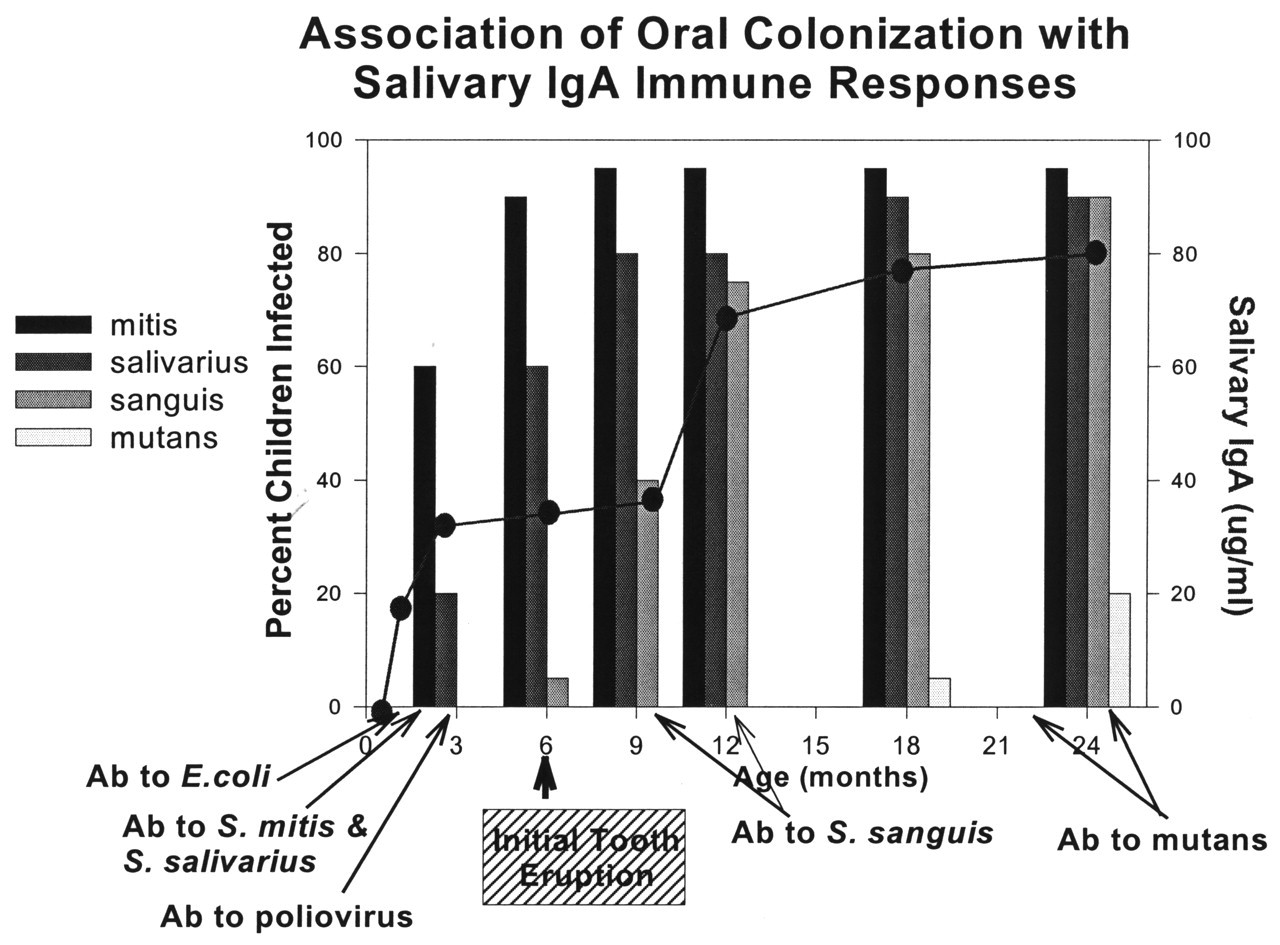
The oral immune environment undergoes rapid, early development (reviewed in Smith and Taubman, 1992, 1993). Although secretory IgA antibody in saliva and other secretions is essentially absent at birth, mature SIgA, i.e., dimeric IgA with a bound secretory component, is the principal salivary immunoglobulin secreted in individuals by one month of age. Consequent to the environmental antigenic challenge, mucosal IgA antibody to pioneer gut (e.g., Escherichia coli) and oral (e.g., Streptococcus mitis and S. salivarius) microbiota appears within weeks of initial exposure (Fig. 2). In the first months of life, an infant’s saliva may contain considerable concentrations of IgM and may, occasionally, be dominated by the IgA1 isotype. However, by six to nine months of age, most children exhibit a more adult-like distribution of salivary IgA1 and IgA2 subclasses. Salivary antibody to oral commensal microbiota can be detected in both subclasses at this time. Taken together, this evidence suggests that significant maturation of the mucosal immune response has occurred by the end of the first year of life.
Natural exposure to mutans streptococci also results in a mucosal immune response to these organisms (Smith et al., 1998a). This response often begins to be observed in the second and third years of life. Western blot and ELISA analyses reveal that the major responses appear to be directed primarily to streptococcal components which are considered to be important in colonization and accumulation, such as antigen I/II, glucosyltransferase, and glucan-binding protein(s). Interestingly, in some children, antibody to mutans streptococcal antigens can also be detected independently of the ability to detect ongoing infection in the second year of life. As is the case with many bacterial challenges throughout the body, the threshold of immunological response is lower than that of persistent infection; therefore it is not surprising to observe antibody to S. mutans antigens in the absence of its colonization. Longitudinal studies suggest that antibody reactive with mutans streptococci results from contact with mutans streptococci, rather than from earlier colonizing oral streptococci, since well-developed salivary IgA antibody to pioneer oral streptococci can be demonstrated prior to the detection of antibody reactive with mutans streptococci. Thus colonization, at least not extensive colonization, with mutans streptococci is apparently not required for the development of salivary antibody to associated mutans streptococcal antigens.
Salivary immune responses to mutans streptococci show significant individual characteristics in early childhood. Children respond at different rates following infection, a condition which may be partly the result of the extent of infection (antigen dose) or age at the time of infection (maturation of immune response). Even siblings may differ in the amounts or kinds of IgA antibody specificities appearing in their saliva. These variations may stem from differences either in the inherent ability of the child to respond or in the characteristics of genetically different strains of mutans streptococci (thus potentially differing antigenic challenge) ultimately colonizing the child. The rate, specificity, and/or extent of the mucosal immune response to previous encounters with the organism may also contribute to the success or failure of permanent colonization.
Thus, the evidence from salivary IgA responses to commensal oral microbiota indicates that the mucosal immune system is relatively well-developed by the period during which children typically become infected with mutans streptococci. Most children apparently respond immunologically to transient infection or ongoing colonization with mutans streptococci in early childhood. Although the distribution and specificity of children’s responses are not identical, antibody to a few major antigens predominates. Analysis of these data suggests the possibility that such responses could be protective if induced prior to critical colonization events.
(III) Molecular Pathogenesis of the Disease
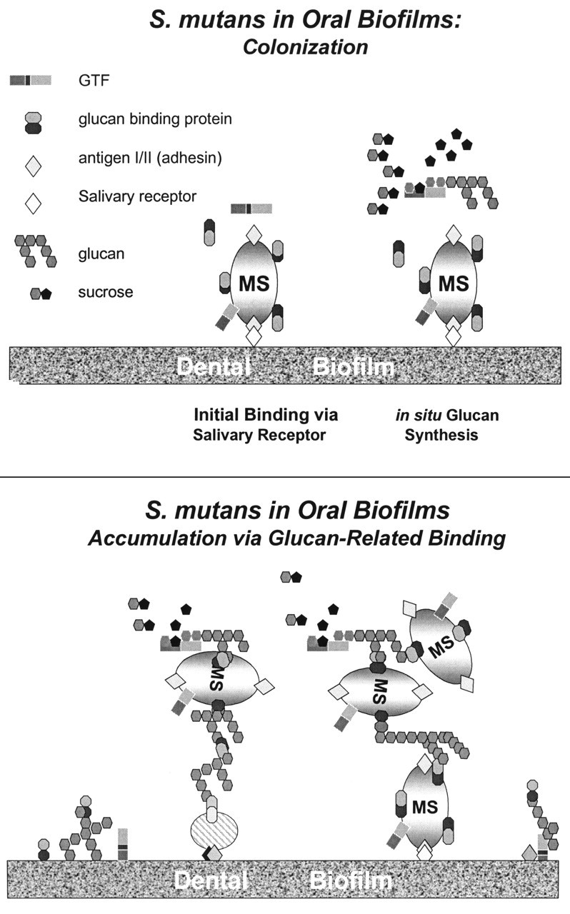
The molecular pathogenesis of mutans streptococci appears to involve several phases (Staat et al., 1980), each of which may offer targets for immunological intervention (Fig. 3). Acidogenic streptococci require the hard surfaces furnished by teeth for sustained colonization and accumulation. Initial attachment to the tooth is achieved via the interaction of bacterial proteins with lectins in the dental pellicle covering the tooth surface. This trait is characteristic of a family of streptococcal adhesins, referred to as antigen I/II or PAc in Streptococcus mutans, which have been demonstrated to bind to salivary components in experimental tooth pellicles (Hajishengallis et al., 1992). Lamont and co-workers (1991) have evidence to suggest that the S. mutans antigen I/II adherence to saliva-coated Streptococcus sanguis and Actinomyces viscosus is mediated through an acidic, mucin-like glycoprotein (agglutinin) found in parotid and submandibular saliva (Demuth et al., 1990). Other binding properties have also been attributed to antigen I/II (reviewed in Hajishengallis and Michalek, 1999). At least 2 binding regions of antigen I/II may be involved in salivary-component-mediated adhesive activities (Crowley et al., 1993; Nakai et al., 1993; Kelly et al., 1995).
The ultimate pathogenicity of mutans streptococci occurs through erosion of the hydroxyapatite-like mineral in dental enamel by lactic acid, a bacterial metabolic end-product. However, significantly destructive concentrations of this acid require the substantial accumulation of these acidogenic streptococci in dental plaque. This accumulation process is initiated by the activity of extracellular glucosyltransferases (GTF), several of which are constitutively secreted by mutans streptococci. In the presence of sucrose, GTFs synthesize several forms of high-molecular-weight branched extracellular glucans. GTFs that synthesize insoluble forms of glucan (S. mutans GTF-B and GTF-C) have been most closely associated with pathogenicity. These glucose polymers provide scaffolding for the aggregation of mutans and other oral streptococci through interaction with bacterial cell-associated glucan-binding proteins. Several glucan-binding proteins have been described in S. mutans and S. sobrinus. Although each of these GBPs has the ability to bind to certain forms of glucan, and some have been shown to be cell-associated, their unique contributions to in vivo plaque development are as yet unclear. GTFs also contain glucan-binding domains which permit binding to glucans. The interactions of glucans with cell-associated glucan-binding domains of GTFs and GBPs combine to cause extensive accumulation of mutans streptococci in the dental biofilm. Since GTFs and GBPs are also secreted into the extracellular environment, their specific or non-specific incorporation into the salivary pellicle would also provide binding sites for mutans streptococci.
Theoretically, the next phase of pathogenesis results from the metabolic activities of these masses of accumulated mutans streptococci (and possibly of other accumulation-associated micro-organisms). Mutans streptococci are the most prolific producers of lactic acid in these accumulations (Gibbons and van Houte, 1975), although other “low pH bacteria” may also contribute (van Ruyven et al., 2000). Dental caries then ensues, because the resulting increase in lactic acid synthesis cannot be sufficiently buffered to prevent enamel dissolution.
(IV) Effective Molecular Targets for Dental Caries Vaccines
Several stages in the molecular pathogenesis of dental caries are susceptible to immune intervention. Micro-organisms can be cleared from the oral cavity by antibody-mediated aggregation while still in the salivary phase, prior to colonization. Antibody could also block the receptors necessary for colonization (e.g., adhesins) or accumulation (e.g., glucan-binding domains of GBPs and GTF), or inactivate GTF enzymes responsible for glucan formation. Modification of metabolically important functions may also be targeted. In addition, the antimicrobial activity of salivary IgA antibody may be enhanced or redirected by synergism with innate components of immunity, such as mucin or lactoferrin. This review will concentrate on adhesins, GTFs, and GBPs as vaccine targets, since most of the recent experimental effort has been directed toward these components.
(A) Adhesins
Adhesins from the two principal human pathogens, Streptococcus mutans (variously identified as antigen I/II, PAc, or P1) and Streptococcus sobrinus (SpaA or PAg), have been purified. Russell and Lehner initially described the S. mutans component in 1978. Antigen I/II (AgI/II) was found both in the culture supernatant as well as on the S. mutans cell surface. This 185-kDa protein is composed of a single polypeptide chain of approximately 1600 residues (Lee et al., 1988). Significant sequence homology (66%) exists between S. mutans AgI/II and S. sobrinus SpaA (Tokuda et al., 1990; LaPolla et al., 1991) as well as with at least one adhesin from Streptococcus gordonii, a non-cariogenic oral streptococcus (Demuth et al., 1990). However, despite the homology between the two mutans streptococcal adhesins, each appears to bind to separate components in the pellicle (Gibbons et al., 1986; Lamont et al., 1991).
S. mutans Ag I/II contains an alanine-rich tandem repeating region in the N-terminal third, and a proline-rich repeat region in the center of the molecule. These regions have been associated with the adhesin activity of Ag I/II. Crowley and colleagues (1993) and Nakai and co-workers (1993) have each described a region within or near the alanine-rich region that can bind salivary components in experimental tooth pellicles. Lehner, Kelly, and co-workers (1994; 1995) suggested that the proline-rich central portion contains an adhesion epitope, basing their conclusions on adhesion inhibition assays involving recombinant fragments of Ag I/II.
Immunological approaches support the adhesin-related function of the AgI/II family of proteins and their repeating regions. For example, abundant in vitro and in vivo evidence indicates that antibody with specificity for S. mutans AgI/II or S. sobrinus SpaA can interfere with bacterial adherence and subsequent dental caries. Antibody directed to the intact antigen I/II molecule or to its salivary-binding domain blocked adherence of S. mutans to saliva-coated hydroxyapatite (Hajishengallis et al., 1992). Furthermore, numerous immunization approaches have shown that active immunization with intact antigen I/II (Lehner et al., 1981; Katz et al., 1993) or passive immunization with monoclonal (Ma et al., 1990) or transgenic antibody (Ma et al., 1998) to putative salivary-binding domain epitopes within this component can protect rodents, primates, or humans from dental caries caused by S. mutans. Immunization of mice with synthetic peptides (residues 301-319) from the alanine-rich region of antigen I/II suppressed tooth colonization with S. mutans (Takahashi et al., 1991). Immunization with S. sobrinus SpaA constructs protected rats from caries caused by S. sobrinus infection (Redman et al., 1995). Protection in these experiments could conceivably occur by antibody blockade of initial colonization events or antibody-mediated agglutination and clearing of adhesin-bearing bacteria from the saliva.
(B) Glucosyltransferases (GTFs)
S. mutans and S. sobrinus each synthesize several glucosyltransferases. The deduced sequences of these enzymes vary from 1400 to nearly 1600 amino acid residues and contain considerable sequence homology, despite differences in the water-solubility and linkages among the glucans synthesized. Genes responsible for glucan synthesis in S. mutans are gtfB (Shiroza et al., 1987), which synthesizes an α-1,3-linked insoluble glucan, gtfC (Pucci et al., 1987), which synthesizes glucan with both α-1,3 and α-1,6 linkages, and gtfD (Honda et al., 1990), which synthesizes a soluble α-1,6-linked glucan. Similarly, the products of gtfI (Russell et al., 1987) and gtfS (Gilmore et al., 1990) genes of S. sobrinus synthesize insoluble and soluble glucan products, respectively. Mutational inactivation techniques have shown that each of these gene products is important to the cariogenicity of the respective mutans streptococcal strain. For example, insertional inactivation of S. mutans gtf genes to replace functional wild-type copies of the gene markedly reduced caries when gtfB and gtfC genes that coded for GTFs synthesizing water-insoluble glucan were inactivated. Similar inactivation of the gtfD gene also resulted in a mutant with lower cariogenicity on smooth surfaces (Yamashita et al., 1993). In addition, the ability of GTF from initially colonizing S. mutans to synthesize water-insoluble glucan has been correlated with caries incidence in young children (Mattos-Graner et al., 2000).
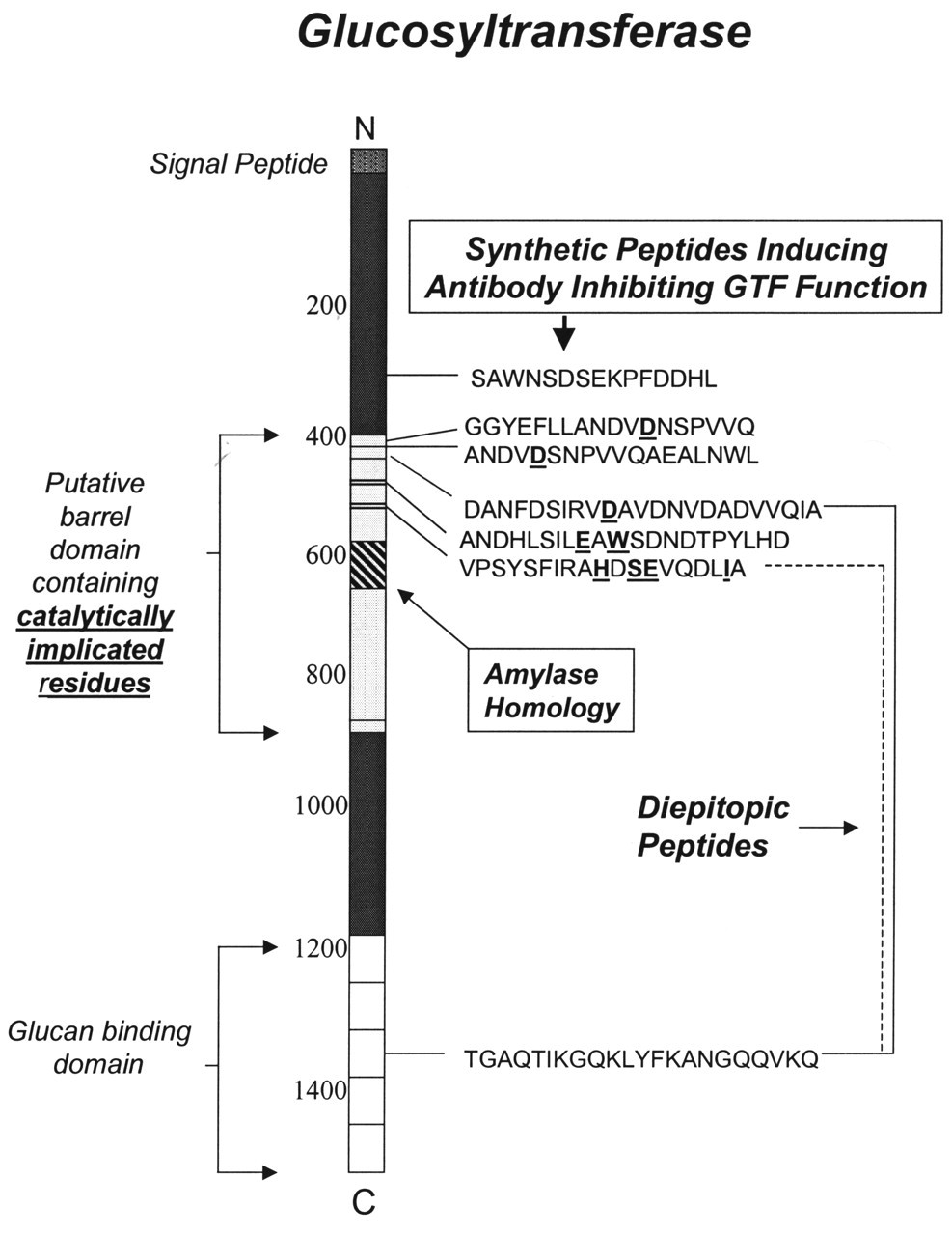
The activity of GTF is mediated through both catalytic and glucan-binding functions. The catalytic activity of GTF appears to be associated with several, sequentially separate, residues in the N-terminal third of the molecule. These residues have been identified by a variety of methods, including the labeling of catalytic intermediates and site-directed mutagenesis (Mooser et al., 1991; Funane et al., 1993; Devulapalle et al., 1997; Tsumori et al., 1997; Monchois et al., 2000). Insight into catalytically important residue identification has been obtained by sequence alignment techniques (MacGregor et al., 1996; Devulapalle et al., 1997), which have revealed significant homology between GTFs and alpha amylase with respect to several invariant residues important to the catalytic activity of the alpha amylase family, suggesting that the amylase (β,α)8 barrel element may also be a feature of the GTF catalytic domain. Collectively, these studies have identified several residues within this putative (β,α)8 barrel element which may be involved in the catalytic activity of GTF. These residues are underlined in the peptide sequences shown in Fig. 4.
The C-terminal region of the GTF molecule contains a pattern of repeating sequences which have been identified in all GTFs from mutans streptococci. Evidence that this region has glucan-binding function is obtained from observations that large C-terminal, tryptic fragments of GTF retain the ability to bind alpha 1,6 glucan (Mooser and Wong, 1988; Wong et al., 1990; Abo et al., 1991), and that recombinant products containing some or all of the C-terminal third of GTF-I of S. downei or S. sobrinus also can bind alpha 1,6 glucan (Abo et al., 1991; Kaseda et al., 2000). The glucan-binding potential of these repeating GTF sequences is also supported by the observations that amino acid deletions in this region remove glucan-binding activity or decrease the efficiency of insoluble glucan synthesis (Konishi et al., 1999). Also, a glucan-binding protein (GbpA) of S. mutans contains reiterated sequences that are very similar to those found in this region of GTF (Banas et al., 1990).
Antibody directed to native GTF or sequences associated with its catalytic or glucan-binding function interfere with the synthetic activity of the enzyme and with in vitro plaque formation (Taubman and Smith, 1972; Smith et al., 1978). Since mutans streptococcal GTFs bear significant sequence homology, active immunization with either S. mutans or S. sobrinus GTFs induced protective immune responses in experimental dental caries rodent models after infection with several mutans streptococcal species (reviewed in Smith and Taubman, 1997). However, a much lower level of mutans streptococcal GTF antibody reactivity was observed with GTFs of other oral streptococci (Smith et al., 1981). Interestingly, at least in the case of S. sobrinus GTF, induction of anti-GTF antibody was sufficient to have a significant effect on initial oral colonization with Streptococcus sanguis or S. oralis (Smith et al., 1983). Induction of SIgA antibody in humans by oral or topical GTF administration is accompanied by interference with accumulation of indigenous mutans streptococci after dental prophylaxis (Smith and Taubman, 1987, 1990). Passive administration of antibody to GTF in the diet also can protect rats from experimental dental caries (Hamada et al., 1991). Thus, the presence of antibody to glucosyltransferase in the oral cavity prior to infection can significantly influence the disease outcome, presumably by interference with one or more of the functional activities of the enzyme.
(C) Glucan-binding proteins
The ability of mutans streptococci to bind to glucans is presumed to be mediated, at least in part, by cell-wall-associated glucan-binding proteins (Gbp). Many proteins with glucan-binding properties have been identified in Streptococcus mutans and Streptococcus sobrinus (summarized in Smith et al., 1998b). Each glucan-binding protein has the ability to bind α 1-6 glucan, although other glucan linkages potentially may impart higher binding constants. S. mutans secretes at least three distinct proteins with glucan-binding activity: GbpA (Russell, 1979), GbpB (Smith et al., 1994a), and GbpC (Sato et al., 1997).
GbpA has a deduced sequence of 563 amino acids (Banas et al., 1990). The molecular weight for the processed protein is 59.0 kDa. The carboxy-terminal two-thirds of the GbpA sequence has significant homology with the putative glucan-binding regions of mutans streptococcal GTFs. This C-terminal region contains 16 repeating units which, together, have been shown to represent the full glucan-binding domain of this protein (Haas and Banas, 2000). GbpA has a greater affinity for water-soluble than for water-insoluble glucan.
The expressed GbpB protein is 431 residues long and has a calculated molecular weight of 41.3 kDa (Mattos-Graner et al., 2001). Its sequence is unrelated to those reported for other S. mutans GTFs or Gbp’s, paralleling the lack of reaction of anti-GbpB antibody with these proteins. The N-terminal third contains several immunodominant regions, which may explain the significant apparent immunogenicity of this protein in humans (Smith et al., 1998a) and animals (Smith and Taubman, 1996). Although the function of this protein in the native environment is as yet unresolved, biofilm formation on plastic surfaces by strains of S. mutans is directly correlated with expression of GbpB (Mattos-Graner et al., 2001), suggesting a role for GbpB in this process.
The third S. mutans non-enzymatic glucan-binding protein, GpbC, is composed of 583 amino acids. This protein has a calculated molecular weight of 63.5 kDa. Although GbpC is unrelated in sequence to GbpA or GTF, it bears some sequence similarity to the AgI/II adhesin family. The GbpC protein, detected when S. mutans cultures are stressed during growth, is associated with dextran-dependent aggregation.
Of the three S. mutans glucan-binding proteins, only GbpB has been shown to induce a protective immune response to experimental dental caries (Smith and Taubman, 1996Smith and Taubman, 1997a). Protection can be achieved by either subcutaneous injection of GbpB in the salivary gland region (Smith and Taubman, 1996) or by mucosal application by the intranasal route (Smith et al., 1997a). Saliva samples from young children often contain IgA antibody to GbpB, indicating that initial infection with S. mutans can lead to natural induction of immunity to this protein (Smith et al., 1998a). Preliminary studies have shown that GbpA appears to be less immunogenic than GbpB and that its ability to induce protective immunity is problematic (Smith et al., 1997a). S. sobrinus Gbps have not been evaluated for their protective potential.
(V) Subunit Vaccines
Subunit vaccines, which contain structural elements of the Ag I/II adhesin family, GTFs or GbpB, have been designed for a variety of reasons. It had been observed that immune responses in animals protected by immunization with intact proteins were associated, at least in part, with in vitro measures of functional inhibition. Thus, more recent studies have attempted to optimize immune responses to functional epitopes associated with salivary binding, catalytic processes, or glucan-binding activities by designing subunit vaccines whose constructs contain single or multiple copies of epitopes from these domains. In addition, potentially enhanced protection could be achieved by including, in the subunit vaccine construct, regions of the virulence component containing strong immunological binding properties for induction of the desired arm of the immune response. Furthermore, multivalent subunit vaccines could be constructed of multiple epitopes which target different functions on the same component (e.g., GTF catalytic and glucan-binding activities) or functions on different components (e.g., AgI/II salivary binding and GTF catalytic activity). Such approaches would address the variability in mucosal response characteristics in the target vaccine population. Conjugation of functionally associated peptides to carbohydrate components (Wachsmann et al., 1985; Dougan et al., 1987; Lett et al., 1994), for example glucan, or to other vaccine proteins (e.g., tetanus toxoid) also would increase the immunogenicity of the peptide and broaden the reach of the vaccine.
Designing vaccines in this way also permits one to eliminate regions which may induce unwanted antibody specificities. The Ag I/II family of proteins shares extensive sequence homology with surface proteins of non-cariogenic S. gordonii (Demuth et al., 1990), S. intermedius, and S. oralis (Ma et al., 1991). These homologous sequences may induce cross-reactive responses that could influence colonization, attachment, or accumulation of commensal microbiota. Also, cross-reacting epitopes on serotype f SR protein and human IgG have been reported (Wachsmann et al., 1989; Gangloff et al., 1992). Given this knowledge, subunit vaccines can be designed to include the salivary-binding domain(s), but exclude sequence bearing the potential for induction of unwanted antibody responses.
Subunit vaccines with inherent adjuvant potential could also be constructed by including some or all of the sequence of effective immunoadjuvants. Furthermore, this approach could be adapted to incorporate such effective vaccine constructs into attenuated expression vectors that have the ability to deliver the antigen to inductive sites for antibody synthesis. Of the designs which include mono- and di-epitopic synthetic peptide constructs, single recombinant peptides, or peptide chimeras with immunoadjuvant sequence, used in a variety of applications or within Salmonella expression systems, each has been reported to induce caries-protective immunity in experimental systems.
(A) Synthetic peptide vaccines
As indicated above, at least two regions of the AgI/II-protein family appear to be associated with salivary-binding functions. Monoclonal antibody, raised by immunization with intact Ag I/II, that reacted with the fragment containing the proline-rich region also inhibited the formation of experimental dental caries (Lehner et al., 1992). Similarly, workers in France (Gangloff et al., 1992; Lett et al., 1994) demonstrated that a 14-mer peptide derived from a proline-rich region of the SR antigen, a member of the S. mutans serotype f Ag I/II family of proteins, was reactive with antibody to the native protein.
Synthetic peptide approaches have also shown the alanine-rich repeat region of Ag I/II to be immunogenic and to induce protective immunity. For example, subcutaneous immunization with a synthetic peptide derived from the alanine-rich region of Ag I/II from S. mutans (residues 301-319: PAcA) induced higher levels of serum IgG antibody reactive with recombinant Ag I/II than a synthetic peptide derived from the proline-rich region (residues 601-629) (Takahashi et al., 1991). Intranasal immunization with PAcA, coupled to cholera toxin B subunit, suppressed colonization of mouse teeth by S. mutans (Takahashi et al., 1991). Fusion proteins containing PAcA also inhibited sucrose-independent adhesion of S. mutans to saliva-coated hydroxyapatide beads (Yu et al., 1997). Thus, this S. mutans adhesin contains multiple functionally based epitopes that are sufficiently immunogenic to be considered for dental caries vaccines.
B- and T-cell epitopes (summarized in Smith and Taubman, 1997) have been found in a cell-associated 3.8-kDa protein component antigen (Lehner et al., 1994). Lehner and his colleagues (Lehner et al., 1989a,b, 1994) applied free synthetic peptides containing immunodominant sequences of the 3.8-kDa antigen of S. mutans to the gingival mucosa of macaques, resulting in the formation of both salivary IgA and gingival IgG antibody. Anti-peptide antibody elicited by this topical application method prevented colonization of the teeth by S. mutans.
The identification of functionally relevant residues/domains in glucosyltransferases has led to the design of several synthetic peptide vaccines. Monoclonal or polyclonal antibody preparations which were directed to one of several N-terminal GTF peptides (Fig. 4), each of which contained different catalytically implicated residues, have been shown to inhibit GTF activity (Dertzbaugh and Macrina, 1990; Chia et al., 1993, 1997; Smith et al., 1994b, 1997b, 1999; Laloi et al., 1996). Several of these synthetic peptides which contained strong B-cell epitopes were synthesized on lysine backbones to contain four or eight copies of the respective sequence. These constructs induced protective immunity against experimental dental caries (Taubman et al., 1995; Smith et al., 1997b).
Synthetic peptide constructs have also been based on sequence derived from the repeating sequences in the C-terminal third of GTF, which has been shown to be associated with primer-dependent glucan binding (Abo et al., 1991; Lis et al., 1995; Konishi et al., 1999). A synthetic peptide associated with a putative glucan-binding site (Fig. 4) was shown to contain both B- and T-cell epitopes, to induce antibody which could inhibit the enzymatic activity of GTF (Smith et al., 1994b), and to induce protective immune responses in the rat caries model (Taubman et al., 1995). Furthermore, di-epitopic constructs of this peptide and a peptide from the catalytic domain could be shown to enhance the protective effect (Taubman et al., 2001), presumably because antibody was raised to two functional targets and because the glucan-binding domain peptide provided additional T-cell help for the B-cell epitopes on both peptides.
All of the GTF synthetic peptide sequences which showed protective effects in the above experiments are highly conserved among S. mutans and, often, among S. sobrinus GTFs as well. Antibody directed to these epitopes could therefore be expected to reduce the activity of many of the GTF isozymes expressed by these mutans streptococci, thus extending the protective effect across species lines. In this regard, protection from dental caries caused by either S. mutans or S. sobrinus infection in the rat model has been demonstrated after immunization with synthetic peptides from either the catalytic or glucan-binding domains of one GTF isozyme (Taubman et al., 1995; Smith et al., 1997b). These studies suggested that protection could be achieved by immunization with discrete epitopes associated with several virulence characteristics. Combining epitopes from adhesins and GTFs into one construct and enhancing the immune response with additional sequences (e.g., cholera toxin subunits) could theoretically increase and possibly extend the protective effect of these subunit vaccines. Some recombinant protein approaches, described below, have used this design.
(B) Recombinant vaccines / Attenuated expression vectors
Recombinant approaches afford the expression of larger portions of functional domains than can be accommodated by synthetic peptides. Also, gene fusions of a functionally relevant sequence linked to a mucosal adjuvant sequence can result in chimeric proteins inherently able to enhance immune responses to the functional epitopes. Furthermore, attenuated mutant vectors such as Salmonella, which contain plasmids expressing recombinant peptides, can target the vaccine to appropriate inductive lymphoid tissue for mucosal responses. Several of these approaches have successfully induced protective immune responses for experimental dental caries in rats or mice by means of chimeric proteins or vectors expressing either adhesin or GTF epitopes. Redman and co-workers have shown (1994; 1995) that oral immunization with recombinant Salmonella typhimurium, expressing surface protein antigen A of Streptococcus sobrinus, was able to induce persistent mucosal immune responses which could confer protection after challenge of Fischer rats with cariogenic S. sobrinus. Hajishengallis and co-workers have genetically linked the 42-kDa salivary-binding receptor (SBR) of S. mutans Ag I/II with the A2 and B subunits of cholera toxin (SBR-CTA2/B) and expressed this chimeric protein in E. coli BL21 (Hajishengallis et al., 1995). Intranasal administration of this chimeric protein with CT resulted in significant reductions in dental caries caused by infection of Fischer rats with S. mutans UA130 (Hajishengallis et al., 1998). The SBR-CTA2/B, expressed in an attenuated S. typhimurium BRD509 vaccine strain containing an nirB promoter (Huang et al., 2000), which was administered intranasally or intragastrically to antibiotic-pre-treated BALB/c mice, resulted in salivary antibody to the SBR and a significant reduction in the number of S. mutans PC3379 recovered from dental plaque after challenge (Huang et al., 2001). Jespersgaard and co-workers (1999) intranasally immunized BALB/c mice with an E. coli-expressed recombinant GTF peptide based on a 290-residue glucan-binding domain sequence, or with a chimeric protein combining this sequence with thioredoxin. Immunization with either peptide resulted in protective effects on experimental S. mutans infection and on resulting dental caries.
Other recombinant strategies involving either adhesin or GTF constructs, with or without mucosal adjuvant sequences, have been shown to induce immune responses to these functional domains which could be ultimately protective in caries vaccine applications. Chimeric proteins, in which short sequences from predicted catalytically active regions of GTF were combined with cholera toxin (Dertzbaugh et al., 1990) or the B subunit of CT (Laloi et al., 1996) and expressed in E. coli HB101, gave rise to immune responses which could inhibit as much as 50% of GTFB activity. Yu and co-workers (1997) designed a fusion protein which contained both a 281-residue saliva-binding alanine-rich region of S. mutans Ag I/II and a 392-residue glucan-binding domain of GTF-I. A recombinant fusion protein, expressed in E. coli XL1-Blue, induced IgG antibody in rabbits or in Holstein cows (Oho et al., 1999) which could inhibit glucan synthesis by GTF and sucrose-independent and -dependent adhesion of S. mutans to saliva-coated hydroxyapatite beads.
Constructs involving the attenuated human S. typhi vector would be expected to have more potential for human vaccine applications than would S. typhimurium, which is a murine pathogen. In this regard, attenuated S. typhi CVD908 strains have been prepared to express peptide chimeras in which GTF sequences, associated with the glucan-binding domain, are combined with tetanus toxin fragment C for immunogenicity (unpublished observations).
(VI) Conjugate Vaccines
Another vaccine approach which may intercept more than one aspect of mutans streptococcal molecular pathogenesis is the chemical conjugation of functionally associated protein/peptide components with bacterial polysaccharides. Added to the value of including multiple targets within the vaccine is that the conjugation of protein with polysaccharide enhances the immunogenicity of the T-cell-independent polysaccharide entity. This principle was first demonstrated by Landsteiner (1936) and Avery and Goebel (1929) and has been applied with great success in the Hemophilus influenzae type b (Hib) conjugate vaccines to induce protective immunity to the capsular polysaccharide of H. influenzae in infants and young children. Two groups have applied this approach to dentally relevant components. Lett and co-workers (1994) covalently coupled an adhesin-associated 14-mer synthetic peptide to the serogroup f polysaccharide of S. mutans strain OMZ 175 by reductive amination. Subcutaneous injection with the conjugate induced systemic IgM and IgG antibody responses to both peptide and polysaccharide which could be boosted upon subsequent injection. The presence of both B- and T-epitopes in the peptide was required for effective responses. Intragastric intubation of the conjugates associated with liposomes induced primary and secondary salivary IgA antibody to both components (Lett et al., 1995). In separate studies, Taubman and co-workers (1998; 1999) have reported that conjugation of either tetanus toxoid or S. sobrinus GTF to the water-soluble glucan synthesized by GTF significantly enhanced serum IgG and salivary IgA antibody levels to the water-soluble glucan and to the conjugated protein. Serum GTF inhibitory activity was also improved by conjugation. The relative protective capacity of either conjugate approach has yet to be tested. Since initial S. mutans infection occurs at an age (< 2 yrs) when children are unable to mount significant anti-polysaccharide responses, these approaches will be especially important if conjugate vaccines are shown to enhance the level of protection significantly over that achieved with protein-based vaccines.
(VII) Routes to Protective Responses
Mucosal applications of dental caries vaccines are generally preferred for the induction of secretory IgA antibody in the salivary compartment, since this immunoglobulin constitutes the major immune component of major and minor salivary gland secretions. Many investigators have shown that exposure of antigen to mucosally associated lymphoid tissue in the gut, nasal, bronchial, or rectal site can give rise to immune responses not only in the region of induction, but also in remote locations. This has given rise to the notion of a “common mucosal immune system” (Mestecky, 1993). Consequently, several mucosal routes have been used to induce protective immune responses to dental caries vaccine antigens.
(A) Oral
Many of the earlier studies relied on oral induction of immunity in the gut-associated lymphoid tissues (GALT) to elicit protective salivary IgA antibody responses. In these studies, antigen was applied by oral feeding, gastric intubation, or in vaccine-containing capsules or liposomes. Although the oral route was not ideal for reasons including the detrimental effects of stomach acidity on antigen, or because inductive sites were relatively distant, experiments with this route established that induction of mucosal immunity alone was sufficient to change the course of mutans streptococcal infection and disease in animal models (Michalek et al., 1976; Smith et al., 1979) and humans (Smith and Taubman, 1987).
(B) Intranasal
More recently, attempts have been made to induce protective immunity in mucosal inductive sites that are in closer anatomical relationship to the oral cavity. Intranasal installation of antigen, which targets the nasal-associated lymphoid tissue (NALT) (Brandtzaeg and Haneberg, 1997), has been used to induce immunity to many bacterial antigens, including those associated with mutans streptococcal colonization and accumulation. Protective immunity after infection with cariogenic mutans streptococci could be induced in rats by the IN route with many S. mutans antigens or functional domains associated with these components. Protection could be demonstrated with S. mutans AgI/II (Katz et al., 1993), the SBR of AgI/II (Hajishengallis et al., 1998), a 19-mer sequence within the SBR (Takahashi et al., 1991), the glucan-binding domain of S. mutans GTF-B (Jespersgaard et al., 1999), S. mutans GbpB (Smith et al., 1997a), and fimbrial preparations from S. mutans (Fontana et al., 1999), with antigen alone or combined with mucosal adjuvants.
(C) Tonsillar
The ability of tonsillar application of antigen to induce immune responses in the oral cavity is of great interest. Tonsillar tissue contains the required elements of immune induction of secretory IgA responses (van Kempen et al., 2000), although IgG, rather than IgA, response characteristics are dominant in this tissue (Boyaka et al., 2000). Nonetheless, the palatine tonsils, and especially the nasopharyngeal tonsils, have been suggested to contribute percursor cells to mucosal effector sites (Brandtzaeg, 1996), such as the salivary glands. In this regard, Fukuizumi and co-workers (1999) have shown that topical application of formalin-killed S. sobrinus cells in rabbits can induce a salivary immune response which can significantly decrease the consequences of infection with cariogenic S. sobrinus. Interestingly, repeated tonsillar application of particulate antigen can induce the appearance of IgA antibody-producing cells in both the major and minor salivary glands of the rabbit (Inoue et al., 1999).
(D) Minor salivary gland
The minor salivary glands populate the lips, cheeks, and soft palate. These glands have been suggested as potential routes for mucosal induction of salivary immune responses (Crawford et al., 1975; Schroeder et al., 1983), given their short, broad secretory ducts that facilitate retrograde access of bacteria and their products (Nair and Schroeder, 1983), and given the lymphatic tissue aggregates that are often found associated with these ducts. Experiments in which S. sobrinus GTF was topically administered onto the lower lips of young adults have suggested that this route may have potential for dental caries vaccine delivery. In these experiments, those who received labial application of GTF had significantly lower proportions of indigenous mutans streptococci/total streptococcal flora in their whole saliva during a six-week period following a dental prophylaxis, compared with a placebo group (Smith and Taubman, 1990).
(E) Rectal
More remote mucosal sites have also been investigated for their inductive potential. For example, rectal immunization with non-oral bacterial antigens such as Helicobacter pylori (Kleanthous et al., 1998) or Streptococcus pneumoniae (Hvalbye et al., 1999), presented in the context of toxin-based adjuvant, can result in the appearance of secretory IgA antibody in distant salivary sites. The colo-rectal region as an inductive location for mucosal immune responses in humans is suggested from the fact that this site has the highest concentration of lymphoid follicles in the lower intestinal tract. Preliminary studies have indicated that this route could also be used to induce salivary IgA responses to mutans streptococcal antigens such as GTF (Lam et al., 2001). One could, therefore, foresee the use of vaccine suppositories as one alternative for children in whom respiratory ailments preclude intranasal application of vaccine.
(VIII) Adjuvants and Delivery Systems for Dental Caries Vaccines
Mucosal routes of antigen delivery often require additional components which can potentiate aspects of the immune response to induce sufficient antibody to achieve a protective effect.
(A) Cholera and
E. coli
heat-labile enterotoxins
Cholera toxin (CT) is a powerful mucosal immunoadjuvant which is frequently used to enhance the induction of mucosal immunity to a variety of bacterial and viral pathogens in animal systems. CT is an ADP-ribosylating bacterial toxin with A and B subunits. The non-toxic pentameric B subunit binds to ganglioside receptors on target cells. The toxic A subunit is then translocated into the cell, where this enzymatically active peptide transfers the ADP ribose group of NAD to a GTP-binding protein, initiating the toxic effects by increasing intracellular levels of cAMP. The adjuvant effects of CT (reviewed in Freytag and Clements, 1999) are broad-based and can include increased mucosal epithelial cell and macrophage production of pro-inflammatory cytokines, up-regulation of B7-2 co-stimulatory factors on APCs, and increased antigen transfer from the mucosal to the systemic compartment. These effects may be, at least in part, locally manifest and may involve dendritic cells as the predominating antigen-presenting cell (APC) population (Porgador et al., 1997).
Mucosal application of soluble protein or peptide antigen alone rarely results in elevated or sustained IgA responses. However, addition of small amounts of CT or the closely related E. coli heat-labile enterotoxins (LT) (Takahashi et al., 1996) can greatly enhance mucosal immune responses to intragastrically or intranasally applied mutans streptococcal antigens (Russell and Wu, 1991; Martin et al., 2000) or to peptides derived from these antigens (Smith et al., 2001b). One approach to reducing or eliminating toxicity while maintaining adjuvanticity was to remove the A subunit from the CT complex. Mucosal immunization with the B subunit of CT, mixed with, chemically conjugated to, or genetically fused to either intact antigen or the presumed functional domains of mutans streptococcal virulence components (Czerkinsky et al., 1989; Dertzbaugh et al., 1990; Takahashi et al., 1991; Wu and Russell, 1993; Laloi et al., 1996; Wu et al., 1997) resulted in significant mucosal responses to the complexed antigen (and to CT), which could be shown to be protective under certain conditions (Katz et al., 1993) and long-lived (Harrod et al., 2001).
Other approaches have been sought to modify CT or LT, since the increase in CTB-assisted immune response was significantly less than that achieved with the CT holotoxin, and since, in some systems, the presence of a small amount of intact CT was required for response enhancement. Hajishengallis and co-workers (1995) were able to improve mucosal adjuvanticity by replacing the toxic A1 (N-terminal portion) of CT with a 42-kDa portion of the AgI/II containing the salivary-binding region (SBR), thereby creating a chimeric protein composed of the SBR linked to the non-toxic C-terminal A2 and B subunits of CT. Similar enhancement, albeit by potentially different mechanisms, occurred after replacement of the toxic A1 subunit of LT (serogroup IIa) with SBR (Martin et al., 2001). Responses were long-lasting (Hajishengallis et al., 1996b), and chimeric SBR-CTA2/B proteins could be expressed in E. coli and in attenuated Salmonella typhimurium expression vectors (Hajishengallis et al., 1996a,b; Huang et al., 2000) which could be shown to induce protective immune responses in BALB/c mice when administered intragastrically or intranasally (Huang et al., 2001).
Alternatively, detoxification of CT and LT has been attempted with the use of site-directed mutagenesis techniques to modify amino acid residue areas of the toxic A1 subunit which are critical to its enzymatic activity or to the required proteolytic cleavage required for activation. Several detoxified CT and LT mutants have been prepared which retain significant adjuvanticity and may have potential for human use. One such mutant, LT [LT(R192G)], obtained by substituting glycine for arginine 192 in the A subunit (Dickinson and Clements, 1995) to interfere with trypsin-mediated cleavage, has been shown to induce mucosal and systemic immune responses to intranasally applied GTF peptides which were generally comparable with those observed with CT (Smith et al., 2001b).
(B) Microcapsules and microparticles
Combinations of antigen in or on various types of particles have been used in attempts to enhance mucosal immune responses. The particularization of antigen achieved by these approaches may increase the association of antigen with M cells overlying inductive regions of the secretory immune system (Neutra and Kraehenbuhl, 1992). Microspheres and microcapsules made of poly(lactide-co-glycolide) (PLGA) have been used as local delivery systems (Eldridge et al., 1990; Ermak et al., 1995) because of their ability to control the rate of release, evade pre-existent antibody clearance mechanisms, and degrade slowly without eliciting an inflammatory response to the polymer. Oral (Challacombe et al., 1992) or topical (Rafferty et al., 1996) immunization with soluble antigen in PLGA microcapsules can lead to enhanced mucosal responses, although the organic solvents required for formulation can diminish the biological activity of the antigen. Denaturation can be significantly diminished by methods which incorporate antigen into microparticles in an aqueous phase (Hsu et al., 1994). Intranasal immunization of aqueously incorporated GTF-PLGA microparticles, which also contained 1% gelatin as bioadhesive, induced long-lasting salivary immune responses (Smith et al., 2000). Starch microparticles can also be used to increase mucosal responses to soluble antigens. Montgomery and Rafferty (1998) have shown that topical administration to the oral mucosa of bioadhesive degradable starch microparticles containing dinitrophenyl-bovine serum albumen, in combination with L-α-lysophosphatidylcholine (as a penetration enhancer) and interleukins IL-5 and IL-6, potentiated long-lived salivary IgA responses, compared with antigen delivered in soluble form.
(C) Liposomes
Liposomes, which are phospholipid membrane vesicles manufactured to contain and deliver drugs and antigens, have been used to enhance mucosal responses to mutans streptococcal carbohydrate (Childers et al., 1990-1991) and GTF (Childers et al., 1996). Liposomes are thought to improve mucosal immune responses by facilitating M cell uptake and delivery of antigen to lymphoid elements of inductive tissue (Childers et al., 1990). Gastric intubation of rats with GTF-liposome vaccines induced responses which diminished dental caries caused by subsequent infection with S. mutans (Childers et al., 1996). Clinical studies comparing intranasal immunization with GTF-liposomes vs. GTF alone showed that both vaccines increased local (nasal) and salivary IgA antibody responses to GTF up to five-fold (Childers et al., 1999). Only the nasal-wash IgA1 antibody response was higher with the liposome-containing, compared with the protein-alone, vaccine.
(D) Other approaches
Other methods to enhance mucosal responses are being adapted for dental caries vaccine use. Monophosphoryl lipid A, when administered intranasally to mice as an aqueous formulation with soluble GTF or incorporated into liposomes containing GTF, induced primary and secondary salivary IgA responses which exceeded those achieved with GTF-liposomes (Childers et al., 2000). Oligodeoxynucleotides containing unmethylated CpG dinucleotides (CpG ODN) induce proliferation of B-cells and activation of macrophages. Intranasal or oral administration of CpG ODN with tetanus toxoid (TT) enhanced murine IgA responses in many mucosal secretions, including saliva (McCluskie and Davis, 2000). Similarly, intragastric administration of CpG ODN with TT- Al(OH)3 enhanced systemic IgG responses to TT, compared with those achieved with TT - Al(OH)3 (Eastcott et al., 2001).
(IX) Past, Present, and Future Human Applications
(A) Active immunization
Few clinical trials have been performed to examine the protective effect of active immunization with dental caries vaccines containing defined antigens. However, several studies have shown that mucosal exposure of humans to immunization with glucosyltransferases from S. mutans or S. sobrinus can lead to the formation of salivary IgA antibody, albeit at modest levels. Childers and co-workers (1994) orally immunized adults using enteric-coated capsules filled with crude S. mutans GS-5 GTF antigen preparations contained in liposomes. Parotid salivary IgA antibody responses, primarily of the IgA2 subclass, were induced in five of seven subjects. Similarly, nasal immunization with dehydrated liposomes containing this GTF preparation induced significant IgA1 antibody response in nasal washes (Childers et al., 1997, 1999). Parotid salivary antibody levels to GTF were of lower magnitude. In earlier studies, this group (Childers et al., 1990-1991) showed that oral administration of capsules containing the purified serotype carbohydrate antigen of S. mutans in liposomes gave rise to low but detectable levels of salivary antibody.
Smith and Taubman (1987, 1990) reported that mucosal immunization with GTF could influence the re-emergence of mutans streptococci in young adults after a dental prophylaxis. Levels of parotid salivary IgA antibody to GTF increased after oral immunization with S. sobrinus GTF in enteric capsules, administered together with aluminum phosphate. Immunization under this protocol delayed the re-accumulation of indigenous oral mutans streptococci, compared with a placebo group given buffer-filled capsules. A delay in mutans streptococcal re-emergence was also observed after topical administration of GTF on the lower lip, although this protocol did not result in a significant detectable increase in antibody to the vaccine (Smith and Taubman, 1990). Taken together, these studies support the hypothesis that mucosal immunization with dental caries vaccines could be protective, especially in pediatric populations where mutans streptococci is not yet a permanent member of the dental biofilm.
(B) Passive immune approaches
Passive antibody administration has also been examined for effects on indigenous mutans streptococci. Mouthrinses containing bovine milk (Filler et al., 1991) or hen egg yolk IgY (Hatta et al., 1997) antibody to S. mutans cells led to modest short-term decreases in the numbers of indigenous mutans streptococci in saliva or dental plaque.
Longer-term effects on indigenous flora were observed after topical application of mouse monoclonal IgG or transgenic plant secretory SIgA/G antibody, each with specificity for Ag I/II (Ma et al., 1990, 1998). In these experiments, teeth were first treated for nine days with chorohexidine. Following anti-bacterial treatment, antibody was topically applied for three weeks. Recolonization with mutans streptococci did not occur for at least two years after treatment of subjects with mouse monoclonal antibody or at least 4 months after treatment with the transgenic antibody to the Ag I/II epitope. In contrast, the teeth of all subjects topically treated with non-specific monoclonals were re-colonized with mutans streptococci by 82 days in the former experiment and by 58 days in the later experiment. Bivalent antigen binding appeared to be required, since Fab fragments did not afford protection. The authors suggest that the secretory form of the monoclonal antibody may be more efficacious because of its apparent increased survival time in the oral cavity, compared with IgG, as well as the increased avidity emanating from its tetravalency (Ma et al., 1998). The explanation for the long-term effects on mutans streptococcal colonization after a relatively short exposure to antibody remains unresolved. Apparently, antibody blockage of an important adhesin epitope during the reconstruction of the dental biofilm following chlorohexidine treatment places indigenous mutans streptococci at an insurmountable competitive disadvantage for recolonization. An interesting parallel may be the observation that young children who do not become naturally infected with mutans streptococci during the “window of infectivity” remain undetectably infected for several years (Caufield et al., 1993; Smith et al., 1998a), potentially because its niche in the dental biofilm has been filled by other indigenous flora.
Experimental passive immune protection could also be achieved with antibody to GTF (Hamada et al., 1991) or GbpB (Smith et al., 2001a). Thus, topical or dietary administration of immune reagents with specificity for epitopes on these proteins may also have potential human application.
(C) Prospects and concerns
Traditional vaccine therapy indicates that immunization should take place prior to infection. Given the apparent pattern of mutans streptococcal colonization and the association of these organisms with disease, this would suggest that immunization for dental caries should begin early in the second year of life for those populations under “normal” risk for infection. Our understanding of the mucosal immune response indicates that such children are competent to mount secretory responses to protein antigens, although their ability to respond to polysaccharide determinants in mucosal applications of conjugate vaccines is as yet unknown. If bacterial colonization of the dental biofilm is complete after eruption of all primary teeth, and if one can, through immunization, prevent mutans streptococcal colonization prior to this period, then the benefit of early immunization might extend until secondary teeth begin to erupt, exposing new ecological conditions. Subsequent immunization could then be initiated.
Clearly, several possible dental caries vaccine approaches may have application in pediatric clinical trials. Several mutans streptococcal components have been shown to protect in animal model systems. Protective epitopes of these components continue to be identified and incorporated into designs to increase their protective value. Several routes of administration can induce protective immune responses, at least in model systems. Also, significant progress is being made in the adjuvant field to remove the toxic properties of powerful mucosal adjuvants while maintaining their adjuvant properties. Animate (attenuated bacterial vectors) and inanimate (liposomes, microparticles) delivery systems have been identified to provide more efficient targeting of the vaccine. Both passive and active immunization approaches have demonstrated success in animal models and human clinical trials.
With all these apparently effective pre-clinical approaches, one may ask, “What is the
If we accept that each approach could give a reasonable level of protection in humans, one still needs to consider for whom and under what circumstances the dental caries vaccine is intended. For example, the ideal vaccine application for a child with asthma, the second most common chronic childhood ailment, may be at a site (e.g., rectal) and with an adjuvant (e.g., detoxified CT or LT) that is quite different from that sufficient to give a protective response (e.g., intranasal) to a healthy child. Also, from economic and societal standpoints, a vaccine strategy for children to whom the full advantages of pediatric care are available may not be the ideal approach for children with limited health care access. For the former group of children, perhaps an intranasal application of a GTF or AgI/II-based vaccine may be best, provided the child is otherwise healthy and given the ability of parents to make the office visits required to receive multiple immunizations with a plethora of vaccines. In contrast, for the child in the barrio, the ideal vaccine may be an attenuated Salmonella vaccine delivery system that contains vaccine epitopes for multiple infectious diseases (e.g., mutans streptococcal, Salmonella, and rotovirus infections) to induce protective responses to several childhood diseases and to provide “herd immunity” by shedding the vaccine strain. Since the thrust of the WHO vaccine effort is to reduce, rather than increase, the number of different immunizations that a child receives, an approach that combines epitopes from several vaccines is likely to be perceived as more desirable for global application. In fact, a “living” Salmonella vaccine choice may also have broader application, given our increasing societal dependence on pediatric daycare and the resulting possibilities for “herd immunity” in this setting. Thus, features inherent in the vaccine content, type, and method of application, in vaccine cost to manufacture and in manpower to deliver, in vaccine acceptance within and caries risk of the target populations, as well as the local realities of vaccine therapy—all suggest that more than one vaccine approach may ultimately be optimal for human use.
What are the unanswered questions? We base our hypotheses about the timing, route, antigen, and delivery vehicles for response on pre-clinical studies and on studies of human mucosal (salivary) immune responses to indigenous flora. However, at present, the nature of the immune response to mucosal immunization with non-replicating antigens by any route in the target pediatric population is essentially unknown, since this is new territory in human vaccine therapy. Dental caries vaccines would be the first non-living vaccine to be applied by any mucosal route during the first three years of life. Given that we would be able to induce a detectable mucosal response in this hypothetical pediatric clinical trial, we do not know what the coverage will be. Longitudinal studies in families suggest that siblings who are presumably challenged with the same parental strains of mutans streptococci can demonstrate different responses to dominant GTF and AgI/II antigens. Thus, a multi-component vaccine may be needed to ensure broadest coverage. Also, high-risk populations who have elevated familial exposure to mutans streptococci, coupled with environmental conditions favoring colonization, may require a different approach than populations with “normal” risk for infection. Such high-risk populations may require both active and passive mechanisms for protection. Pediatric clinical trials in each of these types of populations with, potentially, more than one type of vaccine are likely to be required to provide insights into the best vaccine strategy(ies) for broadest protection of the respective populations. Also, understanding the signals for colonization and growth of cariogenic streptococci in dental biofilms may help us devise more refined and informed techniques to “lock out” those bacteria that can cause us harm.
Mutans streptococcal (MS) colonization of humans in the first three years of life. The percentage of children colonized with mutans streptococci is indicated on the ordinate. Percentages reflect children under modest maternal challenge (approximately 50% colonized) or children exposed to high maternal levels (approximately 90% colonized). If high maternal MS levels (dose) are combined with significant exposure to dietary sucrose, initial dental colonization with MS occurs at a younger age. Appearance of salivary IgA antibody to indigenous oral flora and antigens of immunization. The appearance of salivary IgA antibody (Ab) to micro-organisms associated with indigenous infection or immunization is indicated by arrows. The percentages of children infected with indigenous oral streptococci (S. mitis, S. salivarius, S. sanguis, S. mutans) are indicated by the bars. Data are summarized from Smith and Taubman, 1992, 1993. Models of mutans streptococcal (MS) colonization and accumulation in dental biofilms. Association of synthetic peptides with putative regions of glucosyltransferase function. Lines indicate the approximate location in the GTF sequence of synthetic peptides used in multiple antigenic peptide constructs for immunization and dental caries protection experiments (Smith et al., 1993a, 1994b, 1997b, 1999; Taubman et al., 1995, 2000, 2001). Amino acid residues putatively associated with catalytic functions are underlined and in bold. Di-epitopic peptide constructs containing two copies of each peptide are indicated by the respective line links. The boundaries of the putative (β,α)8 barrel element are indicated by a bracket. The SAWNSDSEKPFDDHL sequence has been shown to inhibit GTF activity (Dertzbaugh and Macrina, 1990).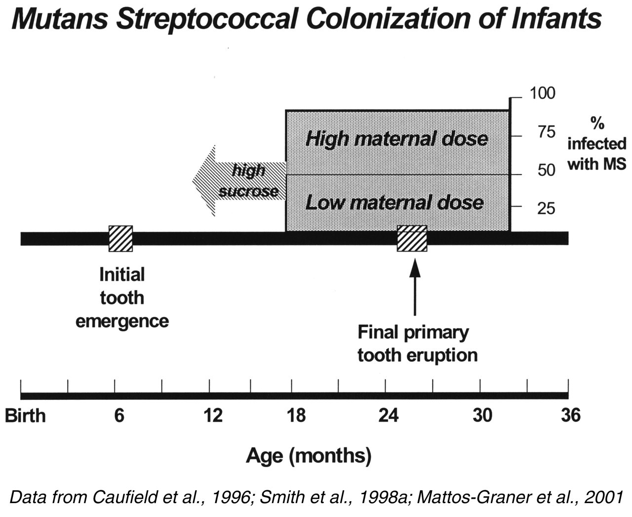
Footnotes
Acknowledgements
The author’s research was performed through the generous support of the National Institute of Dental and Craniofacial Research (DE-01653, DE-04733, DE/AI-12324). The author is also grateful to Leigh Barnes for assistance with manuscript preparation.