
Abstract
Cyclophosphamide (CTX) is a synthetic antineoplastic drug with severe and life-threatening side effects. Studies in search of protective agents, preferably natural products, that can alleviate these side effects are valuable because they can contribute to improve current chemotherapeutic treatment strategies. Curculigo orchioides Gaertn (family Hypoxidaceae) is well known for its medicinal use in the Indian Ayurvedic system of medicine, and various studies have been reported that proved its immunomodulatory and anti-inflammatory properties. In this study, the tumor reduction capacity of CTX in combination with C orchioides methanolic extract was studied using Dalton’s lymphoma ascites-induced solid tumor models. Effect of C orchioides on the reversal of the damage induced by CTX administration (intraperitoneally) was also determined in this study. For this, solid tumor volume, serum cytokine levels, hematolological parameters, intestinal histopathology, and serum and tissue biochemical parameters (Glutathione [GSH], alkaline phosphatase [ALP], glutamate pyruvate transaminase [GPT], lipid peroxidation [LPO]) were analyzed. Immune suppression and increased serum proinflammatory cytokine levels caused by CTX administration (25 mg/kg body weight) were reversed by C orchioides (20 mg/kg body weight). The alcoholic extract enhanced the tumor reduction capacity of CTX and reduced GPT and ALP levels in liver and serum, which were elevated by CTX administration. The LPO level was also lower in the CTX-administered animals when treated with the C orchioides extract. In conclusion, the plant extract when administered in combination with CTX, can result in enhanced anticancer properties; it also ameliorates the toxic side effects of CTX.
Introduction
Cyclophosphamide (CTX) is a nitrogen mustard alkylating agent, which is widely used to treat a broad spectrum of human cancers and some autoimmune disorders. It is mainly used in combination chemotherapy and causes the death of tumor cells by preventing their proliferation. 1 Biotransformation of CTX is catalyzed by hepatic cytochrome P450 to form 4-hydroxycyclophosphamide and its tautomer, aldophosphamide. A small portion of aldophosphamide undergoes a spontaneous elimination reaction to yield phosphoramide mustard and acrolein (causes bladder toxicity). Phosphoramide mustard forms intrachain and interchain cross-links of DNA strands at the guanine N-7 position. This reaction is irreversible and leads to cell death. The remaining aldophosphamide is oxidized by the enzyme aldehyde dehydrogenase to form carboxyphosphamide. 2 CTX exhibits dose-dependent cytotoxicity against rapidly proliferating cancer cells. Hence, the major drawback will be the unprotected state of other rapidly proliferating normal cells, which leads to side effects. Increased doses of CTX treatment result in immune suppression, which is mainly caused by the elimination of T-cells.
The major side effects of CTX administration are cardiac toxicity, hematopoietic depression, hemorrhagic cystis, gonodal dysfunction, nausea, gastrointestinal toxicity, vomiting, alopecia, renal toxicities, and so on. 3 Most chemotherapeutic drugs cause dose-limiting toxicity to bone marrow elements, thus bringing about immune suppression. 4 Recently, it has been found that the use of immunostimulants such as Bacillus Calmette-Guérin (BCG), levamisole, and cytokines 5 along with chemotherapy could reduce myelosuppression and leukopenia induced by chemotherapeutic agents. Several plant extracts such as Withania somnifera, 6 Tinospora cordifolia, 7 and others were shown to enhance the immunity of mice treated with CTX. Herbal preparations that could synergize with the antitumor activity of conventional therapies, provide protection from their side effects, and provide a stimulated immune state will be of great help in improving the current strategies of cancer treatment.
Curculigo orchioides Gaertn (family Hypoxidaceae) is a tiny herbal plant widely distributed in India, China, Malaysia, Japan, and Australia. It is a small annual herb found in subtropical Himalayas from Kumaon eastward and in the Western Ghats from Kinkan southward. 8 It is also known by the name golden eye grass or black musli. 9 The edible tuber is reputed for its various medicinal properties and is the main constituent of one of the important Rasayana drugs used for vigor and vitality in the Ayurvedic system of medicine. Other reported beneficial effects of C orchioides are mast cell stabilization; antihistaminic, 10 immunomodulatory, 11 antioxidant, 12 and antitumor potential 13 ; antiosteoporotic activities, 14 and so on. In the present study, we focus on the synergistic inhibitory effects of C orchioides and CTX against Dalton’s lymphoma ascites–induced solid tumor development and the chemoprotective activity of C orchioides against CTX-induced oxidative damage.
Materials and Methods
Plant Material
C orchioides was locally collected and identified, and a voucher specimen was deposited at the herbarium of Amala Ayurveda Research Centre, Kerala, India (specimen no. 62653). The whole plant was washed, dried at 45°C and powdered. Methanolic extract (70%) of the whole plant was prepared using the Soxhlet apparatus and was evaporated to dryness at 50°C under reduced pressure. The yield obtained was 10.2%. The dried extract was stored at −20°C in amber glass bottles and used per requirement. For in vivo experiments, the required quantity of the dried extract was dissolved in minimum quantity of methanol, which is then suspended in phosphate-buffered saline (PBS) containing 1% gum acacia and the alcohol was evaporated.
Animals
Balb/c and Swiss albino mice (male, weighing 25-28 g) were purchased from the Small Animals Breeding Station (SABS), Government Veterinary College, Mannuthi, Thrissur, Kerala. They were maintained in well-ventilated cages under standard conditions of room temperature, pressure, and humidity and fed with normal mouse chow (Sai Durga Feeds and Foods, Bangalore, India) and water ad libitum. All the animal experiments were carried out with the prior approval of the Institutional Animal Ethics Committee (IAEC) and strictly adhering to the guidelines of the Committee for the purpose of Control and Supervision of Experiments on Animals (CPCSEA) constituted by the Animal Welfare Division, Government of India.
Chemicals and Reagents
CTX (Phosmid-500) was purchased from Neon laboratories Ltd, Mumbai, India. GSH and 5-5′ dithiobis(2-nitrobenzoic acid) were purchased from Sisco Research laboratories Pvt Ltd, Mumbai, India. Pararosaniline and naphthyl acetate were obtained from Loba Chemei, Mumbai, India. Glutamate pyruvate transaminase (GPT) and alkaline phosphatase (ALP) analyzing kits were purchased from SPAN Diagnostics Ltd. All other chemicals used were of analytical reagent grade.
ELISA Kit
Highly specific quantitative Sandwich ELISA kits for mouse granulocyte monocyte colony-stimulating factor (GM-CSF), interferon (IFN)-γ, interleukin (IL)-2, and tumor necrosis factor (TNF)-α were purchased from Pierce Biotechnology, USA.
Cyclophosphamide Administration
CTX was administered intraperitoneally (ip), at concentrations of 25, 20, and 15 mg/kg body weight (0.1 mL) for 10 consecutive days.
Toxicological Evaluation of C orchioides
Five groups of Balb/c mice (n = 8, 6-8 weeks old) were used in this study. The control group received 1% gum acacia in PBS, whereas the experimental groups received C orchiodes extract (25, 50, 100, and 200 mg/kg body weight) ip for 14 consecutive days. All groups were observed for mortality, behavioral changes, and body weight during this period. On the 15th day, all the animals were killed by cervical dislocation. Blood was collected by heart puncture, and serum was separated and used for the analysis of hepatic markers (ALP, 15 GPT 16 ) and renal function markers (creatinine 17 and blood urea nitrogen 18 ). Organs such as liver, spleen, thymus, kidney, and lungs were collected, and weights were recorded.
Determination of Solid Tumor Volume
Swiss albino mice (6-8 weeks old) were divided into 8 groups (n = 8). Solid tumor was induced on the right hind limb of all the animals by injecting Dalton’s lymphoma ascites tumor cells (1 × 10⁶ cells/animal) subcutaneously. Group I consisted of the untreated controls. The animals in groups II, III, and IV received 10 doses of 3 different concentrations of CTX: 25, 20, and 15 mg/kg body weight, respectively. The animals in groups V, VI, and VII received 10 doses of C orchioides extract (20 mg/kg body weight) along with 3 different concentrations of CTX: 25, 20, and 15 mg/kg body weight, respectively. The animals in group VIII received C orchioides extract alone for 10 days. All treatments were started 48 hours after tumor induction, and CTX was administered 2 hours after the administration of extract. Tumor radii were measured using Vernier calipers from the seventh day of tumor inoculation, and measurements were done every third day for a period of 30 days. Tumor volume was calculated using the formula V = (4/3)πr12r2, where r1and r2 are the radii of the tumor along 2 directions.
Hematological Analysis
Three groups of Balb/c mice (n = 8, 4-6 weeks old) were used in this study. Animals in group I were treated with CTX (25 mg/kg body weight, ip) for 10 consecutive days. Animals in groups II and III were treated with 10 mg/kg body weight and 20 mg/kg body weight of C orchioides (ip), respectively, along with CTX, for 10 consecutive days. Blood was collected from the caudal vein prior to CTX treatment and every third day after CTX administration. Parameters such as total white blood cell (WBC) count (hemocytometer method), differential count (Leishman’s stain), and hemoglobin content (cyanmethemoglobin method) were estimated.
Lymphoid Organ Weight, Bone Marrow Cellularity, and Number of α-Esterase-Positive Cells
Three groups of Balb/c mice (n = 24, 4-6 weeks old) were used in this study, and all the animals were administered CTX (ip, 25 mg/kg body weight) for 10 consecutive days. Animals in group I animals were untreated CTX controls, whereas animals in groups II and III were treated with C orchioides extract (10 and 20 mg/kg body weight, ip, respectively) along with CTX for 10 days. The body weights of animals were recorded at different time points (48 hours, 7th and 11th days), and 8 animals from each group were killed humanely at every time point. The weight of lymphoid organs (thymus and spleen) was taken and expressed as relative organ weight with respect to the body weight. Bone marrow cells were collected from both femurs, and the cell number was determined using a hemocytometer 19 and expressed as total live cells per femur. The number of α-esterase-positive cells were determined by the azo dye coupling method. 20 A smear of bone marrow cells from the above preparation was made on clean glass slides, air-dried, stained with α-naphthyl acetate and pararosaniline hydrochloride, counter-stained with hematoxylin and observed under a 100× magnification. The number of α-esterase-positive cells was expressed out of 4000 cells.
Biochemical Analysis
Blood was collected by heart puncture from the animals in the above experiment, and serum was separated out. The liver was collected and homogenized using ice cold Tris buffer (0.1 M, pH 7.4). Serum and homogenate were used for the estimation of ALP 15 , GPT, 16 and lipid peroxidation (LPO). 21 Urea and creatinine levels in the serum were also determined. Intestinal mucosa was used to estimate the levels of GSH by the method of Moron et al. 22
Cytokine Production
Blood was collected from the animals used for the study of hematological parameters at 48 hours and on the 11th day for separating the serum. Estimations of cytokines such as IL-2, IFN-γ, GM-CSF, and TNF-α were performed by the ELISA method using specific antibodies, according to the manufacturer’s instructions.
Histopathological Analysis of the Intestinal Damage Caused by CTX
The jejunal portion of the small intestine from the previous experiment was excised and fixed in 10% formaldehyde. Sections (4 µm) were stained with eosin and hematoxylin and observed under a light microscope.
Data Analysis
All data were expressed as mean ± standard deviation. The mean values were statistically analyzed using 1-way ANOVA using the Graph pad Instat software package. The significant differences between the groups were further analyzed by Bonferroni’s t test. P values <.05 were considered significant.
Results
Toxicological Evaluation of C orchioides
Short-term administration (14 days) of C orchioides extract at doses up to 250 mg/kg body weight did not produce any mortality or change in behavior, body weight, relative organ weight, or hepatic and renal functions when compared with normal untreated animals (Table 1). A dose of 20 mg/kg body weight was selected for further studies based on the preliminary observations on the relative effects of different concentrations on hematological parameters and antitumor studies.
Toxicity Profile of Curculigo orchioides. a

Abbreviations: D/T, dead/treated mice; ALP, alkaline phosphatase; GPT, glutamate pyruvate transaminase.
Values are the mean ± standard deviation. All the treated animals were carefully examined for 14 days for any signs of toxicity (behavioral changes and mortality). “None” means that no toxic symptoms were seen during the observation period.
Reduction of Solid Tumor Volume
The effect of C orchioides on solid tumor reduction in animals treated with CTX is shown in Figure 1. The administered dose of CTX was found to be effective in controlling tumor growth, and the administration of C orchioides along with CTX produced a synergistic effect on tumor growth inhibition. The animal group that was treated with 20 mg/kg body weight of the extract and received 25 mg/kg body weight of CTX exhibited the lowest tumor volume among all the groups. Administration of CTX at a dose of 15 mg/kg body weight along with the C orchioides extract was found to be more effective in reducing the tumor volume than administering the higher dose of CTX (25 mg/kg body weight) alone.

Effect on solid tumor reduction: Balb/c mice were divided into 8 groups. Group I: control (untreated); group II: cyclophosphamide (CTX; 25 mg/kg body weight [B.wt]); group III: CTX (20 mg/kg B.wt); group IV: CTX (15 mg/kg B.wt); group V: Curculigo orchioides 20 mg/kg B.wt; group VI: CTX (25 mg/kg B.wt) + C orchioides; group VII: CTX (20 mg/kg B.wt) + C orchioides; group VIII: CTX (15 mg/kg B.wt) + C orchioides. All the animals were induced with solid tumor by injecting Dalton’s lymphoma ascites tumor cells (1 × 10⁶ cells/animal) subcutaneously on the right hind limb of mice. The radii of the developing tumors were measured every third day, starting from the seventh day of tumor inoculation, for 30 days using Vernier calipers.
Hematological Parameters
CTX administration drastically reduced the total WBC count in mice (Figure 2). Myelosuppression as seen from the WBC count was observed throughout the period of CTX administration. Administration of C orchioides could significantly increase the WBC count to 4500 ± 232 cells/mm3 (20 mg/kg body weight) and 4200 ± 312 cells/mm3 (10 mg/kg body weight) versus the drastic reduction to 3200 ± 230 cells/mm3 by CTX administration alone. The reduced WBC count was observed throughout the period of study, but it was shifted toward normal levels by the administration of C orchioides (20 mg/kg body weight). There was no significant effect on the differential count, hemoglobin level, and body weight (data not shown).

Effect of Curculigo orchioides extract on total white blood cell count. Mice were given 10 doses of cyclophosphamide (CTX), and every 3rd day, blood was collected from the caudal vein in a sterile manner, and total white blood cell count was monitored.
Lymphoid Organ Weight
Table 2 shows the relative organ weights of the thymus and spleen. CTX treatment drastically reduced the relative weights of thymus and spleen in control animals. Administration of C orchioides at 20 mg/kg body weight showed a significant (P < .05) enhancement in the weights of the thymus and spleen.
Relative Organ Weights of the Spleen and Thymus. a

Abbreviations: CTX, cyclophosphamide; b.wt, body weight.
Balb/c mice were divided into 3 groups (n = 24). Control animals were given 10 doses of CTX (25 mg/kg b.wt), and the treated animals were given 10 doses of C orchioides extract along with CTX intraperitoneally. From each group, 8 animals were killed at each time point, and the relative organ weights of the thymus and spleen were determined. Values are mean ± standard deviation.
Normal versus control: P < .05, significantly different from normal.
Control versus control: P < .05 significantly different from control.
Normal versus control: P < .05 significantly different from normal.
Bone Marrow Cellularity and α-Esterase-Positive Cells
There was a drastic reduction in bone marrow cellularity in CTX-treated control animals (6.3 ± 0.228 × 10⁶ cells/femur) after 48 hours, when compared with normal animals (16.00 ± 0.584 × 10⁶ cells/femur). Treatment with C orchioides extract (8.62 ± 1.08 × 10⁶ cells/femur) could significantly increase the bone marrow cellularity on the same day. Afterward, the cellularity was observed to significantly increase gradually, which is a clear indication of the ability of the extract to enhance bone marrow cell proliferation (Table 3).
Bone Marrow Cellularity and Number of α-Esterase-Positive Cells. a

Abbreviations: CTX, cyclophosphamide; b.wt, body weight.
Balb/c mice were divided into 3 groups (n = 24). Control animals were given 10 doses of CTX (25 mg/kg b.wt) and the treated animals were given 10 doses of C orchioides extract along with CTX intraperitoneally. From each group, 8 animals were killed at each time point, and the bone marrow cells were collected from the femur and made into a single-cell suspension. The bone marrow cellularity was determined using a hemocytometer. The α-esterase-positive cells were determined by the azo dye coupling method. Values are the mean ± standard deviation.
Normal versus control: P < .05, significantly different from normal.
Control versus treated: P < .05, significantly different from control.
Normal versus treated: P < .05, significantly different from normal.
The effect of C orchioides extract on the number of α-esterase-positive cells is presented in Table 3. The number of α-esterase positive cells was significantly decreased after CTX administration. C orchioides treatment elevated the α-esterase activity of maturing monocytes. On the 11th day, the number of α-esterase-positive cells of the CTX-only group was 682.50 ± 94.52 cells/4000 cells, which was found to be increased to 925.54 ± 92.56 cells/4000 cells by C orchioides treatment (20 mg/kg body weight).
Cytokine Levels
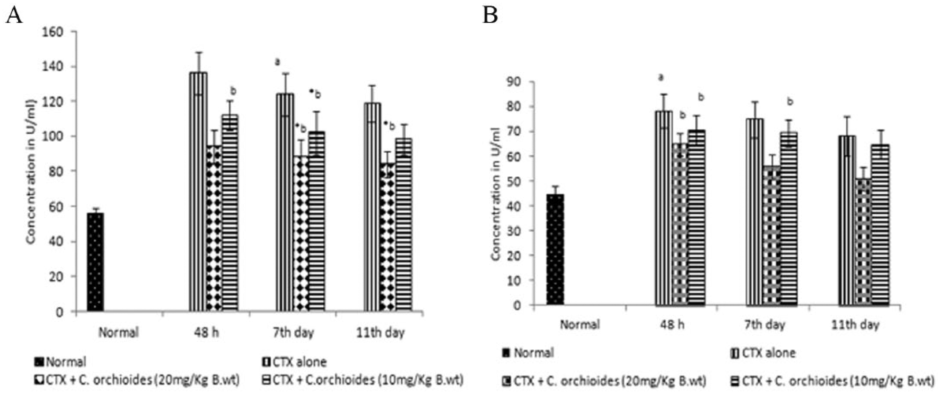
CTX administration resulted in a drastic decrease in the levels of cytokines such as IL-2 (8.40 ± 0.96), IFN-γ (654 ± 43.26), and GM-CSF (22.60 ± 3.60) and a marked elevation in the level of the proinflammatory cytokine, TNF-α (311.8 ± 56.60). All these changes were reversed after treatment with C orchioides. The serum levels of IL-2, GM-CSF, and IFN-γ were elevated to 16.7 ± 2.56, 30.16 ± 4.50, and 1024 ± 123.76, respectively, on the second day, whereas the TNF-α level was reduced to 155.6 ± 27.90 on the same day. Table 4 represents the serum IFN-γ and GM-CSF profiles, and Table 5 shows the data for IL-2 and TNF-α.
Effect of Curculigo orchiodes on IFN-γ and GM-CSF Levels. a
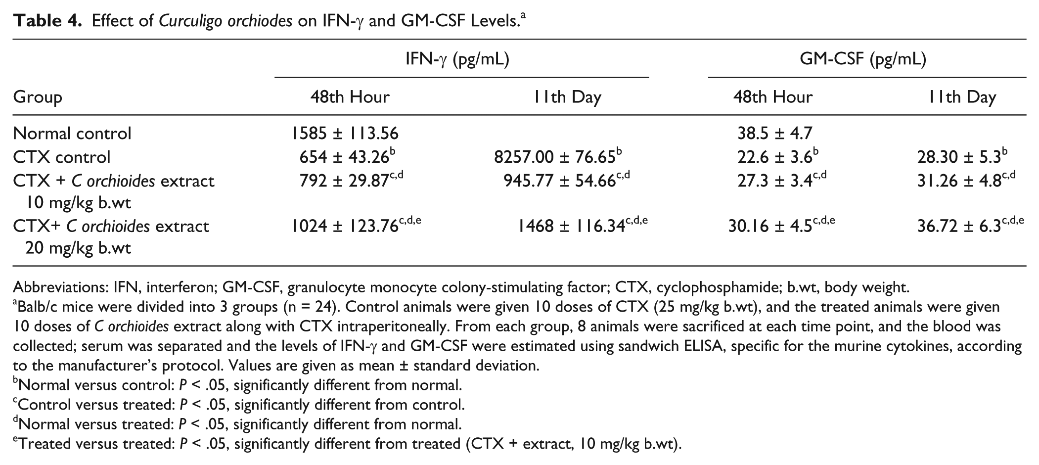
Abbreviations: IFN, interferon; GM-CSF, granulocyte monocyte colony-stimulating factor; CTX, cyclophosphamide; b.wt, body weight.
Balb/c mice were divided into 3 groups (n = 24). Control animals were given 10 doses of CTX (25 mg/kg b.wt), and the treated animals were given 10 doses of C orchioides extract along with CTX intraperitoneally. From each group, 8 animals were sacrificed at each time point, and the blood was collected; serum was separated and the levels of IFN-γ and GM-CSF were estimated using sandwich ELISA, specific for the murine cytokines, according to the manufacturer’s protocol. Values are given as mean ± standard deviation.
Normal versus control: P < .05, significantly different from normal.
Control versus treated: P < .05, significantly different from control.
Normal versus treated: P < .05, significantly different from normal.
Treated versus treated: P < .05, significantly different from treated (CTX + extract, 10 mg/kg b.wt).
Effect of Curculigo orchioides on TNF-α and IL-2 levels. a

Abbreviations: TNF, tumor necrosis factor; IL, interleukin; CTX, cyclophosphamide; b.wt, body weight.
Balb/c mice were divided into 3 groups (n = 24). Control animals were given 10 doses of CTX (25 mg/kg b.wt), and the treated animals were given 10 doses of C orchioides extract along with CTX intraperitoneally. From each group, 8 animals were killed at each time point, and the blood was collected; serum was separated, and the levels of IL-2 and TNF-α were estimated using sandwich ELISA kit, specific for the murine cytokines, according to the manufacturer’s protocol. Values are the mean ± standard deviation.
Normal versus control: P < .05, significantly different from normal.
Control versus treated: P < .05, significantly different from control.
Normal versus treated: P < .05, significantly different from normal.
Treated versus treated: P < .05, significantly different from treated (CTX + extract, 10 mg/kg b.wt).
Reduced GSH, LPO, ALP, and GPT levels
The CTX-only group had a lowered level of GSH in intestinal mucosa (6.88 ± 1.23 nmol/mL) and liver homogenate (2.72 ± 0.25 nmol/mL) after 48 hours, compared with the normal control group (5.68 ± 0.51 nmol/mL). A significantly higher GSH content was observed in the C orchioides–treated group (intestinal mucosa 15.82 ± 2.56 nmol/mL, liver homogenate 3.51 ± 0.29 nmol/mL) on day 11, relative to the CTX control group (Figure 3).

Effect of Curculigo orchioides extract on GSH level. Balb/c mice were divided into 3 groups (n = 24). Control animals were given 10 doses of CTX (25 mg/kg body weight [b.wt]), and the treated animals were given 10 doses of C orchioides extract (10 and 20 mg/kg b.wt) along with CTX intraperitoneally. From each group, 8 animals were killed at each time point, and the GSH levels of (A) intestinal mucosa and (B) liver was estimated by the method of Moron et al. 22 Values are means ± standard deviations. Normal versus control: aP < .05, significantly different from normal; control versus treated: *P < .05 significantly different from control; normal versus treated: bP < .05 significantly different from normal; treated versus treated: cP < .05 significantly different from treated (CTX + C orchioides, 10 mg/kg b.wt).
In the case of serum ALP, the CTX-only group showed an elevated level of 16.40 ± 1.80 U/mL at 48 hours and 17.82 ± 2.62 U/mL on the seventh day. However, administration of C orchioides could lower the elevated level of serum ALP to 14.60 ± 2.58 U/mL by the seventh day. The level of serum ALP in the normal control group was 12.60 ± 1.44 U/mL. In the CTX-only control group, the liver ALP was 19.70 ± 2.80 U/mL at 48 hours after CTX administration, and this level was further elevated to 21.20 ± 3.60 U/mL on the seventh day. On the same day, the elevated level of liver ALP was found to be significantly decreased in the CTX group, which was treated with C orchioides (Figure 4).

Effect of Curculigo orchioides extract on ALP level. Balb/c mice were divided into 3 groups (n = 24). Control animals were given 10 doses of CTX (25 mg/kg body weight [b.wt]), and the treated animals were given 10 doses of C orchioides extract (10 and 20 mg/kg b.wt) along with CTX intraperitoneally. From each group, 8 animals were killed at each time point, and the blood and liver were collected; (A) serum ALP and (B) liver ALP were estimated by King’s method. Values are means ± standard deviations. Normal versus control: aP < .05, significantly different from normal; control versus treated: *P < .05 significantly different from control; normal versus treated: #P < .05 significantly different from normal.
The increased level of serum GPT in the CTX-only treated group (124.00 ± 12.26 U/mL) was lowered after treatment with C orchioides (88.00 ± 10.22 U/mL) on the seventh day after CTX administration, when compared with the normal animals (55.80 ± 3.20U/mL; Figure 5).
Effect of Curculigo orchioides extract on GPT level. Balb/c mice were divided into 3 groups (n = 24). Control animals were given 10 doses of CTX (25 mg/kg body weight [b.wt]), and the treated animals were given 10 doses of C orchioides extract (10 and 20 mg/kg b.wt) along with CTX intraperitoneally. From each group, 8 animals were killed at each time point, and the blood and liver were collected; (A) serum GPT and (B) liver GPT were estimated by Bergmeyer’s method. Values are means ± standard deviations. Normal versus control: aP < .05, significantly different from normal; control versus treated: *P < .05 significantly different from control; normal versus treated: #P < .05 significantly different from normal; treated versus treated: bP < .05 significantly different from treated (CTX + C orchioides, 10 mg/kg b.wt).
CTX administration caused an increase in the amount of LPO products (Figure 6). An increase in the thiobarbituric acid reactive substance level in the serum (3.88 ± 0.32 nmol/mL) and liver (4.12 ± 0.90 nmol/mg/protein formed/min/mg protein) was evident in CTX-only administered animals after 48 hours. It was found that the increased level of serum LPO caused by CTX was significantly reduced by the treatment with C orchioides (3.25 ± 0.36 nmol/mL) after 48 hours. Similarly, the level of liver LPO products was also significantly reduced by the administration of C orchioides (2.98 ± 0.35 nmol/mg/protein formed/min/mg protein) on the seventh day (Figure 6).

Effect of Curculigo orchioides extract on LPO level. Balb/c mice were divided into 3 groups (n = 24). Control animals were given 10 doses of CTX (25 mg/kg body weight [b.wt]), and the treated animals were given 10 doses of extract (10 and 20 mg/kg b.wt) along with CTX intraperitoneally. From each group, 8 animals were killed at each time point, and the blood and liver were collected; (A) serum LPO and (B) liver LPO level were estimated using the TBA (thiobarbituric acid) method of Ohkhawa et al. Values are means ± standard deviations. Normal versus control: aP < .05, significantly different from normal; control versus treated: *P < .05 significantly different from control; normal versus treated: #P < .05 significantly different from normal; treated versus treated: bP < .05 significantly different from treated (CTX + C orchiodes, 10 mg/kg b.wt).
Protective Effect of C orchioides Against the Intestinal Damage Caused by CTX Administration
Histopathological analysis of a jejunal section of the small intestine showed abnormal crypt architecture and damaged intestinal villi in the case of the CTX-only group. Simultaneous administration of C orchioides extract along with CTX could effectively reduce the intestinal toxicity induced by CTX. The crypt architecture was maintained, and the villi damage was reduced (Figure 7).

Histopathological analysis of the small intestine: (A) intestine: normal; (B) intestine: CTX control; (C) intestine: treated with Curculigo orchioides (10 mg/kg); and (D) intestine: treated with C orchioides (20 mg/kg).
Discussion
CTX is a cytotoxic chemotherapeutic drug that acts as an alkylating agent producing reactive carbonium ions that react with DNA. 23 CTX itself has been shown to be inactive in vitro but was found to be activated to cytotoxic metabolites by the mixed function oxidase in hepatic microsomes. 24 The high therapeutic index of CTX was shown to be a result of the metabolite 4-hydroxycyclophosphamide and a phosphoramide derivative. It was found that free radicals are formed during the activation of CTX, which produce tissue injury. 25 Phosphoramide mustard is known to cause myelosuppression. This study was carried out to focus on the immunostimulant and antioxidant activities of C orchioides and its potential to reduce toxicity induced by CTX in order to evaluate its use as an adjuvant during chemotherapy.
In the present study, it was found that the administration of C orchioides extract along with CTX could enhance the tumor reducing capacity of CTX. Even a lower concentration of CTX (15 mg/kg body weight) could give results similar to that of 25 mg/kg body weight of CTX when it was administered along with the C orchioides extract. This implicates the synergistic effect of CTX and C orchioides extract in the reduction of tumor volume. This effect may be partly a result of the immunostimulatory activity of the plant, which stimulates the humoral and cellular arms of the host’s immune system to prevent primary tumor development and shapes tumor immunogenicity. Antitumor activity of the extract13,26 may also have contributed to the synergism, which is very evident when comparing the tumor volume among the CTX-only group and extract + CTX-treated group.
During the study, it was also confirmed that myelosuppression caused by CTX is effectively prevented by C orchioides. The CTX-treated group, which was administered C orchioides, attained a normal WBC count at the end of the treatment, whereas the regenerative capacity in the CTX-only control group was very low and did not return to normal. In the CTX-treated group, administration of C orchioides significantly increased the bone marrow cellularity and α-esterase-positive cells, which signifies an increase in maturing monocytes. This indicates that C orchioides stimulated the hemopoietic system, which is highly sensitive to chemotherapy. This hemoprotective effect can also be attributed to the antioxidant activity of the extract, which may protect the bone marrow cells from the free radical–induced damages caused by CTX. Together, these protective effects could have led to faster recovery from those side effects caused by CTX.
Glutathione metabolism is often correlated with cellular sensitivity to anticancer agents. Glutathione is protective against drug-induced cytotoxicity. It reacts with toxic endogenous and exogenous substances, including free radicals and anticancer agents. 27 The metabolism of CTX in the body produces highly reactive electrophiles. The decreased level of GSH in the CTX-only group may be a result of the electrophilic burden on the cells. C orchioides was found to significantly enhance the level of GSH, which is involved in detoxification.
It has been reported that hydroxyl radicals inhibit ALP activity, 28 and the free radicals generated during the peroxidation reaction will attack the protein molecules in membranes. 29 The liver enzymes, which were moderately increased in the CTX administered group by the seventh day, were lowered after treatment with C orchioides. Similarly, serum and liver GPT levels, which were increased after CTX administration, were significantly lowered in C orchioides–treated groups.
The free radicals generated during the metabolism of CTX can initiate LPO. 30 In the present study, it was observed that CTX administration increased the serum and liver LPO levels, but it was significantly lowered after treatment with C orchioides. Inhibition of LPO in biomembranes can be caused by antioxidants. 31
Cytokines are proteins or glycoproteins that act as cell signaling molecules, and their main function is the modulation of immune responses. Several inflammatory cytokines are induced by oxidant stress, and they themselves trigger the release of other cytokines and also lead to increased oxidant stress. IL-2 is a cytokine secreted by macrophages, necessary for the growth, proliferation, and differentiation of T-cells. Levels of IL-2, which were reduced after CTX administration, were found to be elevated after C orchioides administration. IFN-γ is a cytokine that is critical for innate and adaptive immunity against viral and intracellular bacterial infections and also for tumor control. IFN-γ is an important activator of macrophages. GM-CSF is another immune-stimulating cytokine that functions as a white blood cell growth factor, which stimulates the stem cells to produce granulocytes (neutrophils, eosinophils, and basophils) and monocytes. Administration of CTX drastically reduced the levels of IFN-γ and GM-CSF, whereas the simultaneous administration of the extract enhanced the production of these 2 cytokines. TNF-α is a cytokine involved in systemic inflammation secreted predominantly by macrophages after a variety of stimuli. It is a major proinflammatory cytokine, and its level was drastically elevated after CTX treatment but was observed to be reduced after the administration of C orchioides extract. From these observations, it is clear that the immunomodulatory and anti-inflammatory activity of the plant is responsible for these effects, and one of the mechanisms of action might be the regulation of the production of cytokines by the immune cells.
The small intestine has been reported to have a remarkable capacity for repair. 31 A delay in the rate of recovery after CTX administration could be explained on the basis of high stem cell depletion by apoptotic as well as mitotic death. In the present study, the rate of recovery was faster in the C orchioides–treated group.
CTX dosages can be minimized when it is administered in combination with C orchioides extract, which produces a synergistic effect and reduces tumor development. The results also indicate that administration of the alcoholic extract of C orchioides protected mice from the toxic effects of CTX and promoted the recovery of bone marrow cells and leukocytes. Even though the antioxidant properties of C orchioides are well documented, these effects did not interfere with the tumor-reducing capability of CTX. It is proposed that C orchioides–mediated protection against CTX could be a result of its immunomodulatory activity and by the induction of phase II enzymes involved in the detoxification pathways. These results, thus, provide some insight into the mechanisms involved in the chemoprotective action of C orchioides.
Footnotes
Acknowledgements
The authors are grateful to Dr Ramadasan Kuttan, Director of Amala Cancer Research Centre, for his valuable suggestions and support.
Declaration of Conflicting Interests
The author(s) declared no potential conflicts of interest with respect to the research, authorship, and/or publication of this article.
Funding
The author(s) disclosed receipt of the following financial support for the research, authorship, and/or publication of this article: The financial support from Innovation in Science Pursuit for Inspired Research, Department of Science and Technology, New Delhi, India, to carry out this study is gratefully acknowledged.