
Abstract
This study was designed to investigate the anticancer activity of extracts of the phytomedicine DAS-77. The sulforhodamine B (SRB) in vitro cytotoxicity assay, Sarcoma-180 (S-180) ascites and solid tumor, and L1210 lymphoid leukemia in vivo models were employed. DAS-A001 (ethanol extract, IC50 12 and 13 µg/mL with HCT-116 and PC3, respectively); DAS-A002 (hydroethanol extract, IC50 <5 and 13 µg/mL with HCT-116 and PC3, respectively); DAS-A003 (aqueous extract, IC50 <5 µg/mL with THP-1); and DAS-A004 (dichloromethane:methanol extract; IC50 <5 and 17 µg/mL with HCT-116 and PC3, respectively) demonstrated significant activity in vitro. DAS-A002 and DAS-A003 (80-120 mg/kg) elicited significant (P < .05-.001) dose-dependent inhibition of tumor growth in the S-180 ascites model. Peak effects were produced at the highest dose of 120 mg/kg with inhibition values of 87.50% and 89.23% for DAS-A002 and DAS-A003, respectively, compared with a value of 97.27% for 5-FU (20 mg/kg). As regards the S-180 solid tumor model, inhibition of tumor growth was found to be 52.56% and 37.95%, respectively, for DAS-A002 and DAS-A003. The effect of DAS-A002 was comparable and not significantly different (P > .05) from that of 5-FU (20 mg/kg; 50.18% inhibition). DAS-A003 but not DAS-A002 showed significant activity in the leukemia model with 177.78% increase in mean survival time relative to 211.11% for 5-FU. Findings in this study suggest that the hydroethanol and aqueous extracts of DAS-77 possess significant anticancer activity.
Introduction
Cancer is a complex collection of diseases that can arise in almost any tissue in the body. 1 It has been reported to be a leading cause of death all over the world. 2 Ferlay et al 3 reported that based on current rates in 2008, the worldwide incidence and mortality projection will increase in the next 20 years, almost 21.4 million new cases will be diagnosed annually, and there will be more than 13.2 million deaths from cancer. Based on the high level of morbidity and mortality currently associated with cancer, it certainly represents a major public health burden.
According to Park, 2 natural plant products have been historically used for the treatment of various diseases and still serve as an excellent source for the discovery and development of modern drugs for cancer treatment. Plants contain several classes of phytochemicals that have antioxidative, antimutagenic, and anticarcinogenic effects 4 and numerous clinical trials are ongoing to evaluate their potential as cancer chemopreventive and chemotherapeutic agents.5-7
DAS-77 is an herbal preparation used in the treatment of diverse ailments especially in southwest Nigeria. This phytomedicine is claimed to be effective in the treatment of piles, dysentery, cholera, menstrual pain, stomach disorder, ulcer, diarrhea, and sexually transmitted infections. 8 Antidiarrheal activity associated with inhibition of intestinal motility mediated by muscarinic and α-adrenergic receptors 9 ; gastroprotective property in ulcer models linked with reduction in gastric secretion and acidity, and with antioxidant activity; 10 and antimicrobial effect with greater activity against Gram-negative bacteria than the Gram-positive have been reported. 8 Afolabi et al 11 conducted a 90 day chronic toxicity study of DAS-77 with the conclusion that this phytomedicine is relatively safe with the potential for enhancing in vivo antioxidant activity, and causing reversible electrolyte imbalance and sterility in males. Phytochemical analysis of DAS-77 revealed the presence of tannins, saponins, phenols, flavonoids, and alkaloids.9-11
This study was conducted to investigate the in vitro and in vivo anticancer activity of DAS-77 as part of the broad evaluation of selected Nigerian medicinal plant remedies for antiproliferative property.
Materials and Methods
DAS-77
DAS-77 (Doynik Ventures, Ijoko-Lemode, via Sango Ota, Ogun State, Nigeria) is a phytomedicine composed of equal proportions (1:1) of the milled dried callous bark of Mangifera indica Linn. (Anacardiaceae) and root of Carica papaya Linn. (Caricaceae). The coarse powder is light brown in color with a pungent smell. The product has a solution pH of 8.5.
Extraction
DAS-77 powder was divided into 4 portions of 100 g each. Three portions were separately macerated in about 1500 mL of 95% ethanol (DAS-A001), hydroethanol (1:1; DAS-A002), and distilled water (DAS-A003) for 3 hours with mechanical stirring (Heidolph RZR 2051 Control; 400 rpm). Filtrates were obtained using Whatman filter paper (150 mm). To ensure exhaustive extraction, residues were remacerated (2×). The combined filtrates of DAS-A001 and DAS-A002 extracts were concentrated using Heidolph Rotavapor (LABORATA 4000; 120 rpm and 40°C). The combined filtrate of DAS-A003 was lyophilized. The remaining portion of 100 g of the DAS-77 powder was put into 2000 mL separating funnel with the bottom lined with cotton wool and 1 L of dichloromethane:methanol (DCM:MeOH; 70:30) was put into the separating funnel. After 24 hours, the solvent mixture was drained and the extraction liquid was filtered using Whatman filter paper (150 mm). After a period of 48 and 72 hours, 600 mL of the DCM:MeOH solvent mixture was added to the separating funnel. The solvent was drained 24 hours after and filtered. The combined DAS-A004 extract was concentrated with Rotavapor without vacuum. All dried extracts were weighed and stored in a desiccator. The yield of the extracts was 13.37% (DAS-A001), 22.01% (DAS-A002), 28.46% (DAS-A003), and 10.98% (DAS-A004).
Chemicals
RPMI-1640, minimum essential medium (MEM), fetal calf serum, trypsin, trypan blue, ethanol, penicillin, streptomycin, gentamycin, dimethyl sulfoxide (DMSO), sulforhodamine B, mitomycin-C, paclitaxel, and 5-fluorouracil (Sigma Chemical Co, St Louis, MO, USA); phosphate buffer saline (PBS; Merck, Darmstadt, Germany); trichloroacetic acid (TCA), distilled water, sodium hydroxide, Tris-EDTA buffer, Tris buffer (Hi-Media); acetic acid, sodium bicarbonate, hydrochloric acid (RANKEM, New Delhi, India), isopropanol (SISCO, Mumbai, India), and Tris-acetate-EDTA buffer. All other chemicals used in this study were purchased locally and were of analytical grade.
Cell Lines and Cell Cultures
A549 (lung), HCT-116 (colon), PC3 (prostate), A431 (skin), HeLa (cervix), and THP-1 (leukemia) human cancer cell lines (National Center for Cell Science, Pune, India, and National Cancer Institute, Frederick, MD, USA) were used in this study. These were grown and maintained in appropriate medium, pH 7.4, supplemented with 10% fetal calf serum, glutamine (2 mM), penicillin (100 U/mL) and streptomycin (100 µg/mL). The cell cultures were grown in a carbon dioxide incubator (Heraeus GmbH, Hanau, Germany) at 37°C with 90% humidity and 5% CO2.12,13
Animals
Inbred BALB/c, DBA/2, outbred Swiss albino and F1 hybrid CDF1 mice (18-23 g; about 2 months of age) used in this study were obtained from the Animal Facility of Indian Institute of Integrative Medicine, Jammu, India. The mice were maintained at 22°C ± 2°C with 20 to 25 complete air changes with 100% fresh air, with relative humidity maintained at 50% to 60%. Pelleted feed (Ashirwad Industries, Chandigarh, India) and water ad libitum (autoclaved) were used to maintain the mice, which were housed in transparent polycarbonate filter top cages in animal isolator cabins. All the animals selected for individual experiments were of the same sex and strain. The experimental procedures used in this study were approved by the Institutional Animal Ethics Committee, Indian Institute of Integrative Medicine, Jammu, India.
In Vitro Cytotoxicity Against Human Cancer Cell Lines
The established sulforhodamine-B (SRB) assay12-14 was used to evaluate the in vitro cytotoxicity of the ethanolic (DAS-A001), hydroethanolic (DAS-A002), aqueous (DAS-A003), and DCM:MeOH (DAS-A004) extracts of DAS-77. The extracts at final concentrations of 10, 30, and 100 µg/mL were tested against A549 (lung), HCT-116 (colon), PC3 (prostate), A431 (skin), HeLa (cervix), and THP-1 (leukemia) human cancer cell lines appropriately seeded in 96-well cell culture plates. Subsequent to applicable handling and procedure, the optical density (OD) of the plate wells was recorded with a microplate reader at 540 nm. Inhibition of cell growth was calculated as the percentage survival of treated cells over control cells × 100 (T/C %).
Evaluation of In vivo Anticancer Activity
Sarcoma-180 (S-180) Ascites Model
S-180 cells were collected from the peritoneal cavity of Swiss albino mice used for propagation. A total of 1 × 10⁷ cells/animal were injected intraperitoneally (i.p.) into the peritoneal cavity of BALB/c mice of same sex on day 0. Animals were thereafter randomized and allotted to different treatment groups. The different groups were separately treated i.p. with normal saline (0.2 mL/mouse), 5-FU (20 mg/kg), and DAS-A001, DAS-A002, DAS-A003, and DAS-A004 generally at the dose of 100 mg/kg from day 1 to 9. All the animals were sacrificed under diethyl ether anesthesia on day 12 and the ascitic fluid was collected from the peritoneal cavity for determination of tumor weight, volume, and cell number. 1 The number of cells per milliliter of the ascitic fluid was counted using a hemocytometer (Neubauer’s chamber).
DAS-A002 and DAS-A003 were subjected to graded dose (80, 100, and 120 mg/kg i.p.) effect determination based on the highest value of tumor growth inhibition and least mortality.
Sarcoma-180 (S-180) Solid Tumor Model
The protocol adopted in this model was same as in the S-180 ascites model except that tumor cells (1 × 10⁷ cells/animal) was injected (intramuscularly) into the right thigh of BALB/c mice of same sex on day 0. 1 Based on the results obtained in the S-180 ascites model, the effectiveness of DAS-A002 (125 mg/kg, i.p.) and DAS-A003 (130 mg/kg, i.p.) was investigated in this model. Normal saline (0.2 mL/mouse, i.p.) and 5-FU (20 mg/kg, i.p.) served as control and standard drug, respectively. The longest and shortest diameters of tumors were measured with Vernier caliper on day 13 and tumor volume was determined.15,16

L1210 Lymphoid Leukemia Model
L1210 lymphocytic leukemia cells were collected from the peritoneal cavity of DBA/2 mice. A total of 2.5 × 10⁶ cells were injected i.p. into CDF1 mice of same sex on day 0 of the experiment. Normal saline (0.2 mL/mouse), 5-FU (20 mg/kg), DAS-A002 (100 mg/kg), and DAS-A003 (100 mg/kg) were administered i.p. from day 1 to 9. Mortality was noted and the mean survival time (MST) was calculated.

where, S5 = number of survivors on day 5; ΣS = Sum of daily survivors from day 6 to day 18; and NT = number of no takes (survivors beyond day 18).

where, T = MST of the drug treated mice; and C = MST of untreated control animals. 17
T/C % < 125% = toxic/inactive
T/C % > 125% = significant antileukemic effect
Statistical Analysis
The data obtained in this study are expressed as mean ± standard error of the mean. Results were analyzed using 1-way analysis of variance followed by Dunnett’s multiple comparison test (GraphPad Prism 5; GraphPad Software, Inc, San Diego, CA, USA). As regards comparisons, values were considered significant at P < .05.
Results
In Vitro Cytotoxic Activity
DAS-A001 produced significant cytotoxicity activity in the SRB assay on HCT-116 and PC3 human cancer cell lines with IC50 value of 12 and 13 µg/mL, respectively (Figure 1A). DAS-A002 also elicited significant activity on HCT-116 and PC3 with IC50 values of <5 and 13 µg/mL, respectively (Figure 1B). DAS-A003 was only active on THP-1 human cancer cell line with IC50 value of <5 µg/mL (Figure 1C). As shown in Figure 1D, DAS-A004 was active against HCT-116 and PC3 human cancer cell lines with IC50 values of <5 and 17 µg/mL, respectively.
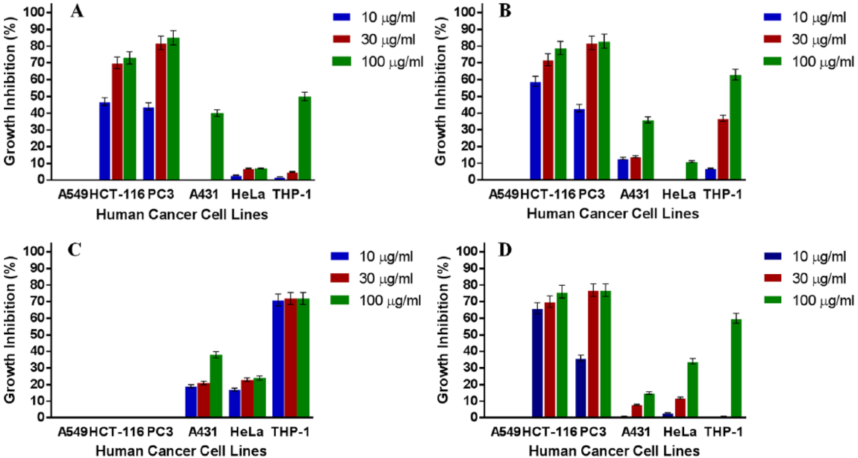
In vitro cytotoxic activity of DAS-A001 against various human cancer cells lines in the sulforhodamine B (SRB) assay. Estimated IC50 values are NA, 12, 13, >100, >100, and 100 µg/mL for A549, HCT-116, PC3, A431, HeLa, and THP-1 respectively. NA implies not active (A); DAS-A002 against various human cancer cells lines in the SRB assay. Estimated IC50 values are NA, <5, 13, >100, >100, and 65 µg/mL for A549, HCT-116, PC3, A431, HeLa, and THP-1 respectively (B); DAS-A003 against various human cancer cells lines in the SRB assay. Estimated IC50 values are NA, NA, NA, >100, >100, and <5 µg/mL for A549, HCT-116, PC3, A431, HeLa, and THP-1, respectively (C); DAS-A004 against various human cancer cells lines in the SRB assay. Estimated IC50 values are NA, <5, 17, >100, >100, and 87 µg/mL for A549, HCT-116, PC3, A431, HeLa, and THP-1 respectively (D).
In Vivo Anticancer Activity of DAS Extracts Against Sarcoma-180 (S-180) Ascites in BALB/c Mice
Results on the effect of DAS extracts against Sarcoma-180 ascites in mice are presented in Table 1. DAS-A001, DAS-A002, DAS-A003 and DAS-A004 (100 mg/kg) produced tumor growth inhibition values of 44.52%, 78.18%, 72.79%, and 0%, respectively, with no mortality recorded in all the groups. The effect of all the DAS extracts was significant (P < .05, .01) compared with control except for DAS-A004. The effects of DAS-A001, DAS-A002, and DAS-A003 were significantly lower (P < .01, .001) compared with that elicited by 5-FU (97.27%).
Effects of DAS Extracts Against Sarcoma-180 Ascites in BALB/c Mice. 1
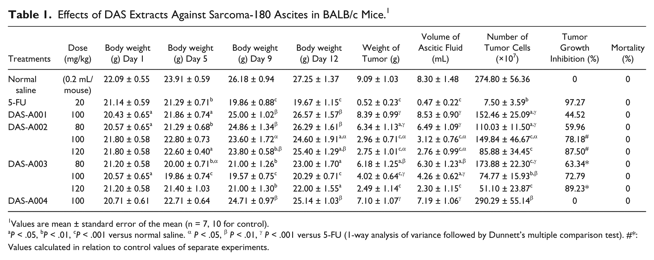
Values are mean ± standard error of the mean (n = 7, 10 for control).
P < .05, bP < .01, cP < .001 versus normal saline. α P < .05, β P < .01, γ P < .001 versus 5-FU (1-way analysis of variance followed by Dunnett’s multiple comparison test). #*: Values calculated in relation to control values of separate experiments.
The effect of graded doses of DAS-A002 and DAS-A003 on Sarcoma-180 ascites is also shown in Table 1. In respect of number of tumor cells, DAS-A002 caused significant (P < .05, .001) and dose-dependent inhibition of tumor growth with values of 59.96%, 78.18%, and 87.50%, respectively, at doses of 80, 100, and 120 mg/kg. No mortality was recorded at these doses as was the case in the control and 5-FU groups. The effects of DAS-A002 at doses of 80 and 100 mg/kg were significantly lower (P < .05, .001) than that elicited by 5-FU (97.27% inhibition). However, the effect of DAS-A002 at the highest dose of 120 mg/kg was comparable and not significantly different than that of 5-FU. At all the doses of DAS-A002 investigated, significant reductions (P < .05-.001) were produced in body weight, and weight and volume of ascitic fluid compared to the control group. DAS-A003 also caused significant (P < .01, .001) and dose-dependent tumor growth inhibition, based on the number of tumor cells, with values of 63.34%, 72.79%, and 89.23%, respectively, at doses of 80, 100, and 120 mg/kg. As was the case in the control and 5-FU groups, no mortality was recorded at all the doses of DAS-A003 investigated. The effects of DAS-A003 at doses of 80 and 100 mg/kg were significantly lower (P < .01, .001) than that elicited by 5-FU (97.27%) but the effect at the highest dose of 120 mg/kg was comparable and not significantly different (P > .05). DAS-A003 also produced significant reductions (P < .05-.001) in body weight, and weight and volume of ascitic fluid relative to the control group.
In Vivo Anticancer Activity of DAS-A002 and DAS-A003 Extracts Against Sarcoma-180 (S-180) Solid Tumor in BALB/c Mice
DAS-A002 (125 mg/kg) and DAS A003 (130 mg/kg) significantly reduced (P < .05, .001) body weight of mice on days 5, 9, and 13, and tumor volume on days 9 and 13 compared with the control group. As regards DAS-A002, tumor growth inhibition reduced from 65% on day 9 to 52.56% on day 13 while in the case of DAS-A003 it reduced from 42.26 to 37.95% on days 9 and 13, respectively, with effects being comparable and not significantly different (P > .05) from those elicited by 5-FU (Table 2).
Effects of DAS-A002 and DAS-A003 Extracts Against Sarcoma-180 Solid Tumor in BALB/c Mice. 1
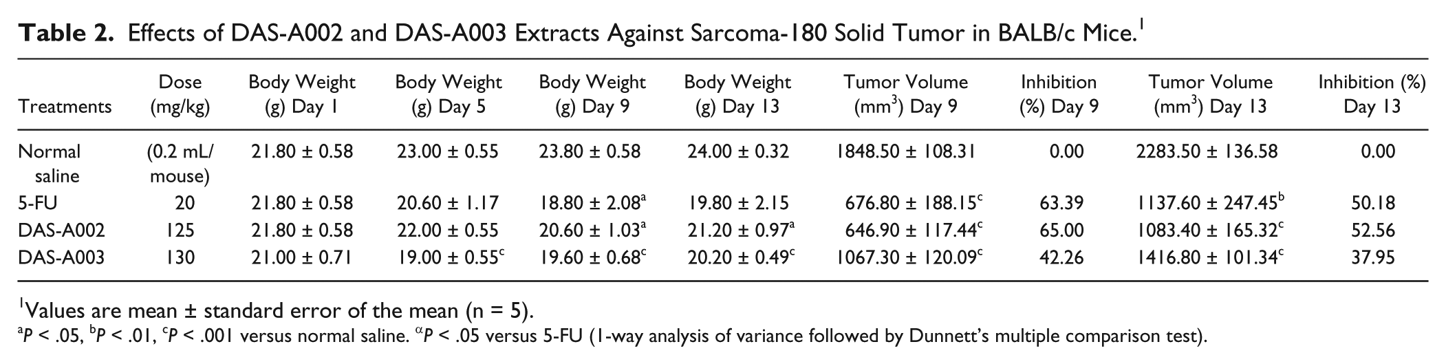
Values are mean ± standard error of the mean (n = 5).
P < .05, bP < .01, cP < .001 versus normal saline. αP < .05 versus 5-FU (1-way analysis of variance followed by Dunnett’s multiple comparison test).
Effect of DAS-A002 Extract Against L1210 Lymphoid Leukemia in CDF1 Mice
The effect of DAS-A002 (100 mg/kg) and DAS-A003 (100 mg/kg) on L1210 lymphoid leukemia model is shown in Table 3. DAS-A002 elicited 11.75 days of MST compared with values of 12 and 19 days for normal saline and 5-FU, respectively. Percentage T/C values were 97.92% and 158.33%, respectively, for DAS-A002 and 5-FU. The MST values for DAS-A003, normal saline, and 5-FU were 16, 9, and 19 days, respectively. Percentage T/C values for DAS-A003 and 5-FU were consequently 177.78% and 211.11%, respectively.
Effect of DAS-A002 and DAS-A003 in L1210 Lymphoid Leukemia in CDF1 Mice. a
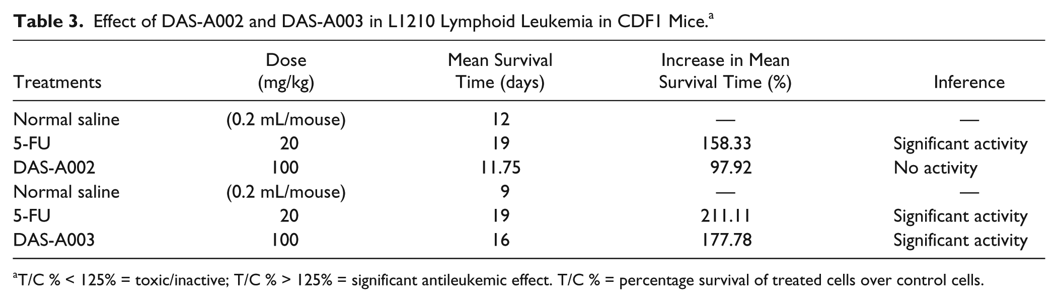
T/C % < 125% = toxic/inactive; T/C % > 125% = significant antileukemic effect. T/C % = percentage survival of treated cells over control cells.
Discussion
The burden of cancer will continue to increase owing to continuing growth and aging of the population of the world. 7 Plant-derived compounds including vinca alkaloids, lignans, terpenoids, quinines, and phytochemicals such as flavones, flavonoids, flavonols have played an important role in the development of several clinically useful anticancer agents. 18 In fact, a good number of the contemporary commercially approved anticancer drugs as well as the natural product–derived compounds in various stages of clinical development as anticancer agents originate from plants. 2 Kosem et al 7 reported that numerous phytochemicals from plants (herbs, fruits, and vegetables) have been reported to interfere with the stages of carcinogenesis by both cell culture and animal studies.
According to the recommendation of the National Cancer Institute, 30 µg/mL is the upper IC50 limit considered promising for purification of a crude extract. 19 In this study, various extracts of the formulation made up of C papaya and M indica plant materials exhibited significant cytotoxic activity against various human cancer cell lines. The ethanol extract (DAS-A001) showed significant activity against HCT-116 and PC3 (IC50 12 and 13 µg/mL, respectively); the hydroethanol extract (DAS-A002) against HCT-116 and PC3 (IC50 <5 and 13 µg/mL, respectively); the aqueous extract (DAS-A003) against THP-1 (IC50 <5 µg/mL); and the DCM:MeOH extract (DAS-A004) against HCT-116 and PC3 (IC50 <5 and 17 µg/mL, respectively).
In the S-180 ascites model, the hydroethanol and aqueous extracts of DAS showed significant anticancer activity at doses of 80, 100, and 120 mg/kg with tumor growth inhibition values of 59.96%, 78.18%, and 87.50%, respectively, for DAS-A002, and 63.34%, 72.79%, and 89.23%, respectively, for DAS-A003 compared with a value of 97.27% for 5-FU (20 mg/kg). Only DAS-A002 showed significant activity at the dose of 125 mg/kg (52.56% tumor growth inhibition) relative to 50.18% for 5-FU (20 mg/kg) in the S-180 solid tumor model. However, this extract was not active in the L1210 lymphoid leukemia model (T/C 97.92% at the dose of 100 mg/kg compared to 158.33% for 5-FU at 20 mg/kg). Although DAS-A003 produced less activity in the solid tumor model (37.95% inhibition) compared with DAS-A002, it was however, significantly active in the L1210 lymphoid leukemia model (T/C 177.78% at the dose of 100 mg/kg compared with 211.11% for 5-FU at 20 mg/kg). This correlates with its significant activity in vitro against THP-1 (human leukemia cell line; IC50 <5 µg/mL).
Otsuki et al 20 investigated the effect of aqueous-extracted C papaya leaf fraction on the growth of various tumor cell lines and on the antitumor effect of human lymphocytes. C papaya extract inhibited the proliferative responses of solid tumor cell lines derived from cervical carcinoma (HeLa), breast adenocarcinoma (MCF-7), hepatocellular carcinoma (HepG2), lung adenocarcinoma (PC14), pancreatic epithelioid carcinoma (Panc-1), and mesothelioma (H2452) in a dose-dependent manner. In addition, C papaya extract inhibited the proliferative responses of hematopoietic cell lines, including T cell lymphoma (Jurkat), plasma cell leukemia (ARH77), Burkitt’s lymphoma (Raji), and anaplastic large cell lymphoma (Karpas-299). It was also found that proliferation of additional cell lines, derived from various malignant tumor cell lines, was similarly inhibited, with no statistically significant differences observed between solid and hematopoietic tumor cell lines. To determine whether the inhibition of proliferative response was associated with decreased cell viability, the authors evaluated potential induction of apoptosis and cell death in the T lymphoma Jurkat cell line. The levels of annexin V- or 7-AAD-positive Jurkat cells were increased in a dose-dependent manner. To confirm the above results, the authors measured the activities of caspase-3 and -7, which are effector caspases in inducing cell apoptosis. Activation of caspase-3/7 on Jurkat cells was induced within 24 hours of C papaya extract treatment. The authors concluded that one of the mechanisms involved in the inhibition of tumor cell proliferation by the aqueous extract of C papaya leaf is associated with the induction of cell death, including apoptosis. Sukardiman 21 evaluated the chloroform fraction from daun papaya (C papaya) in terms of anticancer and induction of apoptosis activity in myeloma cells culture in vitro. Anticancer activity was determined by cell viability method with trypan blue exclusion and apoptosis induction activity was determined by ethidium bromide and acridine orange exclusion and analysis by fluorescent microscope. The result of the research showed that chloroform fraction from C papaya has anticancer activity with LC50 104.4 µg/mL and induction of apoptosis activity in myeloma cells culture in vitro. The authors concluded that the result of the experiment suggested isolation and identification compounds of chloroform fraction (alkaloids) from daun papaya (C papaya), which have anticancer and induction of apoptosis activity. Rahmat et al 22 investigated the antiproliferative activity of pure lycopene compared to both extracted lycopene and juices from watermelon (Citrullus vulgaris) and papaya (C papaya) on human breast and liver cancer cell lines. Papaya juice was found to cause 50% cell death (IC50) of HepG2 cells at 20 mg/mL without significant cytotoxic effect on Chang liver (normal cell line). The juice was found to be more effective than the extracted lycopene sample extracted from C papaya.
Shah et al 23 documented the various reported anticancer activity of M indica in a review. Significant cytotoxic activities have been demonstrated by the stem bark extract of mango against the breast cancer cell lines MCF 7, MDA-MB-435, and MDA-N, as well as against a colon cancer cell line (SW-620) and a renal cancer cell line (786-0). 24 The ethanol/water (1:1) extract of dried aerial parts of mango administered intraperitoneally to mice at a dose of 250.0 mg/kg was inactive on Leuk-P388 model. 25 In vitro, mangiferin dose- and time-dependently inhibited the proliferation of K562 leukemia cells and induced apoptosis in K563 cells line, probably through downregulation of bcr/abl gene expression. 26 Sarkar et al 27 investigated the immunomodulatory activity of alcoholic extract of stem bark of M indica in mice. It was concluded that test extract is a promising drug with immunostimulant properties. Mangiferin mediates the downregulation of NF-κB, suppresses NF-κB activation induced by inflammatory agents, including tumor nuclear factor (TNF), increases the intracellular glutathione (GSH) levels and potentiates chemotherapeutic agent–mediated cell death; this suggests a possible role in combination therapy for cancer. 27 It is likely that these effects are mediated through mangiferin reactive oxygen species quenching and GSH rising; increased intracellular (GSH) levels are indeed known to inhibit the TNF-induced activation of NF-κB. 28 The findings in the Sarcoma-180 ascites and solid tumor models in this study corroborate the previously mentioned reports of anticancer activity for C papaya and M indica as individual phytomedicines.
In conclusion, the results obtained in this study suggest that the phytomedicine DAS-77 possess anticancer activity in vitro and in vivo. The hydroethanolic extract (DAS-A002) was significantly active against HCT-116 (colon) and PC3 (prostate) human cancer cell lines, and Sarcoma-180 ascites and solid tumor models, with no significant activity against L1210 lymphoid leukemia model. The aqueous extract (DAS-A003) was significantly active against THP-1 (leukemia) human cancer cell line, and Sarcoma-180 ascites and L1210 lymphoid leukemia model, with reduced activity in the Sarcoma-180 solid tumor model.
Footnotes
Acknowledgements
The support of Federation of Indian Chambers of Commerce and Industry (FICCI) and Department of Science and Technology (DST), Ministry of Science and Technology, Government of India, in the form of the C.V. Raman Postdoctoral Fellowship award (June 5 to December 1, 2012) given to Dr Abidemi James Akindele is duly acknowledged. The authors also express profound gratitude to Dr Ram A. Vishwakarma, Director of the Indian Institute of Integrative Medicine (IIIM), Council of Scientific & Industrial Research (CSIR), Jammu-Tawi, Jammu & Kashmir, India, for his support in accepting to host the first author for the postdoctoral research. We also express our gratitude to Mr D.O.A. Adesanya for providing DAS-77 for this study.
Declaration of Conflicting Interests
The author(s) declared no potential conflicts of interest with respect to the research, authorship, and/or publication of this article.
Funding
The author(s) disclosed receipt of the following financial support for the research, authorship, and/or publication of this article: AJA was supported by the C.V. Raman Postdoctoral Fellowship award (June 5 to December 1, 2012) from the Federation of Indian Chambers of Commerce and Industry (FICCI) and Department of Science and Technology (DST), Ministry of Science and Technology, Government of India.