
Abstract
Background
SERPINE1 has attracted considerable attention in tumor biology, but its clinical importance in head and neck squamous cell carcinoma (HNSCC) is not yet clear. We therefore examined whether SERPINE1 expression is related to survival in patients with HNSCC.
Methods
We searched three major databases (PubMed, EMBASE, and the Cochrane Library) and identified observational studies reporting survival outcomes in relation to SERPINE1 expression through November 11, 2024. From eligible reports we extracted data on progression-free survival (PFS), overall survival (OS), disease-specific survival (DSS) and disease-free survival (DFS), and calculated pooled hazard ratios (HRs) using random-effects models.
Results
Eleven studies including 733 individuals with HNSCC met the inclusion criteria. Across these cohorts, higher SERPINE1 expression was consistently linked with shorter OS (HR 2.81, P = 0.003) and shorter DFS (HR 1.57, P = 0.004). In contrast, no clear associations were observed for PFS or DSS (P ≥ 0.05).
Conclusion
Current evidence suggests that increased SERPINE1 expression is associated with an unfavorable prognosis in HNSCC, particularly for OS and DFS. Larger prospective studies are needed to confirm these findings and to determine how SERPINE1 assessment might be incorporated into risk stratification and treatment planning for patients with HNSCC.
Introduction
Head and neck squamous cell carcinoma (HNSCC) represents a major global cancer burden and currently ranks seventh among all malignant tumors in terms of incidence. In 2020, it was estimated that about 890 000 individuals developed HNSCC and roughly 450 000 died from the disease worldwide. 1 Its incidence continues to rise and is projected to increase by about 30% by 2030, imposing a substantial global health and societal burden. 1 Major etiologic factors include tobacco and alcohol consumption, betel quid chewing, and infection with oncogenic viruses such as human papillomavirus and Epstein–Barr virus. 2 Consequently, current prevention efforts focus largely on reducing these exposures and promoting healthier lifestyle choices in populations at risk.
The marked biological and molecular heterogeneity of HNSCC underscores the need for robust biomarkers that can improve prognostic assessment and patient risk stratification, yet such markers are still lacking in routine clinical practice. Emerging evidence suggests that SERPINE1 is a promising candidate in this context. SERPINE1, a key regulator of the fibrinolytic system, promotes tumor invasion, angiogenesis, and chemoresistance in part by modulating matrix metalloproteinase (MMP) activity. 3 It also influences immune cell infiltration across multiple cancer types.4,5 Elevated SERPINE1 expression is closely associated with aggressive tumor behavior and unfavorable clinical outcomes. 6 Across several malignancies, including HNSCC, gastric cancer, and non–small cell lung cancer, SERPINE1 contributes to disease progression and metastatic dissemination.7–9 Our previous work has shown that upregulated SERPINE1 expression is a reproducible adverse prognostic factor across neoplasms, including a significant association with poorer overall survival (OS) and disease-specific survival (DSS) in HNSCC cohorts.10,11 Other quantitative syntheses further support the clinical relevance of SERPINE1. In gastroesophageal carcinoma, increased SERPINE1 levels were associated with a 96% higher risk of recurrence (HR = 1.96, P = 0.03) and an 84% higher risk of death (HR = 1.84, P < 0.0001). 12 Another meta-analysis indicated that SERPINE1 promoter variants are linked to breast cancer susceptibility and nodal metastatic progression. 13 However, these analyses do not focus on HNSCC, and meta-analytic evidence specifically addressing SERPINE1 as a prognostic marker in HNSCC is still lacking.
Although several individual studies have reported associations between SERPINE1 expression and survival outcomes in HNSCC, 14 the overall magnitude and consistency of these effects have not been formally quantified. We therefore conducted a PRISMA-compliant systematic review and meta-analysis to evaluate the prognostic value of SERPINE1 for key survival endpoints in patients with HNSCC.
Materials and Methods
This systematic review and meta-analysis was conducted in accordance with the Preferred Reporting Items for Systematic Reviews and Meta-Analyses (PRISMA) 2020 guidelines for reporting systematic reviews and meta-analyses. 15 The protocol was registered with the International Prospective Register of Systematic Reviews (PROSPERO) (CRD420250652840), the international systematic review registry. This systematic review and meta-analysis used only aggregate data extracted from published articles and did not involve any new studies with human participants or animals. Therefore, ethics committee/Institutional Review Board (IRB) approval and informed consent were not required.
Search Strategy
Comprehensive electronic searches were executed in PubMed, EMBASE, and Cochrane Library databases until November 11, 2024. To enhance search sensitivity, Medical Subject Headings (MeSH) and associated entry terms were systematically combined in the search strategy: “Plasminogen Activator Inhibitor 1 OR SERPINE1 protein” with neoplasm terms including “Squamous Cell Carcinoma of Head and Neck”, “Mouth Neoplasms”, “Laryngeal Neoplasms”, “Hypopharyngeal Neoplasms”, and “Oropharyngeal Neoplasms”.
Study Selection
Inclusion criteria required: (a) observational designs (cohort or cross-sectional studies); (b) histologically verified HNSCC involving oropharyngeal, oral cavity, hypopharyngeal, or laryngeal subsites; (c) quantifiable survival metrics including DSS, OS, disease-free survival (DFS), and progression-free survival (PFS). Exclusion criteria encompassed: (a) non-original research (conference abstracts, commentaries, reviews, case reports), preclinical studies, or mechanistic investigations; (b) carcinomas originating from paranasal sinuses, nasal cavity, nasopharynx, or cutaneous sites; (c) non-English language publications; (d) unrecoverable outcome data (directly or indirectly).
Data Extraction and Quality Assessment
Two investigators independently screened titles, abstracts, and full texts, and extracted data from all eligible studies using a standardized form. Disagreements were resolved by discussion with a third reviewer. Extracted variables included study design, study type, sample size, country, tumor subsite, follow-up duration, SERPINE1 assessment method, and survival outcomes.
Hazard ratios (HRs) with 95% confidence intervals (CIs) were preferentially obtained from multivariable models reported in the original articles. When HRs were not explicitly reported but Kaplan–Meier survival curves were available, HRs and corresponding CIs were estimated using the method described by Tierney et al (2007). 16 Because such reconstructed HRs may introduce additional imprecision, sensitivity analyses were performed in which these studies were excluded and the pooled estimates were recalculated.
Methodological quality was assessed with the Newcastle–Ottawa Scale (NOS).
Statistical Methods
All statistical analyses were performed using Review Manager (RevMan) version 5.4. HRs and 95% CIs were pooled for each outcome. Between-study heterogeneity was quantified using Cochran's Q test and the I2 statistic, with an I2 value >50% indicating substantial heterogeneity. Given the clinical and methodological diversity across studies, random-effects models were applied a priori to obtain pooled effect estimates.
Prespecified subgroup analyses according to tumor subsite, study design, or SERPINE1 detection method were not feasible because only two to four studies contributed data for each survival endpoint. To evaluate the robustness of the findings, sensitivity analyses were conducted by excluding studies in which HRs were reconstructed from Kaplan–Meier curves and re-running the meta-analyses. Forest plots were generated for all primary and sensitivity analyses.
Results
Study Inclusion and Data Extraction
The database search initially yielded 151 records. After removing duplicates and screening titles, abstracts and full texts against the predefined eligibility criteria, 11 studies were finally retained for quantitative synthesis.17–27 Figure 1 documents the systematic literature filtration process, demonstrating strict adherence to PRISMA statement standards.

Flow diagram of the screening and selection process.
Study Characteristics and Quality Assessment
The main clinical and methodological features of the included studies are summarized in Table 1. In brief, nine investigations used a retrospective cohort design, one was a prospective cohort, and one was cross-sectional. The studies were conducted in Brazil, Japan, Norway, Australia, China, the Netherlands, Germany and Spain, indicating a broad geographical spread. The sample sizes ranged from 30 to 190 participants, with a median of 52. The carcinoma types studied included oral tongue squamous cell carcinoma (n = 3), oral squamous cell carcinoma (OSCC) (n = 3), head and neck squamous cell carcinoma (n = 4), oral cavity cancer (n = 1), and oropharyngeal cancer (n = 1). SERPINE1 expression was evaluated in different cellular compartments, such as cytoplasm and cell membrane, and quality was assessed using the NOS (Table S1).
Characteristics of Eligible Studies in the Meta-Analysis.

SERPINE1 Expression and Clinical Outcomes
Association of SERPINE1 Expression with PFS
Among the 11 studies analyzed, two studies directly examined the link between SERPINE1 expression and PFS (Figure 2). Random-effects meta-analysis yielded a pooled HR of 1.33 (95% CI: 0.98-1.80; P = 0.07). The confidence interval spanning the neutral threshold (HR = 1.0) indicates an absence of statistical significance. Heterogeneity across studies was assessed with an I2 value of 36% and Cochran's Q statistic (P = 0.21), suggesting moderate heterogeneity. This moderate heterogeneity could be attributed to variations in patient characteristics and study designs, including differences in treatment regimens and study populations. Due to this, a random-effects model was applied to account for study-level variability. Given the small number of studies and the confidence interval crossing unity, the available evidence is insufficient to support a clear association between SERPINE1 expression and PFS.

Forest plots depicting associations between SERPINE1 expression levels and PFS.
Association of SERPINE1 Expression with OS
Three studies explicitly examined the link between SERPINE1 expression and OS (Figure 3). Meta-analysis revealed a combined HR of 2.81 (95% CI: 1.41-5.60; P = 0.003), suggesting that elevated SERPINE1 expression is significantly associated with poorer OS in HNSCC patients. The I2 value for OS was 0%, indicating no heterogeneity across the studies, but a random-effects model was still applied to account for any potential unmeasured variability.
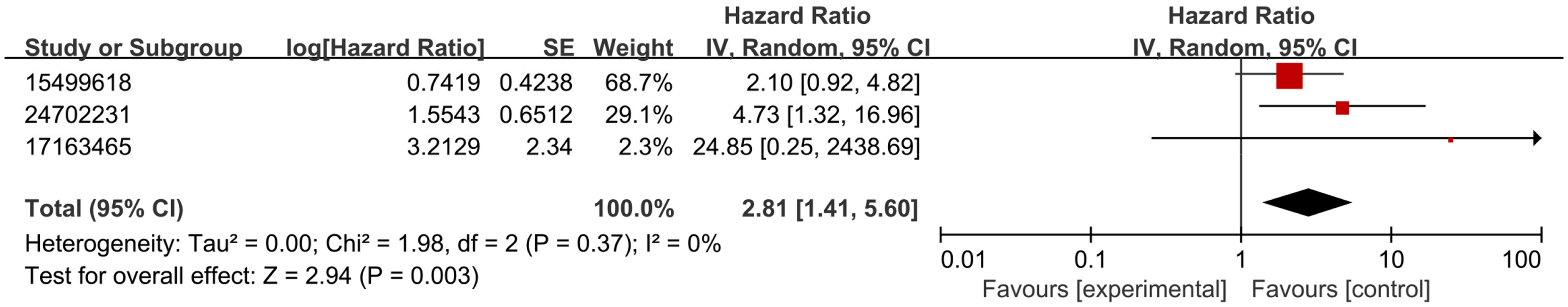
Forest plots depicting associations between SERPINE1 expression levels and OS.
Association of SERPINE1 Expression with DSS
The relationship between SERPINE1 expression and DSS in HNSCC was evaluated across three studies (Figure 4). The pooled HR of 0.90 (95% CI: 0.32-2.54; P = 0.85) failed to demonstrate a significant association between SERPINE1 expression and DSS. The I2 value for DSS was 54%, with a Cochran's Q statistic of P = 0.09, indicating substantial heterogeneity across the studies. This variability may be due to differences in sample sizes, detection methods, or clinical characteristics across studies. A random-effects model was applied to account for this heterogeneity and ensure robust estimates.

Forest plots depicting associations between SERPINE1 expression levels and DSS.
Association of SERPINE1 Expression with DFS
Two studies examined the link between SERPINE1 expression and DFS (Figure 5). The random-effects meta-analysis revealed that elevated SERPINE1 expression correlated with reduced DFS (pooled HR 1.57, 95% CI: 1.16-2.12, P = 0.004). The I2 value for DFS was 0%, with a Cochran's Q statistic of P = 0.44, indicating no significant heterogeneity. But there are potential sources of heterogeneity, such as differences in patient populations, methodological approaches, and sample sizes across the included studies. A random-effects model was applied to account for this variability and to reflect real-world differences in study design and patient characteristics.

Forest plots depicting associations between SERPINE1 expression levels and DFS.
Sensitivity Analysis and Reporting Bias
We performed several sensitivity analyses to examine the stability of the results. First, studies in which hazard ratios had been reconstructed from Kaplan–Meier curves rather than directly extracted from multivariable models were removed. We then recalculated pooled HRs for each endpoint and compared the new forest plots with the primary analyses. Overall, the changes in effect size were small, suggesting that the main conclusions were not driven by these reconstructed estimates.
For PFS, exclusion of the reconstructed study left one cohort, yielding an HR of 1.73 (95% CI 1.02-2.92; P = 0.04). For OS, removing one reconstructed study resulted in a pooled HR of 2.71 (95% CI 1.29-5.68; P = 0.008) with low heterogeneity (I2 = 9%, Cochran's Q P = 0.30). For DSS, exclusion of the reconstructed dataset produced a pooled HR of 1.35 (95% CI 0.77-2.38; P = 0.30) with I2 = 0% (Cochran's Q P = 0.82). DFS estimates were unchanged because none of the contributing studies required HR reconstruction. Detailed forest plots for these analyses are provided in the Supplemental Material (Figures S1–S3).
Due to the limited sample sizes across the subgroup comparisons and the potential influence of small study effects, formal publication bias evaluation was not feasible. However, the results remained stable and supported the original conclusions, indicating a reliable and consistent relationship between SERPINE1 expression and clinical outcomes in HNSCC.
Discussion
In this meta-analysis we brought together data from 733 patients with HNSCC reported in 11 studies to explore whether SERPINE1 expression carries prognostic information. Overall, patients with higher SERPINE1 expression had clearly worse OS and DFS, whereas we did not observe convincing associations with PFS or DSS. These patterns suggest that SERPINE1 may be more closely linked to long-term risks of recurrence and death than to early progression or cause-specific mortality, although the limited number of studies for each endpoint means that these distinctions should be interpreted cautiously. A sizeable proportion of the included cohorts involved oral cancer, and in these series SERPINE1 overexpression was particularly strongly related to poor outcome. This raises the possibility that oral cancer data exerts an important influence on the pooled estimates. At the same time, the biological overlap between oral cancer and other HNSCC subsites implies that the signal is still relevant to HNSCC as a whole.
SERPINE1, located on chromosome 7q22, encodes plasminogen activator inhibitor-1 (PAI-1), a 45-kDa secreted serine protease inhibitor of the serpin superfamily. 28 PAI-1 regulates the urokinase-type plasminogen activator system and fibrinolysis, and is produced by several cell types including endothelial cells, stromal fibroblasts, adipocytes, platelets and tumor cells. 29 Within the peritumoral extracellular matrix it binds to vitronectin and modulates cell adhesion, migration and survival. 30 Through these actions, SERPINE1/PAI-1 integrates extracellular-matrix remodeling, angiogenesis and tumor–stroma interactions. Experimental work in different tumor models indicates that increased PAI-1 promotes tumor growth and neovascularization, whereas genetic or pharmacological inhibition of PAI-1 limits invasion and angiogenesis.31,32 In HNSCC specifically, SERPINE1 overexpression enhances cell migration, supports resistance to apoptosis and is associated with lymph-node and distant metastasis as well as perineural and vascular or lymphatic invasion.21,33,34 Similar findings in gastric and non-small cell lung cancer, where SERPINE1 forms part of hypoxia and epithelial–mesenchymal-transition–related gene signatures,35–38 support a mechanistic explanation for the adverse prognostic effect observed in our pooled analysis. At the regulatory level, SERPINE1 can be induced by hypoxia, TGF-β and inflammatory cytokines, 39 and is a downstream target of tumor-suppressive microRNAs such as miR-30e-3p 7,40; loss of these microRNAs in HNSCC leads to SERPINE1 upregulation and increased invasiveness. Collectively, these data provide a mechanistic basis for the adverse prognostic effect observed in our meta-analysis.
Beyond its role in tumor cell biology, SERPINE1 also influences the immune microenvironment. PAI-1 modulates the differentiation and polarization of monocyte-derived macrophages within tumors.41,42 In OSCC, tumor-derived PAI-1 together with IL-8 drives monocytes toward CD206+ tumor-associated macrophages with an M2-like, pro-tumor phenotype. 18 In HNSCC models, regorafenib-mediated suppression of PAI-1 secretion shifts macrophages toward a more antitumor (M1-like) profile, supporting a causal role of PAI-1 in macrophage plasticity. 43 Concordant findings in gastric cancer, where SERPINE1-dependent exosomal signaling promotes M2 polarization and tumor progression, further reinforce this concept. 8 These observations are consistent with our previous pan-cancer and HNSCC analyses identifying SERPINE1 as an immune-related gene associated with an immunosuppressive microenvironment, poor prognosis and potentially reduced benefit from systemic therapies. Taken together, SERPINE1/PAI-1 emerges as a central regulator of ECM remodeling, angiogenesis and immune escape with consequences for clinical outcome.
Within the broader biomarker landscape of HNSCC, SERPINE1/PAI-1 therefore has both prognostic and potentially predictive value. Other components of the plasminogen activation system and hypoxia-related markers such as carbonic anhydrase IX (CAIX) and vascular endothelial growth factor A (VEGFA) have also been linked to survival in OSCC and early-stage OSCC.44–46 In addition, emerging biomarkers include microRNA signatures and hormone-related pathways: in laryngeal squamous cell carcinoma, specific miRNA expression profiles correlate with tumor progression, 47 and estrogen receptor β2 expression is associated with nodal metastasis and poorer outcomes. 48 Combining SERPINE1/PAI-1 with these markers may improve risk stratification and more precisely capture the biological heterogeneity of HNSCC.
To complement our meta-analysis, we performed an exploratory external validation in the Cancer Genome Atlas–Head and Neck Squamous Cell Carcinoma (TCGA-HNSC) cohort (results provided in the Supplemental material (Figure S4–S7)). In line with our primary findings, high SERPINE1 mRNA expression was associated with worse OS and DSS and with a modestly increased risk of PFS, whereas no clear association was observed for DFS. These post-hoc, transcriptome-based analyses should be regarded as hypothesis-generating rather than definitive, but they provide independent support from a large, well-annotated dataset that SERPINE1 overexpression is consistently linked to adverse long-term outcome in HNSCC. Given that robust, clinically implemented biomarkers in HNSCC remain scarce despite their growing importance for treatment decision-making, SERPINE1 appears to be a credible candidate for further development.
HNSCC constitutes an area that requires in-depth investigation, as there are still no well-established biomarkers. Their importance in guiding treatment decisions is becoming increasingly evident. In this context, the SERPINE1 gene may be a promising candidate. Several limitations of this study should be acknowledged. First, only 11 studies met the inclusion criteria, and some survival endpoints relied on as few as two or three cohorts, limiting statistical power and the generalizability of those estimates. Despite the use of random-effects models and sensitivity analyses, the wide CIs for certain outcomes indicate a non-trivial risk of type II error. Second, the included studies differed in design, detection methods, tumor subsite distribution, treatment protocols and follow-up duration. Although we evaluated heterogeneity using I2 and Cochran's Q and applied random-effects models accordingly, the small number of studies per endpoint precluded robust subgroup analyses and reduces confidence in any subsite- or method-specific conclusions. Third, in three of the 11 studies, HRs were reconstructed from Kaplan–Meier curves rather than directly reported. Although we used standard methods and subjected these estimates to sensitivity analyses, graphical reconstruction inevitably introduces uncertainty and may contribute to wider confidence intervals.
In summary, this work provides the first meta-analysis specifically examining SERPINE1 as a prognostic biomarker in HNSCC and supports its potential clinical relevance. Our findings suggest that SERPINE1 expression may help refine risk stratification and inform prognostic assessment in patients with HNSCC. Prospective, well-designed studies are now needed to validate these associations and to define how SERPINE1 testing could be incorporated into routine clinical practice.
Conclusions
This meta-analysis shows that elevated SERPINE1 expression is consistently associated with poorer OS and DFS in patients with HNSCC, whereas the evidence for PFS and DSS remains limited and inconclusive. These findings support SERPINE1 as a promising prognostic biomarker that may complement existing clinicopathologic factors in risk stratification. If validated in adequately powered, prospective multicenter cohorts, assessment of SERPINE1 expression could help identify patients at increased risk of recurrence and death and may inform the intensity and selection of systemic therapies, including emerging immunotherapeutic approaches.
Supplemental Material
sj-docx-1-tct-10.1177_15330338261435460 - Supplemental material for The Prognostic Value of SERPINE1 in Clinical Outcomes in Head and Neck Squamous Cell Carcinoma: A Systematic Review and Meta-Analysis
Supplemental material, sj-docx-1-tct-10.1177_15330338261435460 for The Prognostic Value of SERPINE1 in Clinical Outcomes in Head and Neck Squamous Cell Carcinoma: A Systematic Review and Meta-Analysis by Shifeng Yan, Xinyu Li, Changyu Zhu and Wei Li in Technology in Cancer Research & Treatment
Footnotes
Abbreviations
Acknowledgments
We would like to express our gratitude to all the researchers whose work contributed to this meta-analysis.
Ethics Statement
The authors have nothing to report.
Author Contributions
Shifeng Yan: Conceptualization, data curation, formal analysis, investigation, methodology, software, writing – original draft, writing – review and editing. Xinyu Li: Conceptualization, investigation, methodology, supervision. Changyu Zhu: Conceptualization, investigation, methodology. Wei Li: Conceptualization, data curation, formal analysis, investigation, methodology, software, supervision, writing-original draft, writing-review and editing.
Funding
The authors disclosed receipt of the following financial support for the research, authorship, and/or publication of this article: This work was supported by the Beijing Science and Technology Planning Project, (grant number Z221100007422026).
Declaration of Conflicting Interests
The authors declared no potential conflicts of interest with respect to the research, authorship, and/or publication of this article.
Data Availability Statement
All data needed to support the conclusions are presented in this paper. Additional data related to this study were obtained from the authors.
Supplemental Material
Supplemental material for this article is available online.