
Abstract
Objective:
To explore the inhibitory and synergistic effects of 5-fluorouracil and curcumin on Hep-2 laryngeal cancer cells and clarify the effect of mesoporous silica nanoparticles as drug carriers.
Methods:
The inhibitory effects of 5-fluorouracil and curcumin on Hep-2 cells were detected using the CCK-8 assay. CompuSyn was used to calculate the synergistic effect of the 2 drugs. Flow cytometry was used to detect apoptosis and cell cycle arrest induced by 5-fluorouracil and curcumin. The drugs were loaded into mesoporous nanoparticles. Western blotting was used to detect the expression of related proteins after treatment. The growth of subcutaneous tumors in BALB/c nude after the intraperitoneal injection with drug-loaded mesoporous silica nanoparticles was recorded.
Results:
5-Fluorouracil and curcumin synergistically induced apoptosis and cell cycle arrest in Hep-2 cells. Mesoporous silica nanoparticles as drug carriers enhanced the therapeutic effects of 5-fluorouracil and curcumin.
Conclusions:
Mesoporous silica nanoparticles are expected to be effective drug carriers that enhance the synergistic effects of 5-fluorouracil and curcumin on laryngeal cancer.
Keywords
Introduction
Laryngeal cancer is a common head and neck cancer malignant neoplasm with a high incidence and mortality rate. An estimated 177,000 new cases of laryngeal cancer and 94,000 deaths worldwide associated with the disease occurred in 2018. 1 Laryngeal cancer accounts for a quarter of all malignant head and neck tumors and approximately 1% of all new cancer globally. Male sex and older age are associated with greater susceptibility to this malignancy. 1,2 In the clinic, the standard treatment for laryngeal cancer remains chemotherapy (alone or combined with radiotherapy) after surgical resection. 3 Nonetheless, given the lack of specificity of traditional chemotherapy, patients have a poor prognosis because of local recurrence and metastasis. 4,5 Thus, improving the chemotherapeutic effect of drugs by endowing them with targeting activity could reduce undesired adverse effects on normal tissues and insufficient dosages in diseased regions. Compared with monotherapy, combination treatments have exhibited promising efficacy in cancer therapy. 6
5-Fluorouracil (5-FU) is a common clinical chemotherapeutic drug for the treatment of laryngeal cancer, and it has been widely used alone or in combination with other anti-cancer drugs to treat many cancers since 1957. 7 The main mechanisms of action of 5-FU include the inhibition of thymidylate synthase activity, DNA synthesis, and DNA repair via the incorporation of its metabolites into the DNA/RNA of cancer cells. 8 All of these activities could ultimately lead to apoptosis and G1 arrest. 9,10 However, the efficacy of 5-FU in the clinic is limited by dose-limiting toxicities and chemoresistance. 11 To overcome these problems, improving the utilization ratio of 5-FU and combining it with other anti-cancer drugs (such as paclitaxel, cisplatin, and curcumin [CRC] 12 -14 ) with different mechanisms of action could be promising strategies to enhance its anti-cancer efficiency.
CRC is a natural extract from the rhizome of Zingiberaceae plants 15,16 that selectively induces apoptosis in cancer cells at the G2 phase via p53 upregulation and mitochondrial apoptosis. 17,18 The compound can also stimulate other extrinsic apoptotic pathways such as TNF-α and Fas signaling. 19,20 Numerous studies have demonstrated the ability of CRC to prevent and treat cancers without cytotoxicity on normal cells. 20,21 Additionally, CRC has been confirmed to have synergistic effects with many anti-cancer drugs, leading to reduced cytotoxicity and increased anti-cancer efficacy against several cancer types. 22 -24 Research on CRC in laryngeal cancer demonstrated that the compound could inhibit cell proliferation and promote apoptosis through the PI3K/Akt and JAK/STAT pathways. 25,26 In addition, CRC enhances the therapeutic effects of other drugs (such as cisplatin and anthocyanin 27,28 ). The combination of 5-FU and CRC has been studied for the treatment of colorectal, breast, liver, and gastric cancers, in which CRC enhanced the therapeutic efficacy and reduced the toxicity of 5-FU. 22 Thus, these two drugs in combination might also have promising effects against laryngeal cancer.
Nanotechnology has been used to develop tumor-targeting drug delivery systems 29 that can reduce the dose and interval of treatment. 30 The functional nanomaterials that have been primarily used as anti-cancer drug carriers include liposomes, polymer nanoparticles, dendritic polymers, and nanomicelles. 29 Mesoporous silica nanoparticles (MSNs) have been proposed as excellent drug delivery systems 31 because they have a relatively large specific surface area and pore volume, an adjustable pore diameter, and easily modified internal and external surfaces. 32 In addition, MSNs has been described as dual-drug carriers for cancer therapy. 33 Compared with normal tissues and the bloodstream (pH 7.2 to 7.4), solid tumors usually have a weakly acidic (pH = 6.0 to 7.0) and hyperthermic environment. Further, cancer cells have a lower pH than the tumor microenvironment. 34 -36 We previously reported dual-responsive MSN-conjugated polymers with thermosensitivity and pH-coupling sensitivity and proved that the drug-loaded polymers had greater anti-cancer efficiency. 32,37 Utilizing these reported MSNs, we examined whether they could be used to carry both 5-FU and CRC as a treatment for human laryngeal cancer. We hope this strategy could maximize both the efficacy and tumor-targeting activity of the combination regimen.
Using Hep-2 human laryngeal squamous cancer cells, we investigated the anti-cancer effects of the combination of 5-FU and CRC and their mechanism of action when loaded into MSNs. Co-delivery of 5-FU and CRC using MSNs resulted in a stronger anti-tumor effect, highlighting the potential of this strategy for treating laryngeal cancer.
Materials and Methods
Cell Culture and Reagents
The human laryngeal squamous cancer cell line Hep-2 was obtained from the Shanghai Institute of Cell Biology, Chinese Academy of Sciences (Shanghai, China). Cells were cultured in RPMI-1640 (Gibco, Thermo Fisher Scientific, Waltham, MA, USA) containing 10% fetal bovine serum (Biological Industries, Beit Haemek, Israel) and maintained at 37°C in a 5% CO2 atmosphere. 5-FU and CRC were purchased from MedChemExpress (Shanghai, China). MSNs conjugated with folic acid and MSNs loaded with 5-FU and/or CRC were provided from Prof. Xiaowei Song of State Key Laboratory of Inorganic Synthesis and Preparative Chemistry, College of Chemistry, Jilin University (Changchun, Jilin, China). 32,37
CCK-8 Assay
Hep-2 cells (1 × 10⁴) were seeded into each well in 96-well culture plates and grown overnight as monolayers. Cells were incubated with different concentrations of 5-FU and CRC for different times. Cell growth and viability were assessed using the CCK-8 assay (MedChemExpress). According to the manufacturer’s instructions, the cell growth rate was calculated as follows: cell viability (%) = (absorbance of the experimental group/absorbance of the control group) × 100. The absorbance was measured using an iMark™ Microplate Reader (BIO-RAD, Kyoto, Japan).
Drug-loaded MSNs
5-FU and CRC were dissolved in distilled water and 80% ethanol, respectively. A UV/Vis spectrophotometer was used to measure the concentrations of 5-FU and CRC at 265 and 425 nm. The standard curves for 5-FU and CRC solutions were established using concentration gradients. MSNs (100 mg) were dispersed in 20-mL solutions of 5-FU (3 mg/mL) and CRC (1 mg/mL). After being shaken for 24 hours at 37°C to reach equilibrium, the drug-loaded MSNs were collected via centrifugation. The encapsulation efficiency was calculated as follows:

where We is the weight of the drug loaded into MSNs and W0 is the initial weight of the added drug.
The loading efficiency was calculated as follows:

where W is the weight of MSNs added.
Drugs Release Experiment
In total, MSNs loaded with 20 mg of 5-FU or CRC were dispersed in 5 mL of PBS at pH 5.0 and 7.2, respectively. The solutions were shaken at 37°C. The 5-FU or CRC content of the solutions was determined using a UV/Vis spectrophotometer at each releasing time point.
Cellular Uptake Capacity of MSNs
For FITC loading, MSNs were mixed with 2 mL of FITC aqueous solution (1 mg/mL). After being shaken for 24 hours at 37°C to reach equilibrium, the FITC-loaded MSNs were collected via centrifugation. Hep-2 cells (2× 10⁵) were seeded onto coverslips in 12-well culture plates and grown overnight as monolayers. The cells were incubated with FITC-loaded MSNs at 37°C for 4 hours. Cells were then washed with PBS three times and fixed with methyl alcohol for 20 minutes. After three additional washes with PBS, cells were stained with Hoechst 33258 for 5 minutes and then washed cells three times with PBS. The coverslips were placed on microscope slides, and the samples were visualized using an FV1000 confocal laser-scanning microscope (Olympus, Tokyo, Japan).
Calculation of the Combination Index (CI)
The synergistic effects of 5-FU and CRC were calculated using the CI and analyzed using CompuSyn software (ComboSyn, Inc., Paramus, NJ, USA). Synergism was indicated by CI < 1.0, additivity was indicated by CI = 1.0, and antagonism was indicated by CI > 1.0. 38
Flow Cytometry
Hep-2 cells (3 × 10⁵) were seeded into each well of six-well culture plates and grown overnight as monolayers. Cells were incubated with 5-FU (20 μg/mL) and/or CRC (20 μg/mL) for 24 hours. Apoptosis was assessed using an Annexin V-FITC/PI apoptosis detection Kit (Wanleibio, Shenyang, China) according to the manufacturer’s instructions. Cell cycle analysis was conducted using PI/RNase Staining Solution (Sungene Biotech, Tianjin, China) according to the manufacturer’s instructions. Both apoptosis and cell cycle analysis were performed using a flow cytometer (guava easyCyte, Merck Millipore, Burlington, MA, USA).
Western Blot Analysis
Western blot analysis was performed as previously described. 39 Cells were harvested and lysed using RIPA buffer after treatment with different drugs. For western blot analyses, 30 mg of total protein from each sample were separated on a 10% SDS–polyacrylamide gel, transferred onto a PVDF membrane (Merck Millipore), and incubated with the indicated antibodies. Protein bands were visualized using a SuperSignal West Pico Chemiluminescent Substrate kit (Pierce Biotechnology, Rockford, IL, USA). Bands corresponding to the proteins of interest were quantified by volume densitometry using ImageQuant software (Molecular Dynamics, Sunnyvale, CA, USA). All experiments were conducted on three separate occasions in duplicate, and the results are presented as the mean ± SD.
In Vivo Animal Studies
BALB/c-nu/nu female mice were purchased from Charles River Laboratories (Beijing, China). All animal care procedures complied with institutional guidelines, and ethical approval was received from the Animal Experimental Ethics Committee of Jilin University (approval number: 2019(53)). Five groups of mice (n = 6 each) were injected subcutaneously in the right flank with 5× 10⁶ Hep-2 cells. Then, the five groups of mice were randomly assigned to receive PBS, unloaded MSNs, FU-loaded MSNs (20 mg/kg), CRC-loaded MSNs (20 mg/kg), and 5-FU- and CRC-loaded MSNs once a day for 12 days. Tumor volume was measured every 2 days according to the following equation: tumor volume = (tumor length) × (tumor width) 2 /2.
Statistical Analysis
The data are expressed as the mean ± SD. For both in vitro and in vivo experiments, an unpaired t-test was used for single comparisons between two groups. All experiments were repeated at least three times. Statistical calculations were performed using SigmaStat software (SPSS v20; IBM, Armonk, NY, USA). P < 0.05 denoted statistical significance.
Results
Synergistic Effects of 5-FU and CRC against Hep-2 Cells
5-FU and CRC inhibit Hep-2 cell growth in vitro
To detect the effects of 5-FU and CRC on cell growth, Hep-2 cells treated with escalating concentrations of the drugs, and cell growth was examined using the CCK-8 assay after 24, 48, and 72 hours. The results revealed a concentration-dependent decline in cell viability (Figure 1A, B, and C). 5-FU had a greater effect on cell viability at lower concentrations (<30 μg/mL), whereas CRC had stronger effects at higher concentrations (>30 μg/mL).
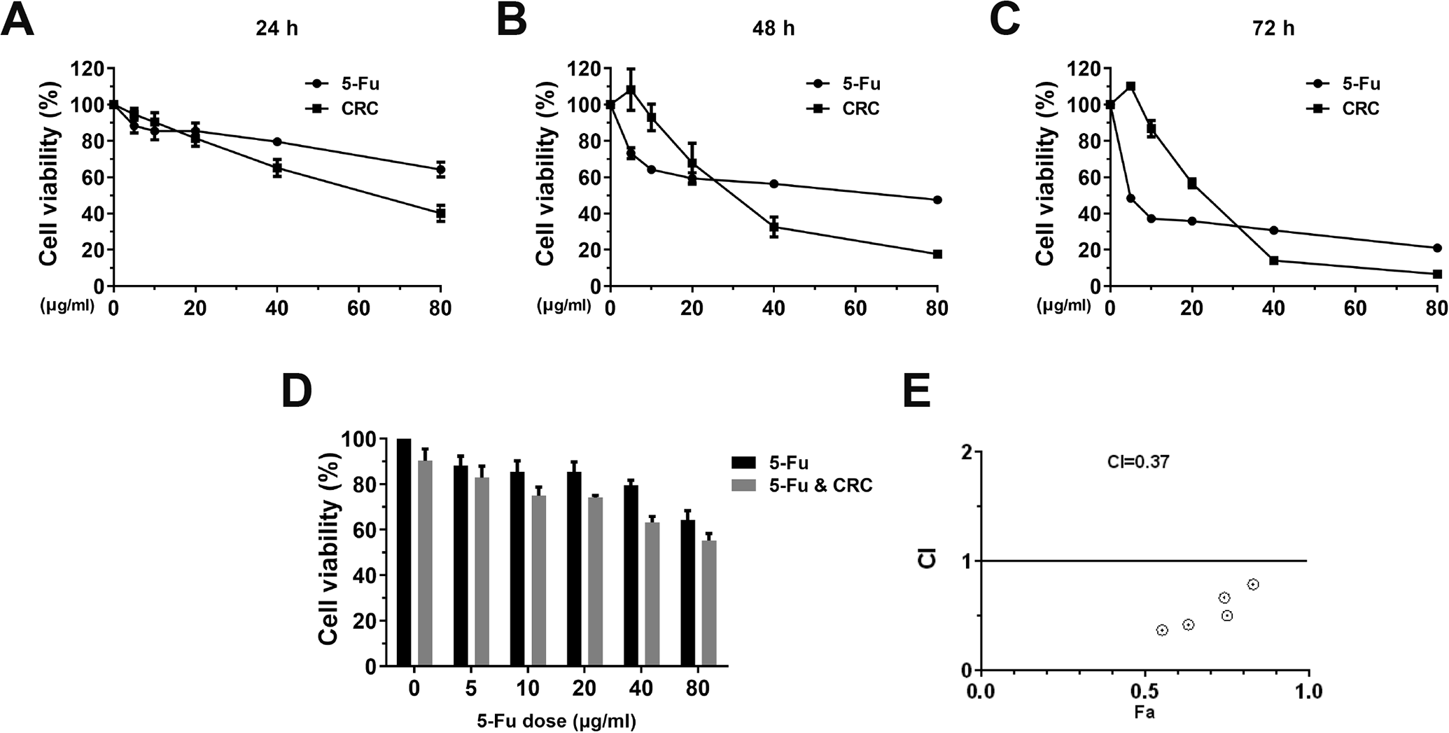
Effects of 5-FU and CRC on Hep-2 cell viability alone and in combination. (A–C) The CCK-8 assay was used to analyze the effects of 5-FU and CRC on cell viability after 24, 48, and 72 hours. (D) Effects of 5-FU (gradient) and CRC (10 μg/mL) on cell viability. (E) The synergistic effects were calculated using CompuSyn. 5-FU, 5-fluorouracil; CRC, curcumin.
Synergistic effects of 5-FU and CRC on viability
We next determined whether 5-FU and CRC could act in a synergistic manner to suppress Hep-2 cell survival. As presented in Figure 1D and E, the inhibitory effects of 5-FU on Hep-2 cell survival were enhanced by 24 hours of co-incubation with 10 μg/mL CRC. Further analysis confirmed the synergistic effects of 5-FU and CRC on Hep-2 cell survival (CI = 0.37).
Effects of 5-FU and CRC on apoptosis and cell cycle progression
Flow cytometry and western blotting were performed to determine whether 5-FU and CRC suppress Hep-2 cell growth and survival by inducing apoptosis and cell cycle arrest. As presented in Figure 2A and C, compared with the findings in the untreated group (apoptosis rate = 8.02 ± 1.23%), Annexin V/PI staining indicated that both 5-FU (apoptosis rate = 13.81 ± 5.66%) and CRC (apoptosis rate = 22.4 ± 5.34%) alone displayed significant pro-apoptotic effects. In addition, the combination of these drugs resulted in a stronger pro-apoptotic effect than noted for either treatment alone (apoptosis rate = 33.98 ± 6.98%). Additionally, the expression of the apoptosis indicator cleaved caspase 3 was also significantly increased after treatment (Figure 4B and D). We next detected the effects of 5-FU and CRC on cell cycle progression. As illustrated in Figure 2B and D, compared with the results in the untreated group (percentage of cells in each phase: G0/G1, 47.51 ± 2.94%; S, 32.17 ± 0.87%; G2/M, 20.32 ± 1.48%), 5-FU arrested cells in G0/G1 (G0/G1, 57.84 ± 0.53%; S, 27.65 ± 0.85%; G2/M, 14.51 ± 1.11%), whereas CRC arrested cells in G2/M (G0/G1, 35.36 ± 1.26%; S, 29.29 ± 1.01%; G2/M, 35.35 ± 2.5%). Meanwhile, cells treated with both drugs accumulated in G2/M (G0/G1, 36.39 ± 1.27%; S, 32.67 ± 0.38%; G2/M, 30.77 ± 0.81%), providing a possible explanation for the synergistic effect of these 2 drugs with different mechanisms of action.
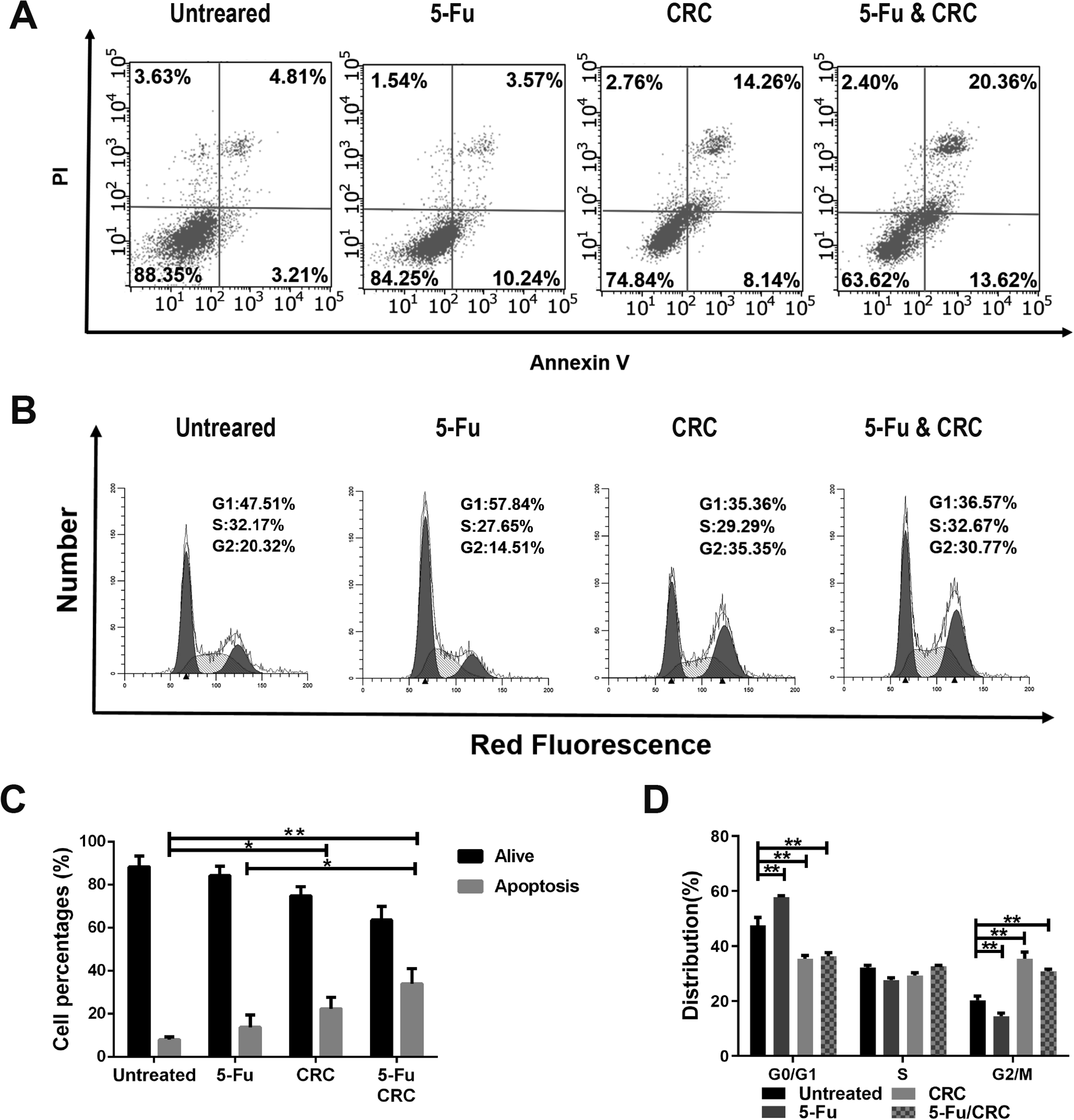
Effects of 5-FU and/or CRC on apoptosis and cell cycle progression. Hep-2 cells were examined using a PI/Annexin V kit to assess apoptosis (A and C) and stained with PI to assess cell cycle progression (B and D) after 24 hours of treatment with 5-FU (20 μg/mL) and/or CRC (20 μg/mL). The results were obtained from three independent experiments and expressed as the mean ± SD. *P < 0.05, **P < 0.01. 5-FU, 5-fluorouracil; CRC, curcumin.
MSNs as Carriers for 5-FU and CRC
Cytotoxicity and loading capacity of MSNs
To evaluate MSNs in drug delivery, we first tested their cytotoxicity and loading capacity. Cells exposed to different concentrations of MSNs for 24 hours displayed no significant difference in cell viability than untreated cells (Figure 3A). We then tested the toxicity of MSNs over time. Similarly, 100 μg/mL MSNs exhibited no cytotoxicity until 72 hours (Figure 3B). To confirm the loading capacity of MSNs, FITC (molecular weight = 389) was used to track the endocytosis and loading activity of MSNs. As presented in Figure 3C, after culture with FITC-loaded MSNs for 4 hours, fluorescence was detected in Hep-2 cells. Therefore, we hypothesized that MSNs can serve as nanoparticles for loading 5-FU and CRC.

Cytotoxicity and drug-loading/release capacity of MSNs. The CCK-8 assay was used to analyze the viability of Hep-2 cells incubated (A) for 24 hours with different concentrations of MSNs and (B) for 24, 48, or and 72 hours with the same concentration of MSNs (100 μg/mL). (C) Confocal laser-scanning microscopy images displaying the cellular uptake of MSNs loaded with FITC. (D and E) The release rate of 5-FU- and CRC-loaded MSNs at different pH values. MSNs, mesoporous silica nanoparticles; 5-FU, 5-fluorouracil, CRC, curcumin.
MSNs can reinforce the effects of 5-FU and CRC on Hep-2 cells
To confirm that MSNs can serve as drug delivery vehicles, we first performed a cytotoxicity assay to determine the 50% inhibitory concentration (IC50) of MSNs loaded with 5-FU and/or CRC (Figure 4A-F and Table 1). The IC50s of both 5-FU and CRC in Hep-2 cells at 24, 48, and 72 hours were both significantly decreased when they were loaded into MSNs.

Effects of 5-FU and CRC were enhanced by MSNs. (A-F) The inhibitory effects of MSNs loaded with difference concentrations of 5-FU (A–C) and/or CRC (D–F) on Hep-2 cells after 24, 48, and 72 hours. (G) The inhibitory effects of unloaded MSNs and MSNs loaded with 5-FU (20 μg/mL) and/or CRC (20 μg/mL) on Hep-2 cells. (H) c-Myc, cyclin B1, survivin, and caspase 3 protein expression in Hep-2 cells treated with 5-FU and/or CRC (free or loaded into MSNs) was examined using western blotting. (I and J) Quantitative analysis of protein expression as measured via the optical density of each band relative to β-actin. The results were obtained from three independent experiments and expressed as the mean ± SD. **P < 0.05, **P < 0.01. 5-FU, 5-fluorouracil; CRC, curcumin; MSNs, mesoporous silica nanoparticles.
Effects of 5-FU and CRC were enhanced by loading into MSNs
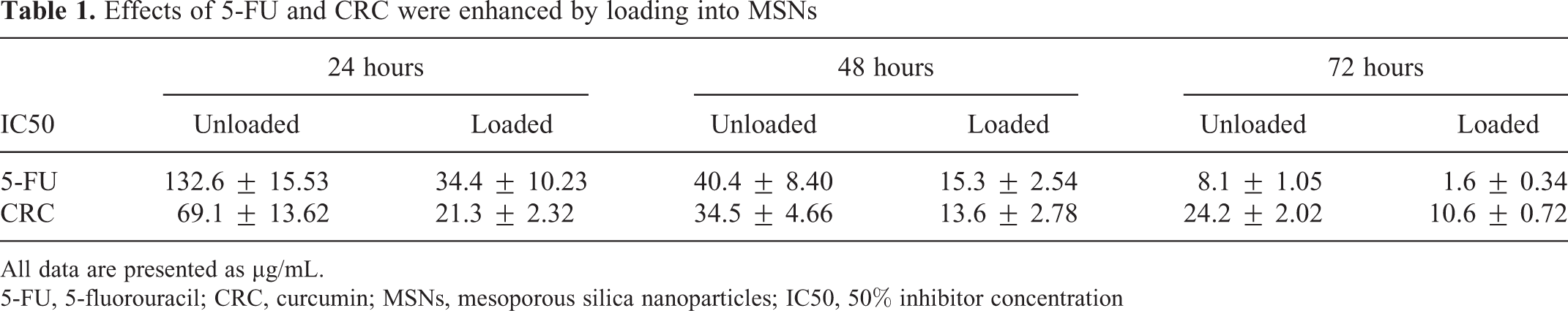
All data are presented as μg/mL.
5-FU, 5-fluorouracil; CRC, curcumin; MSNs, mesoporous silica nanoparticles; IC50, 50% inhibitor concentration
Drug encapsulation and loading efficiencies
The drug encapsulation and loading efficiencies are used to evaluate drug delivery systems. Physio-adsorption of drugs onto MSNs and the electrostatic interactions between the positively charged moieties of polymers contribute to the drug-loading capacity. 30 The loading capacity of MSNs for 5-FU and CRC is presented in Table 2. Using our research conditions, the encapsulation efficiencies of 5-FU and CRC were 30.8% and 67.8%, respectively, and the loading efficiencies for these drugs were 18.53% and 13.56%, respectively.
Loading capacity of MSNs for 5-FU and CRC.

5-FU, 5-fluorouracil; CRC, curcumin
Release rate at different pH values
Solid tumors usually have a weakly acidic (pH = 6.0–7.0) environment. In addition, the pH inside cancer cells is even lower (pH = 5.0–5.5). 34 -36 Because the MSNs we used in this research are pH-sensitive, 32,37 we conducted the release experiments under two pH conditions (PBS buffer at pH = 5.0 and pH = 7.2). The release curves are shown in Figure 3D and E. Both 5-FU- and CRC-loaded MSNs exhibited higher release rates at pH 5.0, indicating that MSNs preferentially release drugs in the tumor environment and cancer cells than in normal tissues.
5-FU- and CRC-loaded MSNs downregulate oncogenes
We found that MSNs loaded with both 20 μg/mL 5-FU and CRC more strongly suppress viability than single drug-loaded MSNs after 24, 48, and 72 hours (Figure 4G). Thus, dual-loaded MSNs can take advantage of both the synergistic effects of drugs and the reinforcement function of MSNs. We then detected the proteins influenced by 5-FU and CRC. As presented in Figure 4H-J, western blot analysis demonstrated that protein expression was altered by different degrees. Similarly intense effects were observed for drug-loaded MSNs, suggesting that MSNs could enhance the effects of anti-cancer drugs on multiple levels.
In Vivo Study of MSNs Loaded with 5-FU and CRC
To investigate the efficacy of drug-loaded MSNs on tumor growth, Hep-2 tumor-bearing nude mice (n = 6) were randomly distributed into five groups: untreated, MSNs, 5-FU-loaded MSNs, CRC-loaded MSNs, and 5-FU/CRC-loaded MSNs. As illustrated in Figure 5A, the weight of the mice was stable following treatment. Alternatively, there was significant suppression of tumor growth in mice treated with 5-FU and/or CRC (Figure 5B and C) compared with the findings in the untreated group. These results indicate that MSNs could be safe chemotherapeutic drug carriers in mice with tumors. Additionally, the smaller tumor volume in the 5-FU/CRC-loaded MSN group also supported the potential of this drug combination.
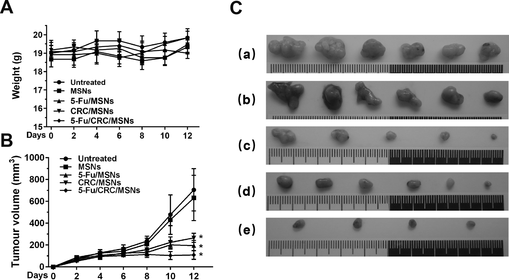
Effects of MSNs loaded with 5-FU and/or CRC on the growth of tumors in nude mice. (A) Mouse weight after treatment with the drugs. (B) Volumes of tumors following different treatments: (a) untreated, (b) MSNs, (c) 5-FU-loaded MSNs, (d) CRC-loaded MSNs, and (e) 5-FU/CRC-loaded MSNs. (C) Tumor growth curves for different treatments. *P < 0.05, compared with the untreated group. 5-FU, 5-fluorouracil; CRC, curcumin; MSNs, mesoporous silica nanoparticles.
Discussion
Laryngeal cancer, a common malignant head and neck tumor with a low survival rate and poor prognosis, accounts for a quarter of all malignant head and neck tumors. 1,2 Laryngeal cancer can be caused by many factors, most of which are related to genetic and environmental factors. Lifestyle habits including smoking and drinking, biliary tract diseases, gastroesophageal reflux, and human papillomavirus infection can all increase the risk of laryngeal cancer in the population. 40,41 At present, surgical resection combined with chemotherapy/radiotherapy is the main therapeutic option for laryngeal cancer. 3 -5 However, patients with advanced laryngeal cancer are prone to recurrence and metastasis after surgery, resulting in poor prognoses. 4,5 In addition, the common side effects of chemotherapy drugs also limit their anti-cancer efficacy. 42
The combination of different chemotherapeutic drugs may reduce the dose of drugs, their toxicity, and multidrug resistance. 43 -45 Recent studies illustrated that the combination of 5-FU and CRC can have great efficacy in the treatment of various cancers, including colorectal, breast, liver, and gastric cancers. 12,23,46 -49 As a widely used anti-cancer drug, the main mechanisms of action of 5-FU include the inhibition of thymidylate synthase activity, DNA synthesis, and DNA repair via the incorporation of its metabolites into the DNA/RNA of cancer cells. 8 However, the efficacy of 5-FU in the clinic is limited by dose-limiting toxicities and chemoresistance. 11 In studies of laryngeal cancer, CRC was revealed to inhibit cell proliferation and promote apoptosis through the PI3K/Akt and JAK/STAT pathways. 25,26 In addition, CRC can also stimulate cisplatin-induced cell death through TRPM2 channel activation. Studies illustrated that the anti-cancer effects of CRC might be related to mechanisms such as proliferation, transformation, invasion, metastasis, and angiogenesis. 22 Meanwhile, extensive research has found that CRC can sensitize various tumors to chemotherapeutic agents including 5-FU. 50 Hence, the combination of 5-FU and CRC might also have promising anti-cancer effects against laryngeal cancer. However, the toxicity and side effects caused by the lack of tumor targeting remain problems to be addressed.
In recent years, the use of nanomaterials to design drug carriers targeting tumor sites has been an important research direction for tumor-targeting drugs. 29 Nanomaterials can prevent drugs from being removed or ingested by the kidneys and liver, permitting them to remain in the blood circulation for a long time and maintain a high concentration in tumor tissues. Tumor-targeting drug carrier nanomaterials include liposomes, polymer nanoparticles, dendritic polymers, and nanoparticles. 29 MSNs, as nanoparticle drug carriers, have a unique structure and remarkable properties. 31
In this study, Hep-2 cells were used to detect the effects of 5-FU and CRC on cell growth at different concentrations and exposure durations. The results demonstrated that 5-FU was more effective at low concentrations, whereas CRC was more effective at high concentrations. In addition, the two drugs produced a synergistic effect on tumor growth, providing a reliable experimental basis for combined drug therapy in the clinical treatment of laryngeal cancer. Given the low targeting potential and high side effects of chemotherapies, in line with previous research, 32,37 we used tumor-targeting MSNs with pH- and temperature-sensitive properties as carriers for 5-FU and CRC. MSNs deliver drugs to tumor sites according to the pH and temperature of the tumor environment. In addition, their pore structure permits them to slowly release drugs. The results illustrated that using MSNs to deliver two drugs to Hep-2 cells significantly improved their anti-tumor efficacy. Moreover, an analysis of protein expression also proved that MSNs loaded with drugs could influence the expression of tumor-related proteins, enhance the anti-tumor effect of drugs, and reduce the dosage of drugs required for efficacy. We also verified the therapeutic effect of MSNs on tumors through animal experiments. In vivo experiments demonstrated that MSNs could deliver drugs to tumors in mice, and no obvious toxicity or side effects were observed. This study demonstrated that 5-FU and CRC have synergistic anti-tumor effects, effectively inhibiting the growth of laryngeal cancer cells. In addition, MSNs as tumor-targeting drug carriers, can further enhance the synergistic effects of 5-FU and CRC on laryngeal cancer.
Footnotes
Abbreviations
Declaration of Conflicting Interests
The author(s) declared no potential conflicts of interest with respect to the research, authorship, and/or publication of this article.
Funding
The present study was funded by the National Natural Science Foundation of China (grant nos. 81773217 and 81572653), the Natural Science Foundation of Jilin Province (grant nos. 20190701065GH and 20160101039JC), and the Health Technology Innovation Project of Jilin Province (grant no. 2019J030).