
Abstract
Purpose:
To develop an infrared optical method of reducing surface-based registration error caused by respiration to improve radiotherapy setup accuracy for patients with abdominal or pelvic tumors.
Materials and Methods:
Fifteen patients with abdominal or pelvic tumors who received radiation therapy were prospectively included in our study. All patients were immobilized with vacuum cushion and underwent cone-beam computed tomography to validate positioning error before treatment. For each patient, after his or her setup based on markers fixed on immobilization device, initial positioning errors in patient left-right, anterior-posterior, and superior-inferior directions were validated by cone-beam computed tomography. Then, our method calculated mismatch between patient and immobilization device based on surface registration by interpolating between expiratory- and inspiratory-phase surface to find the specific phase to best match the surface in planning computed tomography scans. After adjusting the position of treatment couch by the shift proposed by our method, a second cone-beam computed tomography was performed to determine the final positioning error. A comparison between initial and final setup error will be made to validate the effectiveness of our method.
Results:
Final positioning error confirmed by cone-beam computed tomography is 1.59 (1.82), 1.61 (1.84), and 1.31 (1.38) mm, reducing initial setup error by 24.52%, 51.04%, and 53.63% in patient left-right, anterior-posterior, and superior-inferior directions, respectively. Wilcoxon test showed that our method significantly reduced the 3-dimensional distance of positioning error (P < .001).
Conclusion:
Our method can significantly improve the setup precision for patients with abdominal or pelvic tumors in a noninvasive way by reducing the surface-based registration error caused by respiration.
Keywords
Introduction
Rapid evolution of modern radiotherapy technology allows greater radiation dose to be conformed precisely to the target volume while sparing the normal tissues, 1 -6 posing increasingly higher requirements to patient setup accuracy. Immobilization devices such as the thermoplastic mask and vacuum cushion have been used to achieve high positioning repeatability for a long time. 7 -9 However, in clinical positioning practice, mismatch between patient surface and immobilization system (MBSI) is found to cause obvious interfraction setup error. 10
On-board imaging devices such as cone-beam computed tomography (CBCT) and electronic portal imaging device (EPID) have been introduced as image-guided radiation therapy (IGRT) to improve radiotherapy setup precision. 11 -13 X-ray images of patient internal anatomy are registered to the planning computed tomography (CT) scans to determine current positioning error. These methods can detect internal anatomy changes and help correct positioning error. But dose delivered during the imaging can’t be ignored. To avoid extra radiation exposure, radiotherapy positioning systems based on optical surface registration are also developed. Sentinel (C-RAD AB, Uppsala, Sweden) 14 and Catalyst (C-RAD AB, Uppsala, Sweden) 15 take advantage of structured light to reconstruct patient surface in real time for setup error detection. AlignRT (VisionRT Ltd, London, UK) 16 captures a static or respiration-gated 3-dimensional (3-D) surface and registers the acquired surface and the previously recorded reference one for positioning correction. However, for abdominal and pelvic tumors, surface shape changes obviously with respiration, adversely affecting surface registration accuracy.
In this study, a noninvasive and radiation-free method based on optical localization and surface point cloud registration is proposed to reduce MBSI in radiotherapy positioning. By capturing and interpolating the inhale and exhale surfaces, it selects the specific respiratory phase that best matches the planning CT scanning one, thus effectively reduces registration error caused by surface mismatch.
Materials and Methods
Initial setup is based on a rigid transformation matrix
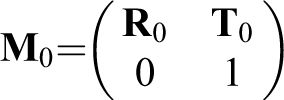

Illustration of initial setup by aligning optical space and planning CT scans by registration based on 4 infrared markers fixed on vacuum cushion. CT indicates computed tomography.
where
Vacuum cushion is shifted according to
The MBSI will cause error between tumor center and LINAC isocenter after initial setup.
10
As a result, another transformation matrix

where
Considering respiration, real-time surface will present continuous deformation. Registration precision will be adversely affected if 2 surfaces are not of the same shape. Therefore, we select the respiratory phase that best matches the surface in planning CT scans by interpolating between exhale and inhale surface.
First, we acquire the exhale surface by asking the patient to exhale and hold on for around 2 seconds. A series of IR markers march through patient surface, and their trajectories will be tracked by Polaris and transmitted to our system. Then, a point set representing exhale surface S ex is acquired. The inhale surface S in is acquired in the same workflow except that the patient is told to inhale.


S x−percentile consists of points that are interpolated at certain percentile on the segment determined by P in−i and its perpendicular foot on S ex as shown in Figure 2.


Respiratory surface interpolation. Nth percentile surface consists of points interpolated at n% of the perpendicular (gray dotted line) from inspiratory surface to the expiratory surface. 50th percentile and 80th percentile interpolation surface are shown as example.
One-norm distance transformation is used to select the S the−percentile that best matches the S CT by aligning the 2 surfaces as much as possible and evaluating the mean distance between them as follows:


where M 1−j is applied to align S j−percentile and S CT to the best, and Δ is the 3-D displacement from P j−percentile−i to S CT. S the−percentile is the S j−percentile with minimum Δ, and its according M 1−j is chosen as M 1 that describes the mismatch between patient surface and vacuum cushion.
So the final positioning error is calculated as the following equation.


where O iso is the LINAC isocenter registered by the IR optical localization system and O tumor is the tumor center determined in the planning CT scans.
In all 15 patients (6 males and 9 females; age range from 49 to 63; median age, 58 years old) diagnosed as abdominal or pelvic tumors and received radiotherapy were prospectively included in our study to evaluate the accuracy of the positioning method mentioned above.
All patients were immobilized with vacuum cushion and were free breathing during simulation and treatment. Elekta kilovoltage (kV) CBCT XVI system integrated in Synergy LINAC (Elekta AB, Stockholm, Sweden) was used for positioning validation. Because the 3-D couchtop used in our study could correct for translations only, no rotation accuracy was evaluated. But our approach is able to provide MBSI correction in 6 DoFs.
Treatment couch was first shifted based on markers fixed on the cushion. To determine initial positioning error E 0, patient underwent CBCT that was performed by a physicist with experience in radiotherapy setup of 6 years. For abdominal tumors, registration between CBCT and planning CT was done by focusing on the soft-tissue gray values within planning target volume. And for pelvic tumors, it was mainly based on pelvis, and soft-tissue gray values would also be considered. Then, treatment couch was further shifted according to the guidance given by our method. The CBCT was performed again by the same physicist to determine the final positioning error E 1. For each patient, positioning errors confirmed by CBCT from 4 fractions were recorded. Thus, in total, setup errors from 60 fractions were analyzed in our study.
Wilcoxon test was performed using the Statistical Package for the Social Science (SPSS, version 22.0 for Microsoft Windows x64,) to compare initial and final positioning errors.
Results
Initial positioning error (presented as mean [standard deviation]) indicated by CBCT was 2.11 (2.43), 3.29 (3.70), and 2.83 (3.26) mm in patient left-right, anterior-posterior, and superior-inferior directions, respectively. After applying our method to further shift the treatment couch, positioning error was reduced to 1.59 (1.82), 1.61 (1.84), and 1.31 (1.38) mm, respectively. Mean positioning error was reduced by 24.52%, 51.04%, and 53.65%. Figure 3 showed the x, y, and z as well as 3-D distance of the initial and final positioning errors in boxplot. The 3-D positioning errors of 59 out of 60 fractions were reduced as for distance. There was a positioning error distance increased from initial 3.15 to 3.23 mm in one fraction. It was mainly due to a failure in x direction, which might be induced by optical block. Wilcoxon test indicated that our method significantly reduced 3-D distance of positioning error after initial setup based on markers fixed on vacuum cushion (P < .001).

Boxplot of initial (blue boxes) and final (red boxes) positioning errors in x, y, and z directions and 3-dimensional distance validated by cone-beam computed tomography (CBCT).
Discussion
Optical surface imaging systems have been investigated to supplement well-established IGRT methods (such as CBCT or EPID), given their special regard to real-time motion as well as being fast, noninvasive, and radiation free. An earlier study performed by Walter et al 15 on Catalyst indicated positioning errors (expressed in mean [standard deviation] in x, y, z with units of mm) of (0.3 [2.2], 2.1 [5.5], 2.6 [1.8]) for patients with abdominal tumors and (−0.9 [1.5], 1.6 [2.2], −1.7 [2.8]) for pelvic targets. Another previous study conducted by Stieler et al 14 on Sentinel showed positioning errors of (−2.6 [4.1], −5.1 [7.4], 4.6 [7.3]) for patients with pelvic tumors. And our approach exhibited positioning errors of (1.6 [1.8], 1.6 [1.8], 1.3 [1.4]). Wiencierz et al 16 conducted a study on two commercial optical surface imaging systems (Catalyst and AlignRT) to validate their positioning accuracy for patients with tumors in various sites including pelvis. Results showed that for patients with pelvic cancers, 50th, 75th, and 90th percentile positioning errors (using an external CT scan as reference image, expressed in x, y, z with units of mm) were (3.5, 3.9, 2.6), (5.9, 5.7, 3.7), and (7.3, 8.6, 6.5), respectively, for AlignRT, and (3.8, 4.5, 3.4), (5.8, 8.8, 5.8), and (6.6, 12.4, 7.2), respectively, for Catalyst. In our study, positioning errors were (1.8, 1.7, 1.2), (2.2, 2.2, 1.8), and (2.8, 2.7, 2.6) for 50th, 75th, and 90th percentile, respectively. Comparison indicated that the method proposed in our study had a competitive accuracy in positioning for patients with abdominal or pelvic tumors, especially in y and z directions.
Such preferable performance of our approach may result from the following facts. For Sentinel and Catalyst, insufficient concern is given to respiratory movement. And for AlignRT, respiration phase is identified by specific point on the surface. In comparison, our method selects surface of specific phase by measuring the similarity of whole 3-D surface and the referenced one, which will help reduce the adverse influence caused by respiration-induced surface deformation during registration.
Besides, previous work of Zhang et al 10 demonstrated the importance of MBSI reduction and proposed a noninvasive method to guide patient setup by integrating CT information and optical surface landmarks to minimize MBSI. However, they didn’t consider that abdominal or pelvic surface suffered obvious deformation from respiration. And precision of tumor localization based on surface registration would be adversely affected by mismatch between real-time and CT scanning surface.
In our study, we hypothesized that the patient surface presented a continuous deformation based on respiratory phase. Thus, we could reconstruct patient surface under any respiratory phase by interpolating between exhale and inhale surfaces, maximizing the accuracy to select the specific surface that best matched the CT scanning one. Result indicated that our method significantly reduced the initial setup error.
Patients with tumors are likely to lose weight during treatment, causing abdominal and pelvic surface to become lower in y direction than thorax when they lie on the treatment couch. As a result, surface deformation caused by respiration will lead to registration error that mainly consists of displacement in y and z directions as shown in Figure 4. Zhang’s previous study 10 showed an average final setup error of 1.30, 2.60, and 1.68 mm in x, y, and z directions, respectively. In comparison, our method exhibited a preferable accuracy in y and z directions, further indicating its effectiveness in reducing registration error caused by respiratory surface deformation. Such analysis is in agreement with previous studies 14,15 on optical surface positioning. They identified larger setup errors in longitudinal and vertical directions. The results were believed to be induced by respiratory movement. A slightly more errors in x direction might result from the absent body side surface, which is hard to capture because of optical block.
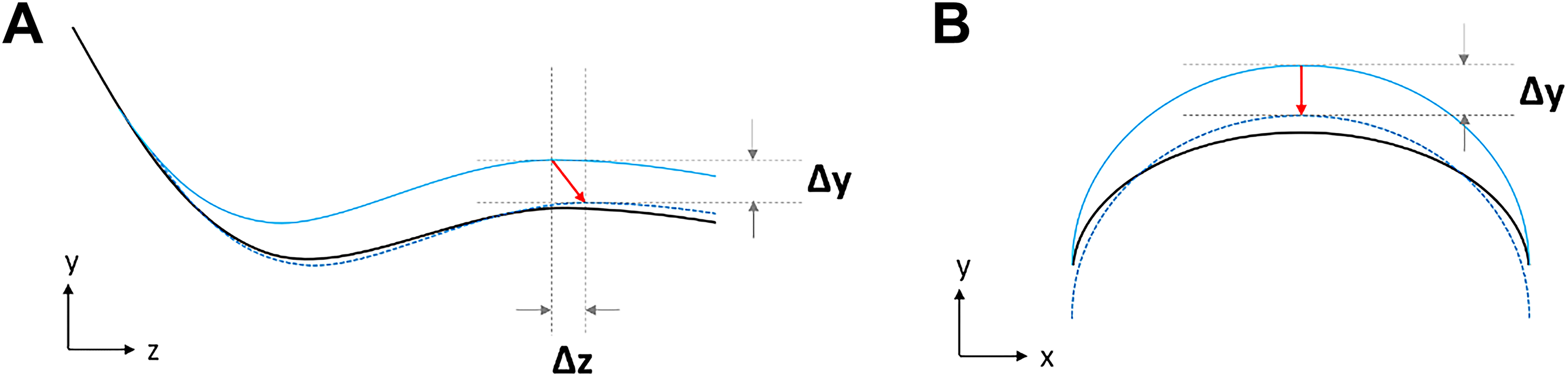
Surface deformation caused by respiration leads to registration error. A, A sagittal illustration of human body from lower thorax to the abdomen. B An axial illustration of human abdomen. Abdominal and pelvic surface is likely to be lower in y direction than thorax when patient lies on treatment couch as result of emaciation. The real-time surface (blue solid line) indicates a higher respiratory phase, but the same patient position (relative to isocenter) as the computed tomography (CT) scanning surface (black solid line) does, mainly causing shift (red arrow) in y and z directions when aligning the 2 surfaces (blue dotted and black solid line).
There is still improvement for our method. The setup correction including surface capture and registration will be done, generally, within 1 minute. It might take longer in consideration of the patient condition. For example, older patients or patients with pain may hard to cooperate and will take longer time with surface capture. Thus, a noncontact method to capture patient surface, such as surface reconstruction based on structured light, is appreciated. It will help the approach with a faster surface acquisition and be applied during treatment. Besides, all points of the surface possess the same weight during registration in our method. A nonuniform weight distribution along the surface, such as higher weights for hard surface around pelvis or rib and lower weights for easy-to-deform soft-tissue surface, may be helpful to further decrease registration error resulted from surface deformation, which requires future exploration.
Conclusion
In our study, we proposed a noninvasive method of minimizing MBSI to improve the tumor localization accuracy. Validation conducted by CBCT showed the ability of our method to improve the setup accuracy to a high level. A preferable positioning precision compared with the result of Zhang’s previous work indicated the surface registration error caused by respiration can be effectively reduced with our method.
Footnotes
Declaration of Conflicting Interests
The author(s) declared the following potential conflicts of interest with respect to the research, authorship, and/or publication of this article.
Funding
The author(s) disclosed receipt of the following financial support for the research, authorship, and/or publication of this article: It is a Research Ethics Board (REB)–approved study. This work was supported by Social Development Program of Primary Research & Development Plan in Jiangsu Province (BE2016733, BE2017679).