
Abstract
In this study, the effect of using an epoxy healing agent containing silica nanoparticles instead of the neat epoxy healing agent on the self-healing ability of glass fibers/epoxy composites under the flexural, tensile and shear loadings was assessed. For doing this, in the first step, the silica nanoparticles (0, 1, 2 and, 3 wt.%) were dispersed into the ethyl acetate/epoxy healing agent. Then, these mixtures were encapsulated with the urea-formaldehyde polymer by an in-situ polymerization method. Finally, the glass fibers/epoxy composite containing 14 wt.% microcapsules along with 2 wt.% NiCl2(imidazole)4 catalyst were fabricated. To understand how the mechanical-healing behaviors of these composites under the various mechanical loads, these composites were destructed with a quasi-static penetration test for the creation of delamination damage. The healing process was done by the thermal activation method. The obtained results showed that the highest healing efficiency under the flexural, tensile and shear loads belonged to the composite containing 1 wt.% nanosilica into the healing agent, which were 89.8, 87.8, and 156.8%, respectively. The hackles of microcapsules, reduction in the thickness of microcapsules, formation of nanocomposite healing shell and the agglomeration of silica nanoparticles on the healed area were the influence factors on the healing ability of these composites. This work can give a view for healing the microcracks into composite structures by nanocomposite healing agents, which have the higher healing ability and better performance under mechanical loads than conventional healing agents.
Introduction
The smart material is introduced as a matter, which can response to the environmental agents like physical, chemical, and bio-chemical agents for using in especial applications.1,2 One of the progressive trends in the smart materials is fabricating the self-healing materials. Having self-healing ability cause, the matter automatically repairs the created microcracks into itself. 3 In order to give this ability to the polymer and its composite, two distinct major viewpoints have been developed. 4 In the intrinsic system, the matter can repair itself by using polymers with the reversible bonds, 5 synthesizing supramolecular polymers 6 and blending with the second polymer. 7 But in the extrinsic viewpoint, the healing agent is stored into the containers, which can be microfibers, 8 vascular systems 9 or microcapsules. 10
Both viewpoints for laminate composites like carbon fibers/epoxy or glass fibers/epoxy composites have been developed in the last years. For examples, Ruan et al. 11 stored the dicyclopentadiene (DCPD) as healing agent into the nano fibers with the poly phthalazinone ether sulfone ketone (PPESK) shell by using the coaxial electrospinning method. The fabricated membranes by this method had the thickness of 50 μm, which were deposited on the surface of carbon fibers. Then, these carbon fibers were used to fabricate the carbon fibers/epoxy composite with the self-healing ability. The obtained results showed, adding these nanofibers caused to 8.97, 26.14 and 62.12% increase of strength, breaking strain, and fracture energy of neat carbon/epoxy composite. Also, the healing capability under the flexural loading indicated, this composite could recover 89.43% of its initial strength after 2 h of damaging at ambient temperature.
Kostopoulos et al. 12 used a bis-maleimide based polymer as a thermally reversible polymer for creating the healing ability into the carbon/epoxy composite. This polymer can recover the mechanical properties of composite by using the Diels-Alder (DA) reaction as intrinsic mechanism. Damaging the composite was done by low velocity impact test and the healing-mechanical ability of composite was characterized by compression after impact test. The obtained results after the healing activation showed that the carbon fibers/epoxy composite could repair the damage area, so that, the residual compression features of composite were significantly improved. Chen et al. 13 used the Diels-Alder healing mechanism for restoring the mechanical properties of glass fibers/epoxy composites. These researchers added the hexagonal boron nitride for improving the thermal conductivity of composite. According to this method, the degrees of mechanical strength recovery in the glass fibers/epoxy composite were about 70–85%.
In some limited cases, the researchers used the nanoparticles for improving the mechanical-healing performance of polymers, and using them into the laminated composites was not reported yet. For example, Sánchez-Romate et al. 14 added the graphene nanoplatelet into the epoxy/poly caprolactone blends for introducing the new healing activation method by Joule’s heating effect. Actually, these researchers applied the voltage into the both sides of this smart polymeric film. Incorporating the graphene nanoplatelet enhanced the internal temperature of polymeric film, which resulted to active the healing process into that. In other interesting work, Sima et al. 15 incorporated the Fe3O4 nanoparticles into the core material for controlling the dispersion of microcapsules into the epoxy coating. By using the magnetic force, these researchers navigated the microcapsules to the near surface of epoxy coating, which resulted to enhance the healing ability of that.
The nanoparticles can incorporate in three sites into these composites, which can be into the polymeric matrix, the surface of microcapsules and into the healing agent. This work aims to investigate the effect of silica nanoparticles incorporation into the healing agent on the healing-mechanical performances of glass fibers/epoxy as laminate composites. After the fabrication of these smart composites, the samples were subjected to a quasi-static penetration test. The healing process was done by putting the damaged composites into oven at the temperature of 130°C for 60 min. After that, the healing-mechanical properties of those were studied by flexural, tensile and interlaminar shear tests. This work can give a view for healing the microcracks into composite structures by nanocomposite healing agents, which have the higher healing ability and better performance under mechanical loads than conventional healing agents.
Experimental
Raw materials
The materials for fabricating the self-healable composites containing microcapsules with the silica nanoparticles/diluted epoxy healing agent can be categorized in three groups. The first group were the raw materials for synthesizing the microcapsules, which were urea (Pardis Petrochemical Company, Iran), ammonium chloride, formaldehyde, and hydrochloric acid (Dr. Mojallali Industrial Chemical Complex Company, Iran), sodium carboxymethyl cellulose (NaCMC), 1-octanol and resorcinol (Merck, Germany), and linear alkyl benzene sulfonic acid (LABSA- Behdash Chemical Company, Iran). The second group can be the healing agent ingredients, which were diglycidyl ether of bisphenol A (epoxy KER 828, Korea), ethyl acetate (Dr. Mojallali Industrial Chemical Complex Company) as diluted agent, and silicon oxide nanoparticles with an average size of 20–30 nm (Platonic Nanotech, India). The third group were the materials for fabricating the composites, which were epoxy resin (KER 828, Korea), triethylenetetramine (TETA) hardener, and glass fabric with the surface density of 400 g/m2 (Lintex, China). According to the previous work,16–18 the NiCl2(imidazole)4 as imidazole-based catalyst was synthesized as second agent of healing process, which can cure the diluted epoxy. All materials for fabricating the composites were commercial grade. But, the used materials for synthesizing the microcapsules and catalyst were the high purity materials (approximately 99.9%).
Fabrication of self-healable composites
The fabrication process and assessment of healing ability of composites have been illustrated as flowchart in Figure 1. The fabrication of composites containing the microcapsules with the silica nanoparticles/epoxy healing agent can be performed in four stages. The first stage can be the dispersion of silica nanoparticles into the epoxy as healing agent. To do so, the epoxy was diluted by the ethyl acetate with the weight fraction of 50:50. Then, the 1, 2 and 3 wt.% silica nanoparticles were added into this mixture. Next, the nanoparticles were dispersed into this mixture by using the magnetic stirrer (400 r/min) and sonication devices (24 kHz, 60% output power of 125 W) at the same time for 45 min.
19
The second stage was the encapsulation of these healing agents, which was done by in-situ polymerization of urea-formaldehyde (UF) monomers. The details of synthesis and confirm of this method were explained in the authors' preceding work.
20
The third and fourth stages were dispersing microcapsules and fabricating the composites. So, firstly, 14 wt.% synthesized microcapsules and 2 wt.% NiCl2(imidazole)4 were dispersed into the epoxy resin by mechanical stirrer to provide the self-healing ability. In the next step, the TETA hardener with the ratio of 10 wt.% was added into this mixture. Finally, the glass fibers/epoxy composites were fabricated by hand-layup method.
21
For reducing the trapped air bulbs, the vacuum bag method was performed. The weight ratio between glass fibers and epoxy matrix containing microcapsules was 50:50. It should be noted that the number of glass layers in the flexural and tensile samples were four layers, whereas, the interlaminar shear strength (ILSS) samples had the 12 layers. The curing process was done at the temperature of 25°C for 24 h. To confirm the proper dispersion of microcapsules into the glass fibers/epoxy composite, the microscopical analysis was used, which can be observed in Figure 2. From this figure, the microcapsules have covered the surface of glass fibers into the composite structure. The average size and core content of these microcapsules were 225 μm and 71.2%, respectively, which were confirmed in previous work.
16
A Flowchart for showing the fabrication stages and assessing the mechanical-healing properties of self-healing glass fibers/epoxy composites containing the capsulated nanosilica-epoxy healing agent. The FESEM analysis from the fracture surface of glass fibers/epoxy composite with the proper dispersion of microcapsules.

Mechanical-healing investigations
To investigate the healing ability of these smart composites, the flexural, tensile and ILSS were chosen. Testing specimens were dimensioned in accordance to ASTM D790 (125 × 15 mm2), ASTM D3039 (250 × 25 mm2) and ASTM D2344 (50 × 20 mm2) standards, respectively. The thickness of flexural and tensile samples was 2.3 mm, whereas the thickness of ILSS samples was 4 mm. It should be noted that using the 12 plies glass fibers for achieving the thickness of 4 mm was due to the size limitation for performing the test according to the mentioned standard. The crosshead speed for performing the flexural, tensile and ILSS tests were 3.5, 2 and 1 mm/min, respectively. The important factor for investigating the healing ability is the healing efficiency of composite under the various loads, which can be calculated according to the equation 1.22,23 The quasi-static penetration test for selecting the various damage forces in the self-healing composites

As per previous works,16,24 the forces of 500, 1050 and 2900 N were selected to create the delamination damage mode into the flexural, tensile and ILSS samples. The third strength belonged to the healed samples. So, after the creating the damages, the composites were healed by setting them into the oven at the temperature of 130°C for 60 min. For more accuracy, each parameter in tensile, flexural and ILSS has been achieved by testing at least five samples.
Characterization
To characterize the microcapsule features, forming the polymeric and nanocomposite healing shells, and the role of nanosilica on the healing-mechanical features, the field emission scanning electron microscopy (FESEM), Mira3 Tescan (Czech) was used. Also, by using the element-distribution map and energy dispersed spectroscopy (EDS) analyses, the role of silica nanoparticle on the related phenomena were confirmed.
Results and discussion
Recovery of flexural strength
Data obtained from the flexural test of smart composites have been illustrated in Figure 4, which are the initial (undamaged), damaged and healed flexural strength of composites. According to the Figure 4(a), the flexural strength of undamaged sample containing the neat healing agent was 303 MPa. It seems that adding 1, 2 and 3 wt.% silica nanoparticles into the healing agent reduced the flexural strength, so that, the flexural strength of those were 294, 270 and 264 MPa, respectively. This shows that the initial flexural strength of composite has reduced. The reason for this behavior can be related to the effect of silica nanoparticles on the strength of microcapsules. In the study of Lee et al.
25
was found that the silica nanoparticles can act as an emulsifier for synthesizing the microcapsules, which resulted to change the features of microcapsules. To confirm this assumption, the FESEM analysis was done to measure the thickness of microcapsules containing the silica nanoparticles into the healing agent, which can be seen in Figure 5. The effect of incorporating the silica nanoparticles into the microcapsules on the healing-flexural properties of the glass fibers/epoxy composite; (a) undamaged composite; (b) damaging behavior; (c) damaged composite; (d) healed composite. The effect adding silica nanoparticles into the healing agent on the thickness of microcapsules; (a) 0 wt.%; (b) 1 wt.%; (c) 2 wt.%; (d) 3 wt.%.

According to this Figure 5, the microcapsules containing the neat healing agent had a thickness of 321 nm. By dispersing the 1, 2 and 3 wt.% silica nanoparticles, the thickness reached to 281, 110 and 100 nm, respectively. This reduction trend in the thickness of microcapsules can reduce the strength of microcapsules' shell, which consequently reducing the initial flexural strength of self-healing composites. This reduction can change the healing behavior into the composites, which will be discussed in the following section.
As previously mentioned, the damage force of 500 N was selected to create the delamination damage mode into the composite with the neat healing agent for comparing with other composites. The results of damaging with this force by quasi-static penetration test have been illustrated in Figure 4(b). As can be seen in this figure, the composite containing the neat healing agent showed the delamination damage mode. Incorporating the silica nanoparticles into the healing agent changed the damage mode of the composites, so that, according to this figure, the damage mode was matrix cracking. Changing the damage mode can reduce the degree of damages into the composite, which can enhance its ability of self-healing.
To have a comprehensive view about the effect of adding silica nanoparticles into the healing agent, it is better to study the flexural properties of damaged composites. Figure 4(c) shows the initial flexural strength and flexural strength after damaging with the force of 500 N. As can be seen, the flexural strength of composite containing the neat epoxy healing agent was 157 MPa. In other words, this composite had the 51.8% of initial flexural strength. By adding silica nanoparticles, the flexural strength of the damaged composites improved, so that, it reached to 237 MPa in the composite with 3 wt.% silica nanoparticles into the healing agent. In this composite, the flexural strength after damaging was about 90% of initial flexural strength. Therefore, it shows that the created damages in this composite can be matrix cracking. This change in the damage mode can be attributed to the interaction between microcracks and microcapsules into the smart composites.
To find the interactions between microcracks and microcapsules, the FESEM analysis from the fracture surface of these smart composites was done, which can be seen in Figure 6. As can be seen in the composite with the microcapsules containing the neat healing agent, the crack deflection, debonding microcapsules were the interaction between microcracks and microcapsules.
20
As previously confirmed, adding the silica nanoparticles reduced the shell thickness of microcapsules, which can be a reason for changing the interaction between microcapsules and microcracks. According to the Figure 6, by increasing the weight percentage of silica nanoparticles, the crack pinning and fracturing the microcapsules were the main interactions between the microcracks and microcapsules. Reducing the shell thickness can be a reason for enhancing the fracturing the microcapsules. The energy of microcracks can be wasted by fracturing the microcapsules. Therefore, the higher energy needs for crack propagations. So, it can be said that wasting the energy of microcracks by fracturing the microcapsules can be a reason for changing the damage mode into the smart composites containing the silica nanoparticles into the healing agent. The effect of incorporating the various weight fractions of silica nanoparticles into the healing agent on the crack propagation path; (a) 0 wt.%; (b) 1 wt.%; (c) 2 wt.%; (d) 3 wt.%.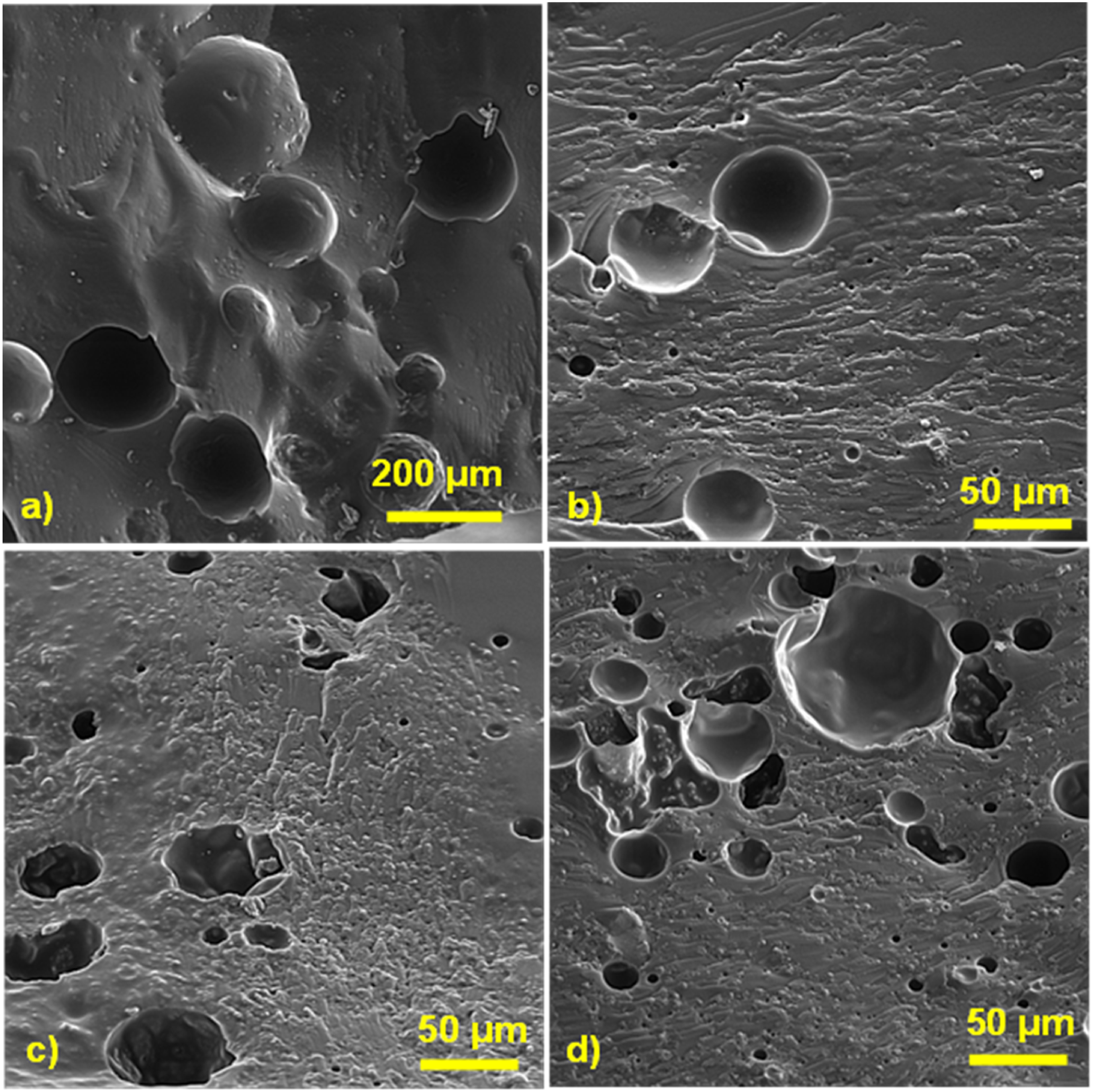
After the healing process, the flexural test of healed composites was performed to characterize the healing ability of composites, which can be seen in Figure 4(d). Although, the composite with the 3 wt.% silica nanoparticles into the healing agent had the highest flexural strength after damaging, but the composite with 1 wt.% silica nanoparticles had the highest healing efficiency, which was 121.6%. Having the higher healing efficiency than 100% means that the healed composite has the higher flexural strength than the undamaged composite. In other words, after the healing process, the initial flexural strength improved. After this composite, the composite containing 2 wt.% silica nanoparticles in the healing agent with the 83% healing efficiency had the highest recovery ability. It should be noted that the composite with neat healing agent had the lowest healing efficiency, which was 67.1%. Reaching the healing efficiency to 121.6% in the composite with 1 wt.% silica nanoparticles and reducing that by increasing the weight fraction of silica nanoparticles can be explained by the various effective factors.
In the propagation path, the microcracks fracture the microcapsules. Then, the healing agent diffuse into the damage area and filled the crack zones. Finally, the diffused healing agent reacts with the catalyst, which results in the formation of a polymeric shell.26,27 These shells can help to close the edges of cracks or fill the crack areas (seen in Figure 7(a)). Dispersing the silica nanoparticles into the healing agent can form the silica/epoxy polymeric shell instead of neat epoxy shell, which can improve the mechanical properties in the damaged areas. For this reason, in the composite with 1 wt.% silica nanoparticles, the healed flexural strength was higher than the initial flexural strength. From Figure 4(d), it can be found that enhancing the weight fraction of silica nanoparticles reduced the healing ability. According to the other sources,28,29 increasing the nanoparticles into the epoxy (here as healing agent) can enhance the viscosity of epoxy, which can influence on the flowability of epoxy resin. So, it can be mentioned that by enhancing the silica nanoparticles, the ability of healing agent for filling the crack zone reduced, which caused to decrease the healing efficiency. Figure 7(b), shows the formed silica/epoxy nanocomposite polymeric shell. By comparing the neat epoxy polymeric shell (Figure 7(a)), it can be found that the thickness of this shell is higher than that of neat epoxy shell. Forming the thicker shell can be attributed to the low flowability of healing agent. The effect of incorporating the silica nanoparticles into the healing agent on the thickness of epoxy healing shell; (a) 0 wt.%; (b) 3 wt.%.
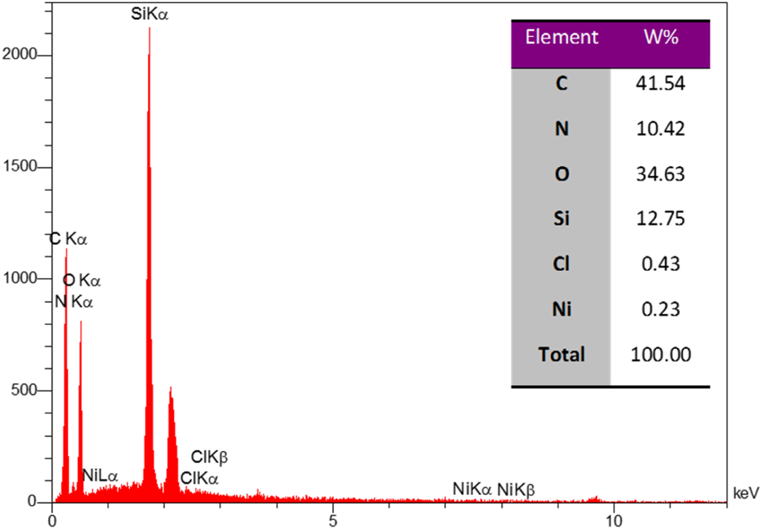
By deeper observing the Figure 7(b), it can be realized that this is not the only influence factor. Agglomeration of silica nanoparticles is another factor, which is clearly seen in this figure. As per previous works,30,31 the agglomeration area might have been weakest area for propagating the microcracks, due to having the low wettability with the healing agent for healing the crack areas. To confirm these agglomeration sites, the element-distribution map and EDS analyses were performed, which can be seen in Figures 8 and 9, respectively. According to these figures, the agglomeration of silica nanoparticles can be easily characterized by distribution of Si and O elements. Also, the Ni and Cl elements shows the remained catalyst into the composite structures. The element-distribution map analysis from the agglomerated silica nanoparticles on the healing area. The EDS analysis from the agglomerated silica nanoparticles on the healing area.
Recovery of tensile strength
Although, it can be predicted that the self-healable composites under the tensile mode are similar to flexural mode, but it is better to characterize the features of these composites under the tensile loading. The results are being presented in Figure 10, which were the tensile strength of undamaged, damaged, and healed composites. Also, the calculated tensile strength after damaging and healing efficiency were reported. According to the Figure 10(a), it can be found that adding the silica nanoparticles reduced the initial tensile strength of composite. This behavior was previously seen in the flexural strength. By comparing the tensile strength of damaged composites under the 1050 N force, it can be realized that it has reducing trend, which has opposite trend in compared with the results in the flexural test. This can be related to the amount of required interlaminar shear stress for creating the damages in the composites. The interlaminar shear stress from the quasi-static penetration test can be calculated according to the equation 2.
32
The effect of incorporating silica nanoparticles into the microcapsules on the healing-tensile properties of glass fibers/epoxy composite; (a) undamaged composite; (b) damaging behavior; (c) damaged composite; (d) healed composite.

The microcapsules can resist against the shear stress up to the certain level by deforming themselves, which absorbed the shear force. If the shear stress be higher, the microcapsules can be deboned from matrix. This phenomenon is named as hackles phenomenon, which was firstly characterized by Gazali et al. 33 In the tensile and shear loadings, the hackles phenomenon can occur. Therefore, the composite cannot absorb the energy of shear stress from quasi-static penetration test. So, it can be expected that the level of damage in the composite containing silica nanoparticles in the healing agent can be higher than the composite containing neat healing agent. The results obtained from quasi-static test confirms this assumption (seen Figure 10(b)). As can be seen in figure, all composites have the similar damage behaviors under the quasi-static penetration test. Like the flexural test results, the maximum healing efficiency was seen in the composite with the 1 wt.% silica nanoparticles, which can be due to the formation of nanocomposite shell into the damage zone. By increasing the weight fraction of silica nanoparticles in the healing agent, the recovery ability reduced, which can be due to the increment of healing agent viscosity and agglomeration of silica nanoparticles. Similar results about the effect of high content nano materials on enhancing the viscosity of epoxy resin and reducing the mechanical features of composites have been reported by other researches.34,35
Recovery of interlaminar shear strength
The summary of data from undamaged, damaged and healed composites under ILSS test have been reported in Figure 11. The shear performance of composites was similar the tensile performance of those. In other words, by adding the silica nanoparticles into the healing agent, the ILSS of undamaged sample showed the reducing trend, so that, the composite with the 3 wt.% silica nanoparticles in the healing agent had 10.7% reduction in the ILSS, which can be attributed to the microcapsules’ strength and effect of that on the ILSS of composites. Also, the shear strength after damaging had a reducing trend, as well as, no change was seen in the damage mode of composites (seen Figure 11(b)). The sample with the 1 wt.% silica nanoparticles had the highest recovery, which was 156.8%, followed by sample with the 2 and 3 wt.% silica nanoparticles, which were 129.2 and 94.3%, respectively. The effect of incorporating the silica nanoparticles into the microcapsules on the healing-shear properties of the glass fibers/epoxy composite; (a) undamaged composite; (b) damaging behavior; (c) damaged composite; (d) healed composite.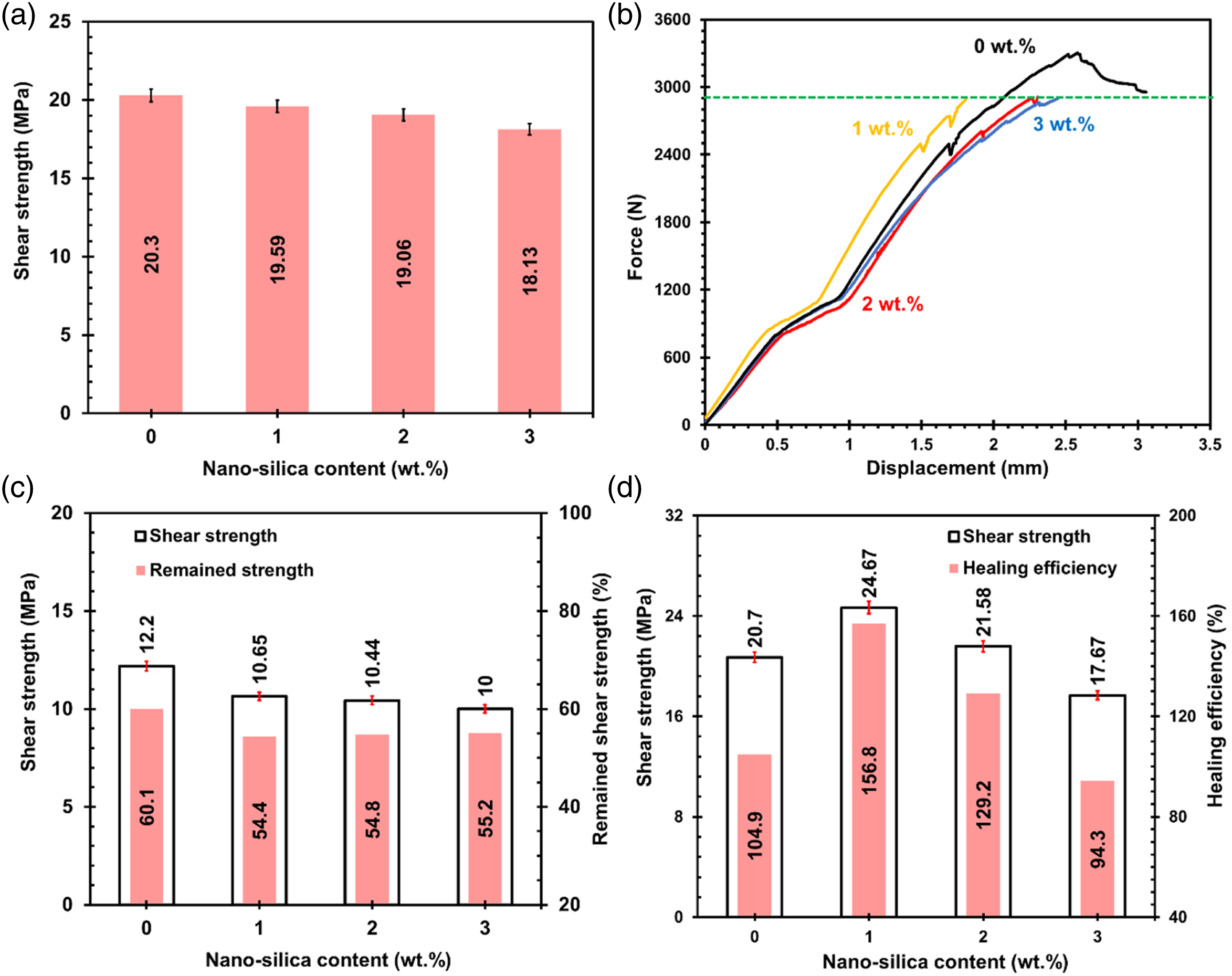
Conclusion
In this study, the silica nanoparticles with the weight fraction of 0, 1, 2 and 3 wt.% were dispersed to the ethyl acetate/epoxy healing agent and encapsulated these mixtures for adding into the glass fibers/epoxy composites to create the healing ability into those. The outcome of this research work can be summarized as follows: 1. Incorporating the silica nanoparticles into the healing agent reduced the thickness of microcapsules, which caused a slight reduction in flexural strength. Nevertheless, this improved the flexural strength of damaged composite and healing ability of that. The maximum healing efficiency and flexural strength after damaging were 121.6 and 89.9%, which belonged to the composite with the 1 and 3 wt.% silica nanoparticles. 2. The sample with the 1 wt.% silica nanoparticles had the highest healing efficiency under the tensile and shear loads, which were 87.8 and 156.8%, respectively. Forming the nanocomposite healing shell instead of neat healing shell were the reason of this improvement in the healing ability of glass fibers/epoxy composite. 3. The hackles of microcapsules, agglomeration of silica nanoparticles on the healed area, and change the crack propagation path were other characterized effective factors on the healing properties of the glass fibers/epoxy composite.
Footnotes
Declaration of conflicting interests
The author(s) declared no potential conflicts of interest with respect to the research, authorship, and/or publication of this article.
Funding
The author(s) received no financial support for the research, authorship, and/or publication of this article.