
Abstract
Polyethylene terephthalate (PET) fiber with four T-lobe cross section (4T-fiber) has been applied to textile products due to the structural characteristic of micro-grooved channels to provide the high wicking, warming, and elastic properties. In this study, nonwoven (NW) filters composed of one layer of 4T-PET NW (containing 65% 4T-fibers) with one, three, or five layers of polypropylene (PP) NW (4T-PET NW + PP NW × 1, 3, or 5) were proposed for blood filtration to remove leucocytes to reduce blood transfusion-related adverse reactions. The removal rate of leucocytes could be enhanced when the PP NW layers were increased. The presence of the 4T-PET NW on the surface of five layers of PP NW (4T-PET NW + PP NW × 5) filters resulted in a higher erythrocyte recovery rate without losing the capability to remove leucocytes and platelets. Based on the theoretical analysis, leucodepletion filters were further prepared by stacking four 4T-PET NW + PP NW × 5 units together. The optimum filtration results of the filters for filtrating 110 mL of red blood cell concentrates could be prepared when four units of 4T-PET NW + PP × 5 NW placed in a series combination.
Introduction
Clinically, the use of leucocyte-depleted blood products is to prevent febrile nonhemolytic transfusion reaction (FNHTR) [1]. Since the development of removing leucocytes by Fleming in 1926 [2], multiple methods were developed to remove leucocytes from blood products such as filtration, centrifugation, sedimentation, cell-washing, freezing, and thawing [3,4]. At present, filtration is the most widely used method to separate leucocytes from other cells [5]. Basically, blood filtration provides a fast, convenient, and inexpensive way with loss of a very small amount of erythrocytes to reduce immunological transfusion reactions [3].
Cotton wool fibers were first used as the filter material to remove leucocytes from blood [6]. Subsequently, different synthetic material fibers were developed for the leucocyte filtration technique, which includes cellulose acetate [7], polyethylene terephthalate (PET) [5], polyurethane [8], polybutylene terephthalate [9,10], polypropylene (PP) [11], polyethylene vinyl alcohol (EVAL) [12], etc. In addition, recent studies of leucocyte depletion techniques revealed that many physical and chemical methods were used for modifying material surface characteristics. For example, PET nonwovens (NWs) with different fiber diameters have been used in the current leukocyte filtration filters [3,13].
Generally, the shape of the nonwoven fibers was cylindrical. In this study, PET NWs made of four T-lobe cross section (4T-shaped) fibers without further modification were applied to the blood filtration. We hypothesized that the 4T-shaped NWs combining multilayer propylene (PP) NWs can be used to increase the efficiency of the leucodepletion with higher leucocyte removal rate, without losing erythrocyte recovery rate and extending filtration time. In this study, 110 mL of saline and red blood cell concentrates (RBCs) were used to test our hypothesis.
Materials and methods
NW materials
PET NWs with 100% circular fibers (fiber diameter: 10 µm, fabric mass: 55 g/m2, fabric thickness: 0.4 mm, Kang Na Hsiung, Taiwan), 4T-PET NWs (fabric mass: 55 g/m2, fabric thickness: 0.3 mm, Kang Na Hsiung, Taiwan) containing 65% 4T-shaped fibers (fiber diameter: 16.4 µm) and 35% circular fibers (fiber diameter: 10 µm), and PP NWs (fiber diameter: 5–20 µm, fabric mass: 20 g/m2, fabric thickness: 0.1 mm, Kang Na Hsiung, Taiwan) containing 3% IRGASURF HL 560 (Ciba, Switzerland) were used in this study. Fiber and NW structure were examined using a scanning electron microscope (SEM) (Hitachi S-4800). Different single or multilayers of NWs with the diameter of 9 cm were used to filtrate 110 mL saline or RBCs.
Water absorbency and vertical wicking tests
A piece of 0.5 g NW was immersed in saline for 15 min, and then the NW was removed from the saline and weighed after dripping for 10 min at room temperature. Water content rate was expressed as below and this test was specified in JIS L 1913: 2010.

Ten cm long NW was suspended vertically with bottom end in the saline. After 30 min, the water wicking height of the NW was measured and expressed as the height (mm)/time (min), as specified in AATCC Test Method 197.
Pore size characterization
An automated capillary flow porosity meter (iPore 1100, Porous Materials, Inc.) was used to measure pore size characteristics as specified in ASTM E1294. The NW was wetted with isopropyl alcohol (IPA) and then extruded under increased air pressure. The mean flow pore diameter, mean flow pressure, bubble point pressure, and bubble point pore diameter were measured by detecting 40 points.
Blood preparation
In this study, human blood was collected using blood bags containing citrate-phosphate-dextrose adenine (CPDA) and approved by the Institutional Review Board of the National Taiwan University Hospital (NTUH-REC No. 201008025R). After centrifugation at 4200 r/min for 5 min and removal of the plasma, the RBCs with buffy coat were pooled and mixed with saline to a hematocrit of 50–60% for the subsequent leucodepletion filtration tests.
Blood filtration analysis
Single or multilayers of NWs with the diameter of 9 cm were assembled to a filter by fixture of the blood filtration system. The difference in height between blood and filter was 3 cm to create hydraulic pressure by gravity and filtration was done at room temperature. After filtration of 110 mL of RBCs, the following equations were used to calculate the effectiveness of blood filtration through the calculations of pre- and post-filtration blood by using the automated hematology cell counting device (XE-2100, Sysmex) and the correction of the Nageotte counting chamber (Tiefe, USA) by a microscope [9,14,15].


Filtration cake analysis
After filtration, 1 cm2 of the nonwoven material containing the filtration cake was extracted and dissolved in 1.5 mL of saline. Through the calculations of blood cells in saline by using the automated hematology cell counting device (XE-2100, Sysmex), blood cell numbers were measured as shown above.
Statistical analysis
All data were expressed as the mean ± standard deviation (SD) from at least three independent experiments. Statistical significance was determined using one-way analysis of variance (ANOVA) followed by t-test and Duncan’s test. p-Values of less than 0.05 and 0.01 were regarded as significant.
Results
Characterization of 4T-PET NW
PET fibers with 4T-shaped morphology and traditional circular contour are shown in Figure 1(a) and (b). The 4T-fibers exhibited microscale grooves between four T-shape lobes. In this study, two PET NWs were made of these two fibers. One PET NW contained 100% circular fibers and the other one was composed of 65% 4T-fibers and 35% circular fibers (Figure 1(c)).
Scanning electron micrographs of PET fibers with: (a) 4T-shaped morphology, (b) circular morphology, (c) 4T-PET NW with 65% 4T-shaped fibers and 35% circular fibers. (d) Water absorption test and (e) vertical wicking test of 4T-PET NW and PET NW by using 110 mL saline. Data are presented as the mean ± SD from three or four different experiments. **p < 0.01.
Figure 1(d) and (e) shows the results of water content and vertical wicking tests of 4T-PET NW and PET NW. The water content was measured after the immersion of NWs in the saline solution for 15 min. As shown in Figure 1(d), the water content of PET NW was significantly increased from 640 ± 8% to 737.3 ± 25.8% (p < 0.01) by the addition of 4T-fibers into the NW. Similarly, the difference of the velocity of vertical wicking between 4T-PET NW (0.70 ± 0.15 mm/min) and PET NW (0.02 ± 0.005 mm/min) was also significant (p < 0.01). Therefore, the 4T-PET NW might be useful for the filtration application when it needs more and rapid water absorbency.
Pore size characteristics of NWs
The mean flow pore diameter of 4T-PET NW and PP NW were 44.7 µm and 25.3 µm, respectively. There are significant difference between them, indicating that PP NW is denser than 4T-PET NW and theses two NWs can play different roles in blood filtration.
Post-filtration volume and filtration time
In order to further understand the basic filtration performance of 4T-PET NW, multilayers of nonwoven were stacked together for filtering 110 mL RBCs. Figure 2(a) shows the post-filtration volume and filtration time of one, three, and five layers of 4T-PET NW (4T-PET NW × 1, 3, or 5). The post-filtration volume slightly decreased from 107.7 ± 0.58 mL to 102.2 ± 2.05 mL with increasing 4T-PET NW layers. Similarly, the filtration time slightly increased with increasing 4T-PET NW layers. Thus, increasing the layers of 4T-PET NW would not seriously influence the blood flow characteristics.
Post-filtration volume and filtration time of: (a) 1,3,5-layers of 4T-PET NW, (b) 1,3,5-layers of PP NW, and (c) 4T-PET NW with 1,3,5-layers of PP NW. Samples are 110 mL RBCs to filtrate. Data are presented as the mean ± SD from three or four different experiments.
PP NW has been widely used for leucocyte filtration due to the good physical and chemical properties [16,17]. The aim of this study was to investigate whether 4T-PET NW could enhance the leucodepletion efficiency of PP NW by designing a series of 4T-PET/PP NW filters for leucocyte filtration. Firstly, the post-filtration volume and filtration time of one, three, and five layers of PP NW (PP × 1, 3, or 5) were evaluated for filtering 110 mL RBCs (Figure 2(b)). Conversely, the post-filtration volume and the filtration time were significantly varied when the PP NW layers were increased. The post-filtration volume was decreased from 89.8 ± 1.82 mL to 77.5 ± 4.85 mL, i.e. a decrease of 12.3 mL, which was two times of that of 4T-PET NW. More notably, the filtration time was increased from 9.1 ± 0.34 min to 48.6 ± 6.05 min. Thus, increasing the layers of PP NW would cause serious obstruction for blood flow to reduce the post-filtration volume and extend the filtration time.
Comparison of post-filtration volume and filtration time of materials by using saline and RBCs to filtrate.

PET: polyethylene terephthalate; NW: nonwoven; PP: polypropylene; RBCs: red blood cell concentrates.
All samples are 110 mL. Values shown are mean ± SD, n = 3.
**p < 0.01; comparison of PP NW × 5 and 4T-PET NW + PP NW × 5.
Filtration cake
Because the filtration time of five layers of PP NW rapidly increased, it is reasonable to assume that the filtered blood cells formed a cake layer on the top surface of NW stacks, which contribute to the retention of more blood cells. Figure 3 shows, in comparison with the original PP NW surface with thin and long fibers, obvious cake formation almost covering the whole surface of PP NW after filtering 110 mL RBCs. In contrast, filtration cake scattered on the surface of 4T-PET + PP NW × 5 filters after filtration, which would not seriously increase the filtration resistance. This indicates that the use of 4T-PET NW on the multilayered PP NW surface could restrict the aggregation of blood components to interfere blood flow through PP NW. Table 2 shows the composition of three blood cells from these two filtration cakes. It was found that the erythrocyte amount exhibited a significant difference between these two cakes (p < 0.01), so it is reasonable to predict that a PP NW filter with 4T-PET NW could develop higher erythrocyte recovery rate.
Scanning electron micrographs of: (a) surface of PP NW before filtration, (b) surface of PP NW after filtration, (c) surface of 4T-PET NW before filtration, and (d) surface of 4T-PET NW after filtration with filtration cake (short arrows) and without cake (lone arrows). Comparison of blood cell count in filtration cake of PP NW × 5 and 4T-PET NW + PP NW × 5. PET: polyethylene terephthalate; NW: nonwoven; PP: polypropylene. Filtration cakes were took from first layer of materials. Samples are 110 mL RBCs. Values shown are mean ± SD, n = 3. *p < 0.05.

Blood filtration
Figure 4(a) shows the capability of one, three, and five layers of 4T-PET NW for the removal of leucocytes from 110 mL RBCs. No obvious effect on the removal of leucocytes was observed on increasing the layers of 4T-PET NW. In addition, the recovery rate of erythrocytes was always more than 90%, and the removal rate of platelets only increased from 18.7 ± 3.25% to 25.1 ± 9.82%. Conversely, the removal rate of leucocytes and platelets could be obviously increased to 86.8 ± 0.84% and 77.8 ± 0.94%, respectively, when the use of five layers of PP NW to filtrate 110 mL RBCs (Figure 4(b)). However, the erythrocyte recovery rate dropped to 69.5 ± 4.38% simultaneously. Based on the different properties of 4T-PET NW and PP NW, filters composed of one layer of 4T-PET NW with one, three, or five layers of PP NW (4T-PET NW + PP NW × 1, 3, or 5) were proposed in this study. As shown in Figure 4(c), when the PP NW layers were increased, the erythrocyte recovery rate was decreased from 96.4 ± 0.91% to 87.2 ± 3.31% only, and both leucocyte and platelet removal rate still could be increased from 39.6 ± 1.97% to 79.9 ± 4.17% and from 32.8 ± 5.15% to 71.3 ± 1.45%, respectively. Thus, the presence of the 4T-PET NW on the surface of multilayered PP NW filters maintained the erythrocyte recovery rate and increased the capability to remove leucocytes and platelets.
Leucocyte removal rate, erythrocyte recovery rate, and platelet removal rate of: (a) 1,3,5-layers of 4T-PET NW, (b) 1,3,5-layers of PP NW, and (c) 4T-PET NW with 1,3,5-layers of PP NW. Samples are 110 mL RBCs to filtrate. Data are presented as the mean ± SD from three or four different experiments.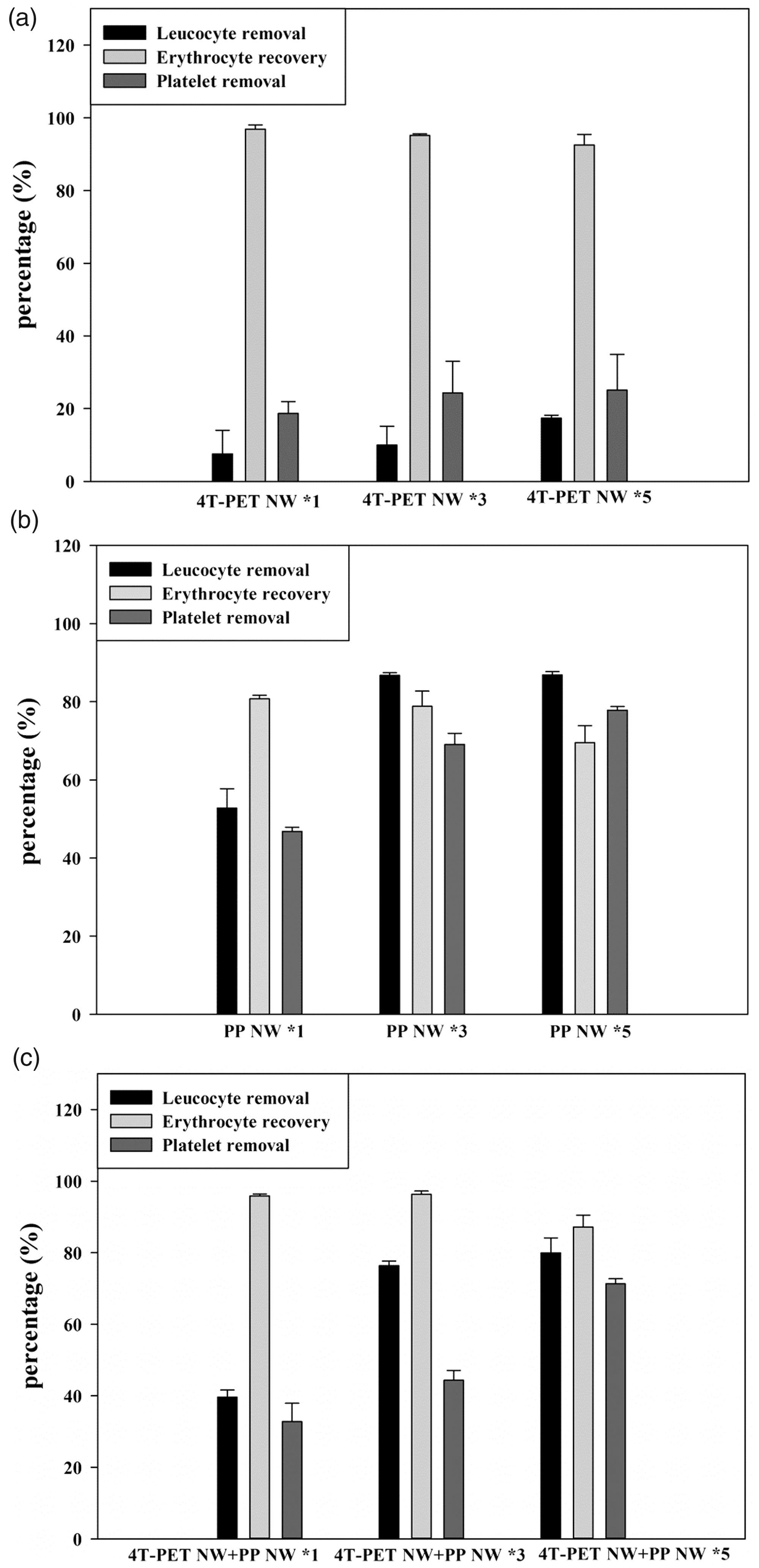
Finally, filters were prepared by stacking four 4T-PET NW + PP NW × 5 units in series (Figure 5(a)). Figure 5(b) shows that when the thickness of filters was increased from one to four units, the erythrocyte recovery rate slightly dropped from 87.2 ± 3.31% to 80.2 ± 2.86%, but both the removal rate of leucocytes and platelets could be obviously increased from 79.97 ± 4.17% to 99.98 ± 0.0058% and from 71.31 ± 1.45% to 94.39 ± 0.91%, respectively, which conformed to the transfusion requirements. Since the thickness of the filter was gradually increased to trap more blood, the post-filtration volume of blood decreased from 96.67 ± 3.88 mL to 88.53 ± 3.14 mL (Figure 5(c)). The filtration time was not noticeably delayed by the increased NW layers, i.e. it just increased by 1 min approximately, indicating 4T-PET NW did not adversely affect the blood filtration process. Table 3 summarizes the optimum filtration results of the filters for filtrating 110 mL RBCs in this study, which consisted of four units of 4T-PET NW + PP × 5 NW placed in a series combination.
Filtration results of multiunits of 4T-PET NW + PP NW × 5 in series. (a) The diagram of four 4T-PET NW + PP NW × 5 units in series; (b) leucocyte removal rate, erythrocyte recovery rate, and platelet removal rate; (c) post-filtration volume and filtration time. Samples are 110 mL RBCs to filtrate. Data are presented as the mean ± SD from three or four different experiments. (d) Comparison of leucocyte removal rate of multiunits of 4T-PET NW + PP NW × 5 in series and their theoretical results. Filtration results of four units (4T-PET NW with five-layer PP NW). Samples are 110 mL RBCs. Values shown are mean ± SD, n = 3. PET: polyethylene terephthalate; NW: nonwoven; PP: polypropylene.

Discussion
The aim of this study is to make use of the structure characteristic of 4T-PET NW to combine with traditional PP NW as an alternative to the existing leucocyte filtration technologies. The efficiency of a leucocyte filter is primarily based on its ability to remove leucocytes and platelets while retaining red blood cells as much as possible. As shown in Figure 4, it could be found that 4T-PET NW, regardless of one, three, or five layers, did not have high performances in the removal of leucocytes and platelets, but possesses high recovery rate of erythrocytes. In contrast, five layers of PP NW had obvious effect on the removal rate of leucocytes and platelets, but the erythrocyte recovery rate dropped significantly. In addition, since PP NW extended the filtration time and reduced the post-filtration volume with increasing nonwoven layers, and 4T-PET NW could maintain high post-filtration volume and low filtration time (Figure 2), a filter composed of one layer of 4T-PET NW with five layers of PP NW were proposed to enhance the performance of leucodepletion efficiency of PP NW filters. As expected, 4T-PET NW on the surface of multilayered PP NW filters enhanced the overall filtration performance with high erythrocyte recovery rate and high removal rate of leucocytes and platelets (Figure 4(c)).
In this study, 4T-PET NW was made of 35% typically circular fibers and 65% 4T-shaped fibers with microscale grooves between the T-shaped structures (Figure 1(c)). Compared to traditional PET NW with 100% circular fibers, 4T-PET NW exhibited more and rapid water absorbency (Figure 1(d) and (e)), which were reasonably attributed to the special 4T-shaped structure. Due to 4T-PET NW with large pores between fibers and small grooves along fibers simultaneously, it is also reasonable to assume that the blood cell cake formed on the dense structure of PP NW might be collapsed and separated by 4T-PET NW (Figure 3). Therefore, 4T-PET NW located on the filter surface could destroy the cake structure to reduce filtration resistance, which is especially useful for the filtration application with high concentration filtrate, such as blood filtration.
Finally, we would like to further increase the removal rate of leucocytes to 99.9% without losing the erythrocyte recovery rate below 80%. Filters were prepared by stacking some (4T-PET NW + PP NW × 5) units in series to repeatedly remove leucocytes to increase the filter efficiency. Since the removal rate of leucocytes of one unit of (4T-PET NW + PP NW × 5) was about 80% (Figure 5(b)), the theoretical value of two units could be calculated by 80% + (1–80%) × 80% =96%. Similarly, the theoretical values of three and four units are 99.2% (96% +(1–96%) × 80% = 99.2%) and 99.84% (99.2% + (1–99.2%) × 80% = 99.84%, very close to 99.9%), respectively. Interestingly, Figure 5(d) shows the experimental leucocyte removal rate of some units stacked in series agreed well with the theoretical calculation results based on the performance of single (4T-PET NW + PP NW × 5) unit. Therefore, the removal rate of leucocytes was successfully increased from 79.97 ± 4.17% to 99.98 ± 0.0058% with increasing from one to four units. In addition, the four-unit filter still maintained the appropriate erythrocyte recovery rate, post-filtration volume, and the filtration time (Table 3). More importantly, we are able to predict how many (4T-PET NW + PP NW × 5) units are needed if more rigorous standard is requested.
Conclusion
A filter composed of the structure characteristic of 4T-PET NW and the performance of the multilayered PP NW in the removal of leucocytes provided an alternative to the existing leucocyte filtration technologies. Due to the special 4T-shaped fibers with microscale grooves between the T-shaped structures, 4T-PET NW on the multilayered PP NW surface could restrict the aggregation of blood components to interfere blood flow through PP NW. The optimum filtration results of the filters for filtrating 110 mL of RBCs could be prepared when four units of 4T-PET NW + PP × 5 NW placed in a series combination.
Footnotes
Acknowledgement
The authors thank the staff of the Third and Eighth Core Lab, Department of Medical Research, National Taiwan University Hospital for technical support during the study.
Declaration of conflicting interests
The author(s) declared no potential conflicts of interest with respect to the research, authorship, and/or publication of this article.
Funding
The author(s) disclosed receipt of the following financial support for the research, authorship, and/or publication of this article: This study was funded by the Ministry of Science and Technology of the Republic of China.