
Abstract
The aim of the present work was to develop and evaluate a controlled drug delivery wound dressing based on polymeric porous microspheres, which are also termed as microsponge drug delivery system. For this purpose, turmeric (drug)-based polymeric porous microspheres formulations were developed by quasi-emulsion solvent diffusion method. The production yields, actual drug content, drug entrapment efficiency, particle size, formulation of the material, and in vitro release were studied. On the basis of maximum drug released, a formulation was selected to incorporate into wound dressings with the help of a binder by spraying technique. Finally, the selected formulation and samples of wound dressings containing microsponges were subjected to scanning electron microscopy and drug release analyses. The result of in vitro release shows that microsponge drug delivery system is a versatile tool that has a potential to convert any wound dressing into a controlled drug delivery wound dressing.
Introduction
A wound dressing can use textile structure (woven, knitted, nonwoven, braided, composites) to cover and protect the wound from infection and microorganism [1]. A wound dressing plays an effective role to provide moist wound environment, protect the wound from the environmental microorganisms, and facilitate the wound healing process by introducing a drug [2]. Wound dressings can be classified into three groups based on their function (debridement, antibacterial, occlusive, absorbent, and adherence), material type (e.g., hydrocolloid, alginate, and collagen), and physical form (ointment, film, foam, gel). Broadly, they are classified as traditional dressings, moist wound management/modern dressings, medicated or drug delivery dressings, and controlled drug delivery (CDD) dressings [1,3]. A CDD dressing delivers the drug to wound sites in a consistent and sustained manner over a long period of time without involving any frequent change. Mostly it could be applied to a wound site in the form of polymers films or polymeric porous structures such as scaffolds [3].
A controlled release drug delivery system is a system that delivers the drug systematically at a predetermined rate for a specified period of time to its site of action [4]. There are many CDD systems used in pharmaceutics, medical, and health care sector that are incorporated into textile structure [5] such as cyclodextrins [6–8], aza-crown ethers, fullerenes [9,10], fibers (electro spun biodegradable solid and core spun hollow fibers) [11–20], and particulate (micro and nano) system [21–23]. A polymeric porous microsphere (PPM) is a new and versatile controlled delivery tool that consists of porous microspheres [24–26]. This system provides outstanding properties such as controlled drug release, site-specific action, and high drug load capacity along with thermal-, chemical-, and physical stability. They are also nontoxic, nonmutagenic, nonallergic, nonirritating, cost-effective in addition to the ease with which they can be developed [26–28]. Its outstanding properties make it an effective tool to entrap a number of active agents for topical treatment (sunscreen, antiacne, anti-inflammatory, antifungal, antidandruff, antipruritics, skin depigmenting, and rubefacients agents) [29–40], oral administration (fluribiprofen, paracitmol, etc) [41–45], vascular wall reconstruction, cardiovascular engineering, biopharmaceuticals, bone substitution, and diagnoses of diseases [46–49]. The microsponges release their active ingredients on a time mode and also in response to other stimuli such as rubbing, temperature, and pH etc [50–52].
The aim of the present work was to develop a CDD dressing using polymeric porous particulate system or microsponge drug delivery system (MDS). For this purpose, the turmeric-based microsponge formulations were developed by quasi-emulsion solvent diffusion (ESD) method and incorporated into cotton (CN) (traditional) and calcium alginate (AN) moist management dressing by spraying technique. In microsponge formulations, turmeric was chosen as a drug that contains a variety of chemically rich compounds (curcumonoids). It also has many biological, pharmacological, and wound healing properties [53–59]. To the best of our knowledge, PPM or MDS-based CDD dressing is being developed and evaluated for the first time.
Materials and methods
Materials
Eudragit RS 100 (copolymer of ethyl acrylate, methyl methacrylate, and trimethylammonioethyl methacrylate chloride) and turmeric were supplied by LAB ERA Scientific Store, Multan, Pakistan. Polyvinyl alcohol (PVA) 30,000–70,000, sodium hydroxide (NaOH), sodium chloride (NaCl), calcium chloride (CaCl2), and dialysis tubing (40 kD) were from Sigma Aldrich. Ethanol, methanol, and monobasic potassium phosphate (KH2PO4) were from BDH laboratory, Riedel-deHean, and E. Merck, respectively. All chemicals used in this study were analytical grade. The nonwoven calcium alginate wound dressing (Kaltostat) from Convatec and traditional gauze dressing having the specifications plain weave, 15 tex warp, and weft linear density, 19 ends per inch, 15 picks per inch, and 20 gsm weight were purchased from the local medical store. Printofix LTC binder (low temperature curable [LTC]) was supplied by Archroma, Pakistan.
Method for preparation of microsponges
Production parameters of quasi-emulsion solvent diffusion method.
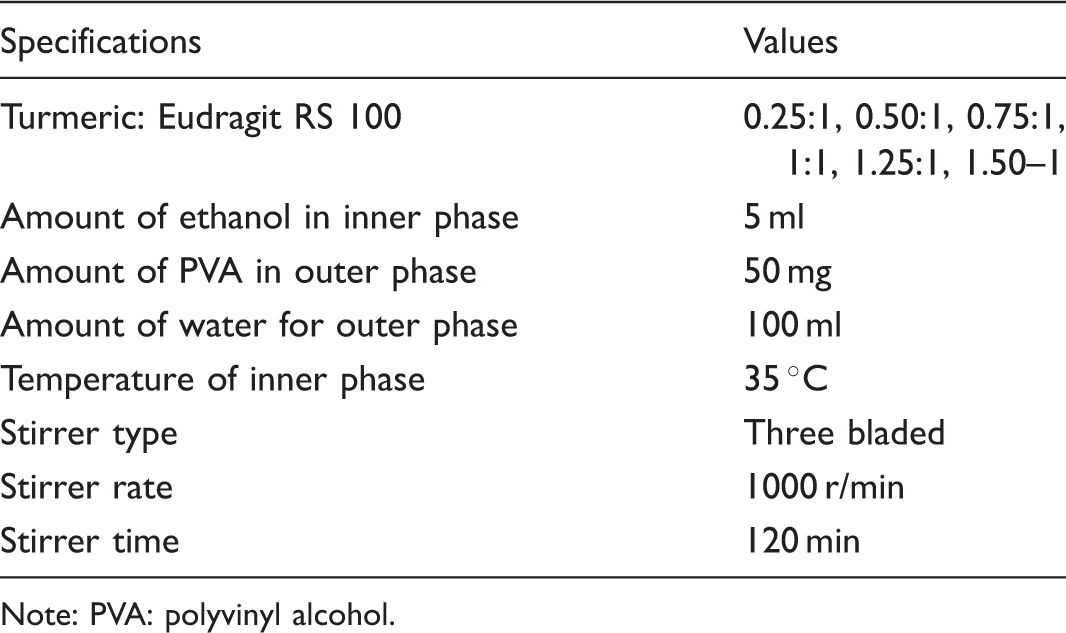
Note: PVA: polyvinyl alcohol.
Characterization of microsponges
Determination of production yield
The production yield that is the ratio of weight of microsponges obtained to the initial weight of the raw materials (polymer + drug) expressed in percentage is calculated by using the following equation (1).

Determination of actual drug content
The weighed samples of drug loaded microsponges (30 mg) were crushed and dissolved in methanol (10 ml). Each sample was then diluted into 10 times. All the samples were filtered and their absorbance values were measured at 424 nm using UV visible spectrometer (Perkin Elmer Model Lambda 25, USA). The actual drug content of these formulations was calculated from the standard calibration curve as shown in the Figure 1.
Standard calibration curve for turmeric in methanol.
Determination of entrapment efficiency
Encapsulation efficiency was calculated from actual drug content by using equations (2) and (3).


Particle size analysis
The particle size of the microsponges was measured by using Zeta sizer (ZEN 3600, Malvern Instruments, Worcestershire, UK).
Fourier Transform Infrared (FTIR) analysis
The FTIR Spectra of turmeric, Eudragit RS 100, and turmeric loaded microsponge formulation (FM-1 to FM-6) were measured in potassium bromide disks using a Perkin-Elmer Model 1600 FTIR spectrometer (USA).
In vitro drug release analysis from microsponges
The microsponges of 50 mg were put into 5 ml potassium phosphate buffer solution (PBS) having a pH 7.4, which is usually the condition for a chronic wound. The PBS containing drug-loaded microsponges was introduced in dialyzer tube (40 kD molecular weight cut off [MWCO] membrane). The dialyzer tube was then placed into a 45-ml cylinder also containing potassium phosphate buffer of 7.4 pH. Finally, cylinder containing release media (PBS) was stirred at a speed of 300 r/min by using a small magnetic stirrer bar under constant temperature (37℃).
The drug release in the outer solution as a result of diffusion from membrane was assessed by sampling the solution (PBS) at specific intervals of time. The drug content release from membrane as the results of diffusion was measured at 424 nm wave length by using UV visible spectrometer from the standard calibration curve as shown in the Figure 2.
Standard calibration curve for turmeric in PBS.
In order to understand the release kinetics of drug, results of the in vitro drug release study were fitted with various kinetic models like zero order (cumulative percent drug released vs. time), first order (log cumulative percent drug retained vs. time), Higuchi (cumulative percent released vs. √time), and Hixson-Crowell’s cube root model ((percentage retained)1/3 vs. time). The kinetic model that best fits the dissolution data was evaluated by comparing the regression coefficient values obtained in various models.
Incorporation of microspheres into textile dressings
The selection of a formulation to incorporate into wound dressings is based on highest cumulative drug release rate. The selected formulation was incorporated into calcium sodium alginate (AN) and cotton (CN) based wound dressings by spraying technique. The aqueous solution composed of a plurality of microsponges and an LTC binder was sprayed on samples. Finally, these samples were dried at room temperature and curing was carried out at 60℃ for 20 min in order to achieve the fixation of the microspheres.
Characterization of selected microsponge formulation and wound dressing containing microsponges
Surface morphology
The surface morphology of the selected microsponge formulation and wound dressings containing microsponges were studied by scanning electron microscopy (SEM) using a Quanta 250 FEI (USA).
In-vitro drug release analysis
Drug release from these dressings treated with microsponges was measured by placing a piece of 2.5 × 2.5 cm into 20 ml of 7.4 pH PBS solution under constant stirring at 35℃. The 2 ml sample from buffer solution was taken after specific intervals of time and replaced with the fresh buffer solution. The drug releases from dressing were measured at 424 nm wave length using UV visible spectrometer by using standard calibration curve as shown in the Figure 2.
Results and discussion
Characterization of microsponges
Optimization of drug: polymer for preparation of turmeric-loaded microsponges.

The particle size of turmeric-loaded microsponges was decreased by increasing the drug amount as compared to polymer. This decrease in particle size depends on the viscosity of organic phase. The viscosity of organic phase decreased due to decrease in polymer amount from FM-1 to FM-6. This decrease in viscosity results in small-sized emulsion droplets and consequently small-sized microsponges achieved [32,60,61].
The FTIR spectra of turmeric, Eudragit RS 100 and turmeric-loaded microsponge formulations (FM-1–6) are given in Figure 3. In FTIR spectra of turmeric, a hydroxyl stretching peak at 3303 cm−1, a CH3 band at 2950 cm−1, a carbonyl stretching band at 1630 cm−1, a C=C at 1540 cm−1, were seen as reported in literature [61,62]. Eudragit RS 100 also showed a CH3 band at 2950 cm−1, an ester C=O stretching peak around 1734 cm−1 and a C–O peak at 1150 cm−1as reported [44].
FTIR spectra of (a) turmeric (b) Eudragit RS 100, (c) FM-1, (d) FM-2, (e) FM-3, (f) FM-4, (g) FM-5, and (h) FM-6.
The turmeric-loaded microsponge formulations (FM-1–6) spectra shows all prominent peaks of polymer and drug at 3320 (phenolic OH), 2950 (alkane CH3 stretching), 1720 (C=O ketone), 1540 (C=C), 1150 cm−1 (C–O). It shows that drug was simply entrapped in the microspheres without any chemical reaction with the polymer.
In vitro drug release analysis
In vitro drug release study of microsponges was performed in phosphate buffer solution having a pH of 7.4. Figure 4 represents the release behavior of six microsponge formulations. The results show that the drug release from FM-1 is high within 24-h time period as compared to remaining formulations. It means that low drug to polymer ratio formulation, which has low drug content has released the drug quickly as compared to those formulations that have high drug content.
In vitro drug release of turmeric from microsponge formulations.
Evaluation of release kinetics of turmeric-loaded microsponge formulations.

On the basis of highest cumulative drug release, FM-1 was selected to incorporate into calcium AN and CN-based wound dressings.
Characterization of selected microsponge formulation and wound dressing containing microsponges
Surface morphology study
The surface morphology of turmeric-loaded microsponge formulations (FM-1) prepared by ESD method and wound dressings containing microsponges were investigated by scanning electron microscope as shown in Figure 5(a) to (c). It was observed by SEM analysis that FM-1 formulation has uniform porous spheres [37,44,61] and has successfully incorporated into wound dressing.
SEM images of (a) FM-1, (b) AN dressing, and (c) CN dressing.
In vitro drug release analysis from wound dressings containing microsponges
The in vitro release studies were performed in phosphate buffer solution having a pH 7.4. The cumulative drug releases from two samples are shown in Figure 6. The results show that the drug release from both samples was 85% up to 8-h time period. Drug release data from both samples fitted into four mathematical models named as zero order, first order, Higuchi model, and Hixson Crowell models. The regression coefficient of calcium alginate dressing is 0.99 and CN dressing is 0.98 showing that the drug releases from these samples follow zero order.
In vitro drug release of turmeric from microsponge treated dressings.
Conclusion
In this study, turmeric-based micro porous spheres (microsponges) were developed, characterized, and incorporated into wound dressings. The micro porous particulates were developed by ESD method. The effect of drug to polymer ratio on yield, drug content, entrapment efficiency, particles size, and drug release was studied. It was found that drug content and entrapment efficiency were increased by increasing the drug amount as compared to polymer. Then, FM-1 formulation was incorporated into calcium alginate and cotton-based wound dressings by spraying technique. The surface morphology of the FM-1 formulation and wound dressing containing microsponges and drug release from these wound dressing were also studied. The results show that the drug release from both samples was more than 85% within 8 h. The study shows that microsponge drug delivery system (MDS) is a versatile tool. It has a potential to convert a traditional or a modern wound dressing into a CDD wound dressing. This system also has greater stability (physical, chemical, and thermal), ease in production, and much fever in cost as compared to cyclodextrins-based drug delivery system.
Footnotes
Declaration of Conflicting Interests
The author(s) declared no potential conflicts of interest with respect to the research, authorship, and/or publication of this article.
Funding
The author(s) received no financial support for the research, authorship, and/or publication of this article.