
Abstract
Silk fibroin (SF), a naturally occurring protein polymer, has several unique properties such as biocompatibility, biodegradability, minimal inflammatory reaction, and endowed with excellent mechanical properties and process ability. The existing scaffolds used in medical industry lack the degradation and/or slow in healing practice, so clinically there is a need for the development of efficient and reliable biomaterial scaffolds for wound healing. Silk fibroin has shown greater potential for tissue engineering applications. This work is focused on designing biomaterial with silk as raw material and wound healing effect of SF scaffolds were also tested. The silk blended scaffolds were prepared by using SF as a vehicle with dextrin, other healing agent of cactus and epidermal growth factor (rhEGF) are used as the drug releasing model. Scanning electronic microscope (SEM) was used to observe the morphology of prepared scaffolds for process versatility and the highly specific surface area. The structure was studied by Fourier transform infrared. The SF was treated at different concentrations of cactus and rhEGF, to investigate the growth inhibition effect of bacterial growth. The SF scaffolds show favorable stability by their structural integrity, morphology, mechanical properties, and powerful antibacterial efficacy up to 100% to Escherichia coli and Staphylococcus epidermidis significantly intended to provide improved environments for the zone of incubation when compared with normal scaffolds without rhEGF. Therefore, the results give the evidence for the application of SF blended scaffolds in the treatment of wound.
Introduction
Skin, one of the sensitive organ in the body, mainly serves as a protective barrier against the environment [1]. Most of the skin wounds can heal naturally, but chronic wounds are difficult to heal [2,3]. The healing reconstruction of damaged tissue needs to produce effective biomaterial that can be utilized as safe scaffolds/matrices for a variety of applications and also for the growth of tissue cells [4–6]. The consumer demand for safe eco-friendly medical textile products has been growing day by day. At present mostly synthetic-based polymers are used to dress the wounds, which are non-biodegradable in nature. Biodegradable polymers have the advantage of delivering biological molecules at controlled rates and it reduces the number of repeated treatments [7,8].
Cotton, silk and wool are natural fiber groups, which are commonly used as biodegradable and biocompatible materials for wound dressing across countries [9–12]. From many years the silk fiber is used as silk blend for many applications, ranging from textiles to biomedical application. Medical textile is one such category of biomedical field. Silk is used in the form of sutures and artificial ligaments in medical applications due to its favorable properties like natural slow biodegradation, superior mechanical properties, biocompatibility and processability [13–15]. Many researchers found and even used silk as ideal platform for drug delivery in different forms like films, hydrogels, fibers and 3D scaffolds in biomedical field [16–18].
Natural product-based medicines are used for the treatment of different diseases [19] and are found to have less toxic effects [20]. Researchers [21–23] found cactus and cactus products to be antiinflammatory, immunomodulatory and anti-oxidative. Cactus has been extensively used in Chinese traditional medicine for treatments of lung disorders, skin diseases and blood circulation diseases. In this work, we also aim to investigate the wound-healing effect of cactus. Hence, cactus also added as one of the ingredient in the process of silk scaffold preparation.
Dextrin is also widely used in a variety of material applications, such as adhesives, food industry and even in textiles [24–26]. This dextrin combination scaffolds has been found to have excellent biocompatibility and degradability [27,28]. Biodegradability and reabsorption are highly desirable features for tissue engineering and for other biomedical applications to avoid further surgery.
The dextrin–rhEGF (recombinant human epidermal growth factor) blends were found to have high potential to treat chronic wounds [29–31]. Hence, current study uses rhEGF along with the combination of dextrin and cactus in the preparation of silk fibroin drug-loading scaffolds.
Materials and methods
Materials
Bombyx mori silk cocoon was obtained in the raw form, from the farmers of Erode district. The rhEGF as REGEN-D™ was purchased from Bharat Biotech International Limited, Hyderabad, India. Cactus pear fruits (Opuntia lindheimeri) were obtained from a plantation located in Namakkal district. Ethanol (absolute GR for analysis) was obtained from Merck (Darmstadt, Germany). Deionized water was used for all experiments.
Preparation of silk fibroin solution
Raw silk fibers were degummed thrice with 0.5% (W/W) NaHCO3 solution at boiling temperature for 30 min and then washed with distilled water. Degummed silk (SF) was dissolved in a ternary solvent system of CaCl2/CH3CH2OH/H2O (1:2:8 in molar ratio) at 80℃ for 6 h. After dialysis with Himedia tubular dialysis membrane-50 in distilled water for 3 days, pure silk fibroin solution was filtered.
Cactus extract preparation
The fruits (6 kg) were manually peeled after washing and pulped with a 2 mm screen, obtaining 5.5 kg of pulp. The pulp was then placed in stainless steel plate and dried in hot air oven at 90℃ and cactus powder that was extracted was used in preparation of scaffolds.
Preparation of silk scaffold
Preparation data of silk fibroin/dextrin/cactus scaffold.
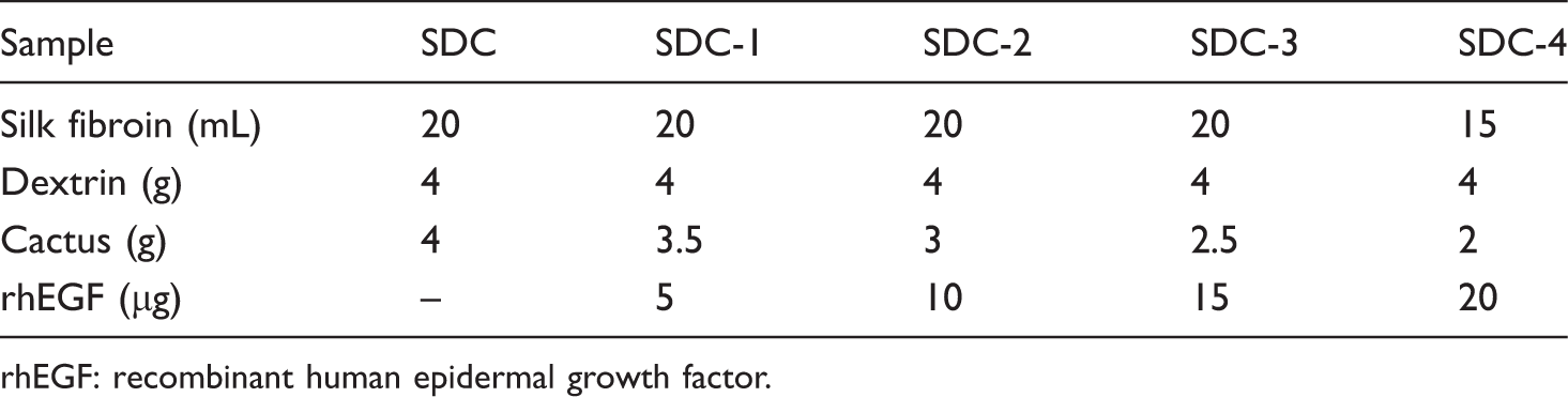
rhEGF: recombinant human epidermal growth factor.
Bacteriological study
The antibacterial properties of prepared scaffold were tested by using disc diffusion method. The discs with the prepared scaffolds were applied to the surface of an agar plate containing bacterial species to be tested. The line of incubation of antimicrobial agent was shown by the presence of growth inhibition zones measured by using Muller-Hinton (HiMedia) as described by Fingold and Baron [32]. The zones of inhibition appear as clear areas surrounding the disc from which the substances with antimicrobial activity diffused. All antibacterial tests were performed by using a Gram-negative (Escherichia coli) and a Gram-positive bacteria (Staphylococcus epidermidis). Amoxycillin antibiotic was chosen as the positive control. After the 24 h incubation, INT (tetrazolium dye) solution was sprayed over the surface of an agar plate to ensure visible indication of inhibition. INT reacts with the metabolites produced by the microorganisms and the surface with the living microorganisms turning pink in color.
Mechanical properties
The compressive mechanical properties of scaffolds were tested by using Universal Testing machine, Housfield-H25KS equipped with a 0.1 kN load cell at ambient room temperature using modification of ASTM method F451-95. The samples were dipped in phosphate buffered saline (PBS) for 2 h and were examined with crosshead speed of 1 mm/min. At least five specimens were tested for each sample, and the averages and standard deviations were reported.
Water uptake and weight-loss-related tests
The water uptake and degradation behavior of the silk fibroin scaffolds were assessed after immersion in an isotonic saline solution (ISS; 0.154 M sodium chloride aqueous solution, pH 7.4) for time periods ranging from 1 day to 30 days. All experiments were conducted at 37℃ and dynamic condition in controlled condition. After each time period the specimens were removed from the ISS and the weights were determined immediately after adsorption of the excess of surface water using a filter paper. The water uptake was calculated using the following expression:

The water stability of regenerated silk fibroin protein based scaffolds was determined by calculating the weight loss in water. Weight loss was tested by immersing different scaffolds (approx. 10 g) in PBS at 37℃ for 7 days at room temperature. After incubation, samples were dried at 65℃ and the remaining mass was measured. Mass loss was expressed as percentage of the original mass of each sample. Weight loss was calculated using the formula:

Morphology
Morphologies of the surface and the cross-section of the lyophilized scaffold samples were studied by scanning electron microscope (SEM, JEOL, JSEM-6390LV, Japan) with an accelerating voltage of 20 kV after gold coating. SEM observations were performed using the followings magnifications: 100×, 200×, 500× and 2000×.
Fourier-transform infrared spectroscopy
The Fourier-transform infrared spectroscopy (FTIR) spectra of silk fibroin/cactus blend ratios were recorded with Spectra 100 FTIR spectrophotometer (Perkin Elmer India Pvt. Ltd.). The IR spectra in the absorbance mode were obtained in the spectral regions of 400–4000 cm−1. Each spectrum of the sample was acquired by accumulation of 32 scans with a resolution of 4 cm−1.
Results
Preparation of silk scaffolds
The silk scaffolds were prepared using cactus and dextrin with rhEGF at various contents (Table 1), which were uniform and tuft materials. The developed scaffold sample exhibited brown color. All the samples were easily removed from the glass plate (model of silk scaffold as an example shown in Figure 1).
Silk scaffold model.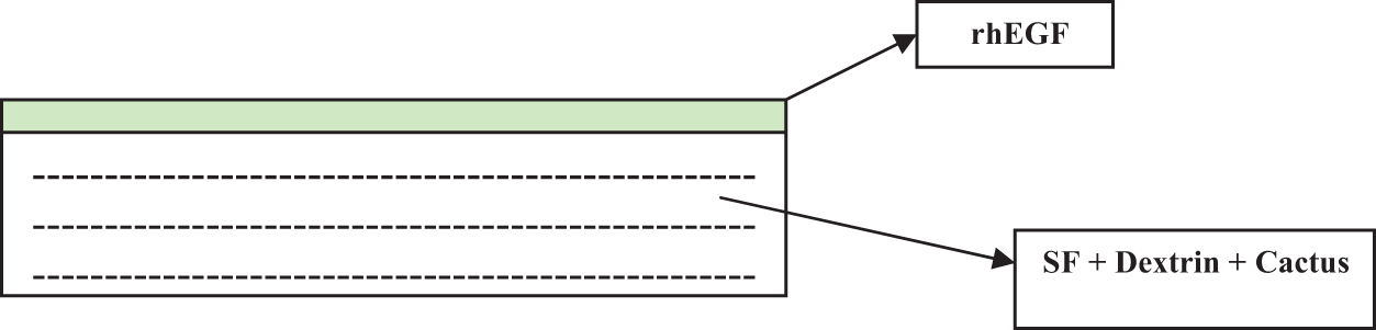
Water uptake and degradation-related properties
The ability to uptake fluids from the surrounding medium plays an important role in tissue engineering. Figure 2(a) shows that water uptake ratios of the scaffolds decreased with decreasing cactus concentration. The water uptake ratio of all the scaffolds reached equilibrium after only 7 h of immersion in aqueous solutions, and can be maintained for up to 15 days. This result shows that the scaffolds possess a good hydration capability and are able to maintain their structural integrity. The differences in water uptake can be attributed to different porosities of the scaffold. It was observed that for the scaffolds with higher porosity, the water uptake ratio increased. Even after 7 days of immersion in PBS, the samples remained stable and swollen. All the scaffolds after soaking in aqueous solutions for 7 days continue to degrade slowly, which is one of the advantages of any biodegradable product (Figure 2(b)).
Water uptake and degradation.
Mechanical strength determination
The compressive strength and modulus of SF dextrin-based scaffolds are shown in Figure 3(a) and (b), respectively. The compressive strength and modulus gradually decreased when the concentration of rhEGF was increased and cactus amount was reduced. In this experiment scaffolds were compressed to 80%. The compressive strength of SDC and SDC-1 was maximum followed by SDC-2, SDC-3, SDC-4 blended scaffolds and SF. SDC and SDC-1 showed significantly higher modulus as compared to pure silk fibroin. These results indicate that there may be formation of compact structure due to ionic interaction, which probably leads to the increase in modulus and strength of blended scaffolds.
Compressive strength and modulus of silk scaffolds. Values are average ± standard derivation of five samples (n = 5).
Morphologies and porosities of the composite scaffolds
In this study, the morphology of the prepared silk fibroin scaffolds was investigated using SEM. This observation was conducted to examine the surface and internal microarchitectures of porous SF–dextrin/cactus scaffolds. The microstructure and architecture of the scaffolds are crucial parameters for tissue engineering applications since they can affect the final outcome of the tissue regeneration. Silk blended scaffold was observed by the SEM observations and it was clearly seen that the scaffold was highly porous with interconnected pores that could be suitable for tissue engineering applications. Figure 4 shows SEM micrographs of the composite scaffolds with cactus and dextrin. The pure SF scaffold exhibited a macroporous structure with interconnected open pores, and pore sizes varied from 100 to 200 µm. In the 3D scaffolds, the minimum pore size must exceed 100 µm, otherwise, the tissue can only penetrate the surface of the scaffolds and cannot easily diffuse sufficient nutrients to the inner tissue. After compounding with cactus and dextrin, the macroporous structure of the scaffold was still maintained, and based on SEM result it was suggested that there was no change in the pore structure incurred by adding both (cactus and dextrin) into the system.
SEM images of silk scaffold prepared with combination of cactus and dextrin only: (a) ×100, (b) ×500, (c) ×200, (d) ×2000.
FTIR analysis
In order to confirm the structure change, prepared scaffolds with different composition were investigated by FTIR spectra. The FTIR spectra of SDC, SDC-1, SDC-2 and SDC-3 scaffolds without rhEGF are shown in Figure 5. Silk fibroin had characteristics of vibration bands in their FTIR spectra, including 1630–1650 cm−1 for amide I (C=O stretching), 1540–1520 cm−1 for amide II (secondary N\H bending) and 1270– 1230 cm−1 for amide III (C–N and N–H functionalities). Further, the bands’ positions also indicated the conformations of the protein material, which is described as follows: 1650 cm−1, 1520 cm−1 as well as 1230 cm−1 representing random coil, and 1630 cm−1, 1520 cm−1 representing ß-sheet structure, for amide I, II and III separately. In Figure 5 the characteristic vibration bands of silk particles were obviously shifted to other positions where indicated ß-sheet structure, such as absorption peaks of 1650, 1540 and 1230 cm−1 moved to 1648, 1509 and 1277 cm−1, correspondingly. The spectra of dextrin presents bands at 3416 (O–H), 2927 (C–H), 1155 (C–O), 1016 (C–C) cm−1. The characteristic vibration bands of dextrin were obviously moved to 3420, 2935, 1162 and 1020 cm−1. Transform infrared (FTIR) analysis showed no change in silk structural composition between cactus and dextrin.
FTIR spectra of different combination scaffolds.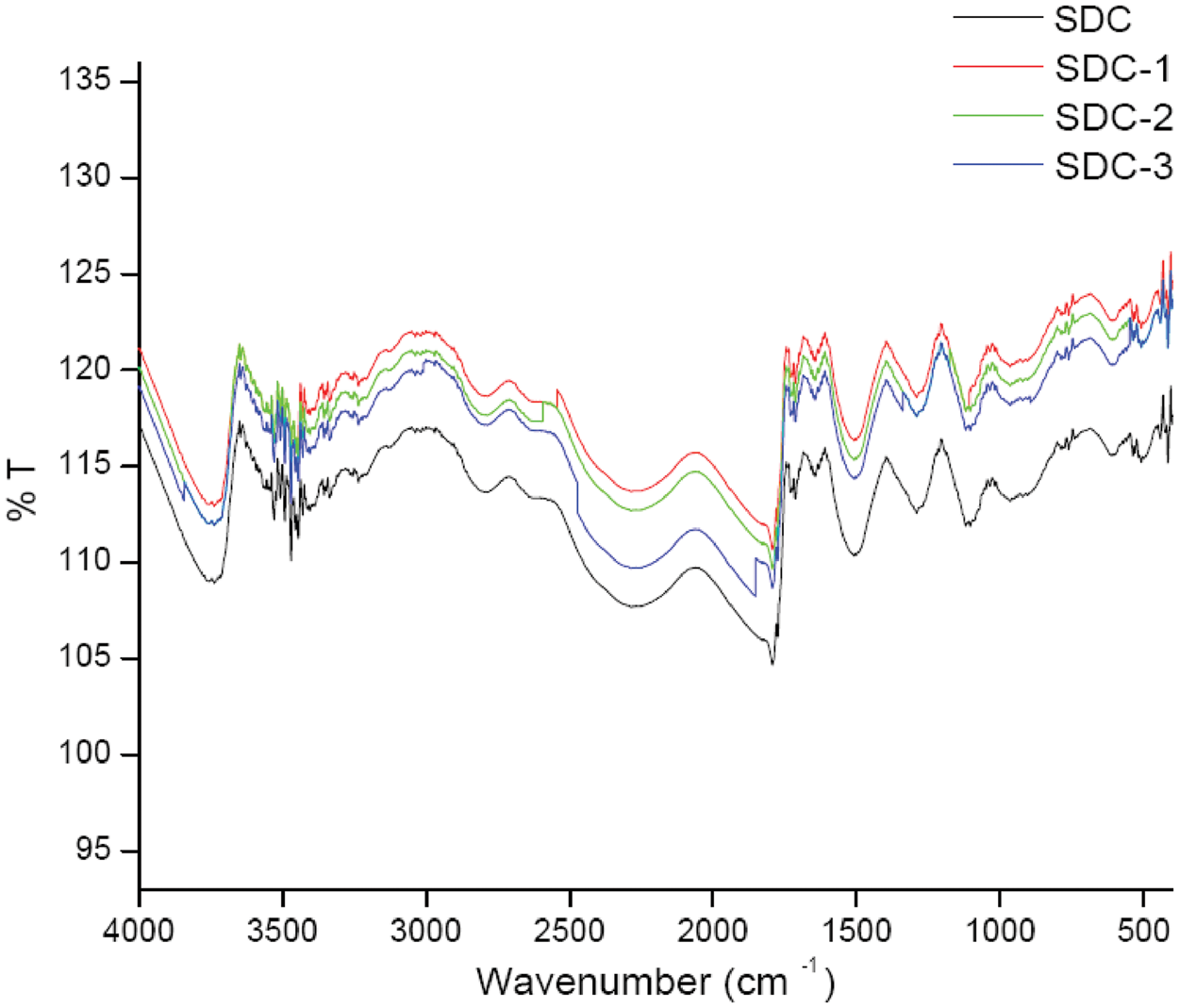
Antibacterial ability
In this study E. coli and S. aureus were used as the test bacteria to examine the antibacterial properties of prepared silk protein based dextrin and cactus scaffolds. The results of antibacterial activity of this study are shown in Figure 6. The inhibition zone is higher in E. coli than S. aureus, which indicates higher susceptibility of Gram-negative bacteria. The zone of inhibition is higher in sample SDC-4 about 27 mm and 30 mm after 24 h of inhibition against S. aureus and E. coli. It is observed that as the concentration of rhEGF increases, the zone of inhibition also increases. This shows that the antibacterial activity is due to the presence of dextrin and rhEGF in silk scaffolds. These results also indicate that the developed scaffold and developed silk blend scaffolds may be suitable to be used as wound barrier with antibacterial properties.
Antibacterial ability of silk composite scaffolds against E. coli and S. aureus.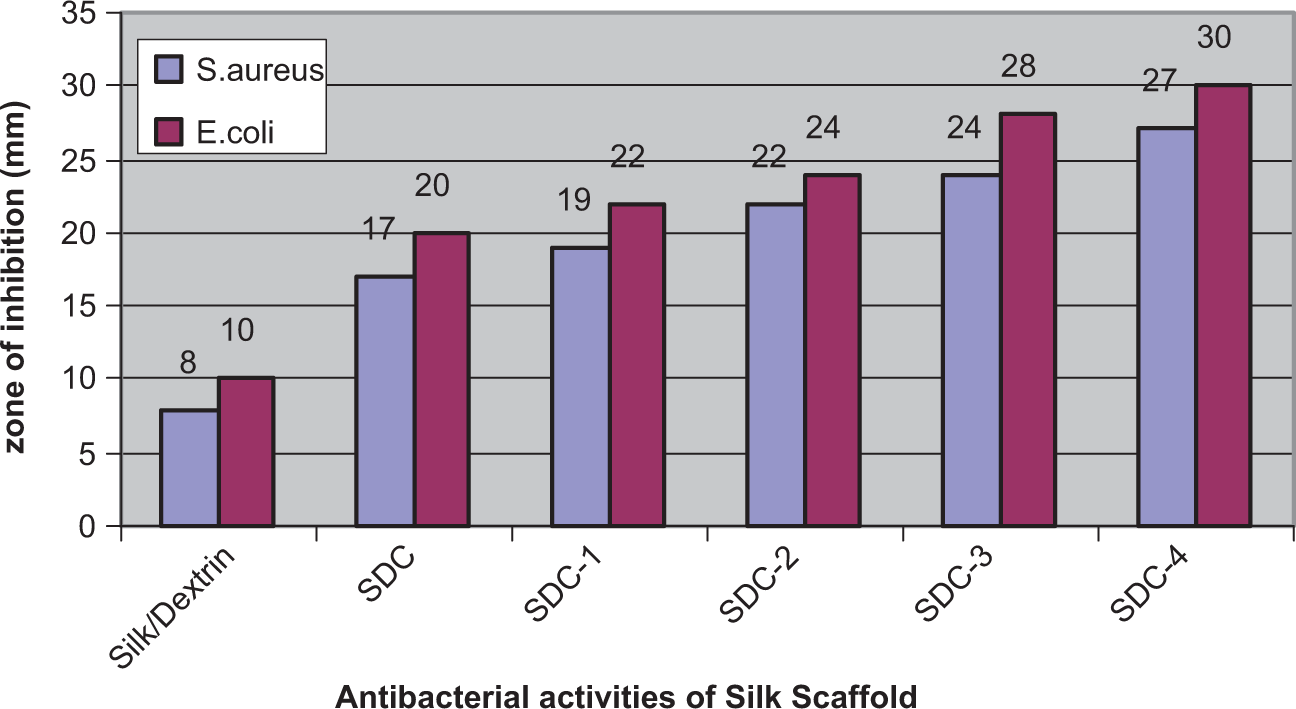
Conclusions
In this work, silk fibroin composite scaffolds were synthesized using dextrin, cactus and rhEGF and characterized using SEM and FTIR. The analysis showed that structural and physical characteristics of silk fibroin were not affected while blending with cactus and dextrin.
The mechanical prosperity is important for any material, which is subjected to stress during use. The developed scaffold also tested to prove the mechanical compatibility while blending with cactus and dextrin. The compressive strength and compressive modulus of silk fibroin has been increased while blending with cactus and dextrin. Therefore the developed scaffold has good mechanical property to be used as a biomaterial.
The swelling property of the developed silk scaffold was tested to measure the degree of absorbing capacity. Since the wound normally produces large amount of fluid (exudates), it has to be absorbed for quick healing and avoid infection. The results show that the developed scaffold swelled quickly in PBS within a day in solution (shown in Figure 2(a)). The capacity of water uptake declined after 4th day. In fact, swelling and degradation of all the samples concurred in water and attained balance at some period of time.
The developed scaffold was found to have higher antibacterial efficacy against both E. coli and S. aureus, which was evidenced by the antibacterial test results. The result indicated that, this scaffold may act as a preventing barrier for wound infection. Hence the developed silk scaffold may be used as a supporting material for wound dressing to avoid infections and further spreading. It may also avoid delayed wound healing and promote the growth of epithelial cells of wound.
The developed scaffolds of this study can be further extended in vitro (with cell lines, primary cells) and in vivo studies are envisioned in order to fully evaluate the biological performance. Cactus is highly used in the context of diabetic control [33] and its extract is capable of rapidly returning blood glucose to nondiabetic levels [34]. Hence, this developed scaffold can also experimented in diabetic wound dressing applications. This study can be extended in the context of various types of wounds and also the mechanisms behind the antibacterial effect may be addressed for broader perspective.
Footnotes
Funding
This research received no specific grant from any funding agency in the public, commercial, or not-for-profit sectors.