
Abstract
Objective:
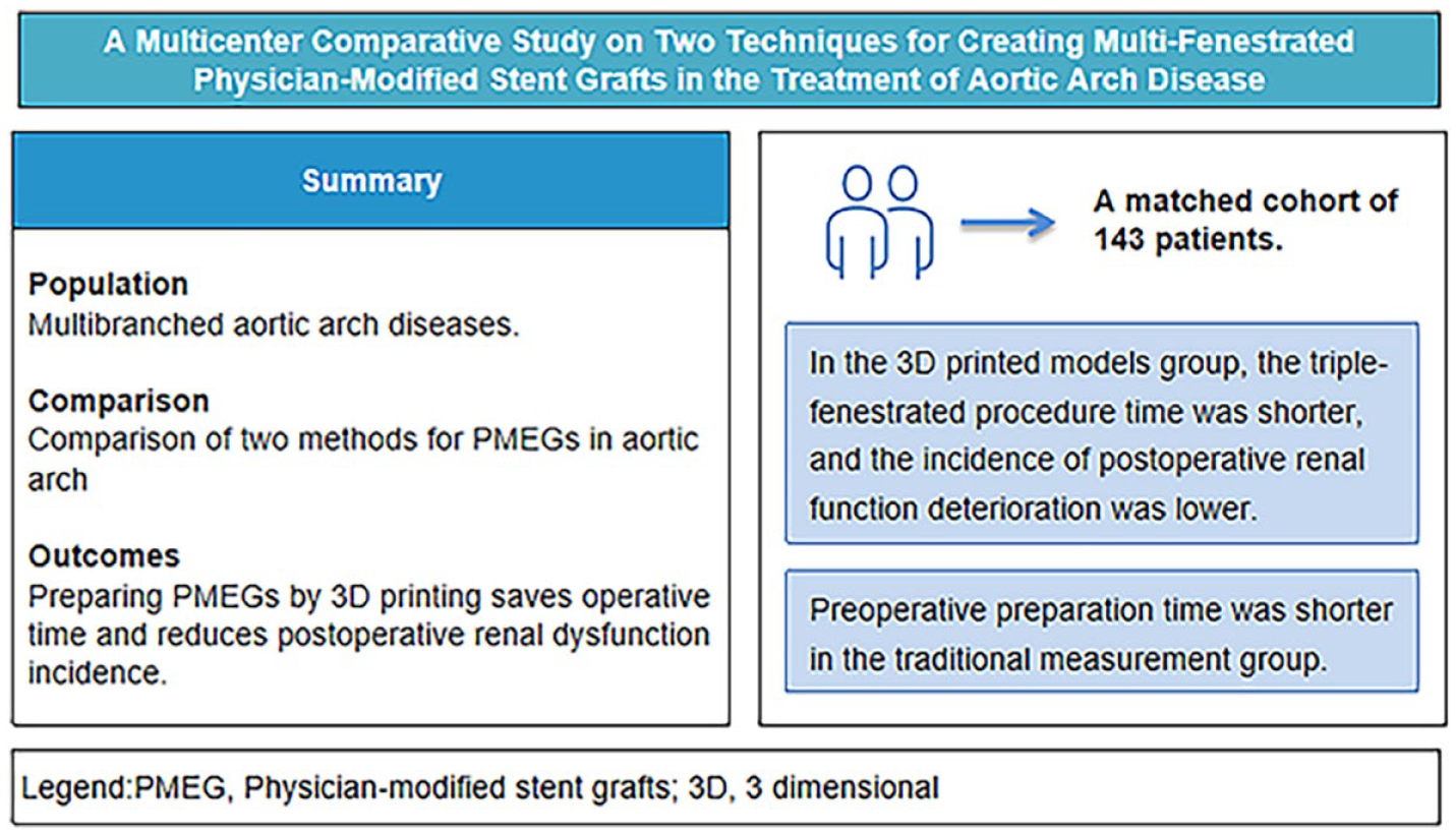
Physician-modified stent grafts (PMEGs) are widely used to treat complex pathologies in the aortic arch, but featured by varied preparation methods. This study was conducted to investigate the differences in operative characteristics and postoperative outcomes of PMEGs prepared using 2 methods: 3D printed models (Group A) and traditional measurement (Group B).
Methods:
This retrospective study included 201 patients treated with multiple fenestrated/branched endovascular repair in the aortic arch at 3 vascular centers in China. This study was designed to establish 2 groups based on different preparation methods, including Group A (3D printed models) and Group B (traditional measurement). Both groups matched (1:3) using propensity scoring for subsequent analysis based on age, history of aortic surgery, type of aortic lesion, number of stent fenestrations, and brand of aortic stent. This study further clarified differences in demographic characteristics, procedure characteristics, 30-day outcomes, and 1-year outcomes between 2 groups.
Results:
A total of 143 patients were enrolled and matched from February 2018 to February 2024, with 91 in Group A and 52 in Group B. Group A had significantly shorter duration of intervention, less contrast use, and lower rate of postoperative renal dysfunction than those in Group B. In addition, no significant differences were observed in the rest of the operative characteristics and postoperative outcomes.
Conclusion:
The use of 3D-printed models may contribute to reducing endovascular intervention time and contrast use, and decreasing the incidence of postoperative renal dysfunction. However, traditional measurement remains a necessary skill for surgeons in case of emergency procedures with short preparation time.
Clinical Impact
This study demonstrates that 3D-printed model-assisted PMEGs preparation significantly reduces endovascular operating time and contrast volume compared to traditional measurement, particularly in triple-fenestrated aortic arch repairs. For clinicians, it provides a validated approach to minimize renal impairment risk and enhance procedural efficiency in elective cases. The innovation lies in leveraging patient-specific 3D anatomy to optimize fenestration positioning. While traditional methods remain essential for emergencies, this technique standardizes complex endovascular reconstruction, potentially reducing reintervention rates.
Get full access to this article
View all access options for this article.