
Abstract
Introduction:
This study aimed to evaluate the protective effects of curcumin on angiotensin-converting enzyme (ACE) gene expression, oxidative stress and anti-oxidant status in thioacetamide (TAA)-induced hepatotoxicity in rats.
Materials and methods:
Total 32 albino Wistar rats (male, 200–250 g) were divided into six groups (n=8). Group 1: untreated controls; Group 2: received TAA (200 mg/kg body weight (b.w.); i.p.) for 12 weeks; Group 3: received curcumin (75 mg/kg b.w.) for 24 weeks; Group 4: received TAA (200 mg/kg b.w.; i.p.) for 12 weeks+curcumin (75 mg/kg b.w.) for 12 weeks.
Results:
A significantly higher ACE gene expression was observed in TAA-induced groups as compared with control, indicating more synthesis of ACE proteins. Treatment with curcumin suppressed ACE expression in TAA liver and reversed the toxicity produced. TAA treatment results in higher lipid peroxidation and lower GSH, SOD and CAT than the normal, and this produces oxidative stress in the liver. Cirrhotic conditions were confirmed by serum enzymes (ALT, AST and ALP) as well as histopathological observations.
Conclusion:
Curcumin treatment reduced oxidative stress in animals by scavenging reactive oxygen species, protecting the anti-oxidant enzymes from being denatured and reducing the oxidative stress marker lipid peroxidation. Curcumin treatment restores hepatocytes, damaged by TAA, and protects liver tissue approaching cirrhosis.
Introduction
Thioacetamide (TAA) is a model hepatotoxic agent having deadly effect on hepatic cells, thus leading to centrilobular liver cirrhosis. 1 It is one of numerous agents that provoke structural and functional changes not only in liver, but also in other tissues such as kidney, thymus, spleen and lungs. 2 It is reasonably anticipated to be a human carcinogen. TAA produces selective renal tubular necrosis, restricted to that portion of the proximal convoluted tubule located in the inner half of the renal cortex. In the development of TAA-induced toxicity, free radicals are thought to play a critical role. 3 Therapeutic herbs and their principles of action have become a focus of attention due to their anti-oxidant properties. 4 Curcumin is a polyphenolic compound, a yellow pigment extracted from turmeric, from the powdered rhizome of Curcuma longa. It is a commonly used food-stabilizing agent and spice in the countries of Far East. 5 It is renowned for its medical properties and as a cure for several types of diseases. It has been extensively studied for its anti-oxidant, anti-inflammatory, anti-carcinogenic and wound-healing effects.6,7 It scavenges and prevents the formation of reactive oxygen species (ROS). 8 Curcumin can repress tumor initiation, promotion and metastasis. 9
Angiotensin-converting enzyme (ACE), encoded by the ACE gene, is a prime catalytic protease in the renin–angiotensin–aldosterone system (RAAS) that converts angiotensin (Ang) I to II. 10 Raised levels of renal ACE have been comprehensively studied in connection with the development of cardiovascular disease in both humans and rodents. 11 Susceptibility to end-organ damage caused by hypertension has been associated with elevated renal ACE. In the case of TAA-induced liver cirrhosis, other parameters such as oxidative stress have been postulated to be major molecular mechanisms basic to tissue alteration. 12
Based on the multifunctional properties of TAA and the anti-oxidant properties of curcumin, this study describes the hepatoprotective effect of curcumin on TAA-induced liver cirrhosis, by estimating serum enzymes (alanine aminotransferase (ALT), aspartate aminotransferase (AST) and alkaline phosphatase (ALP)), oxidative stress (malonyldialdehyde (MDA)), and anti-oxidant status (superoxide dismutase (SOD), glutathione peroxidase (GSH) and catalase (CAT)) to identify ACE gene expression in TAA-induced hepatotoxicity and its possible treatment with curcumin.
Materials and methods
Animals and diet
For this study, 32 male albino rats (Wistar strain) with body weights ranging from 200–250 g were obtained from the animal facility of ICCBS, University of Karachi. These animals were housed in a polypropylene cage and the temperature of the animal room was maintained at 30°C and at 50% humidity with an alternative 12 hours light/dark cycle. The animals were provided with standard food and water.
Study design
The experimental period of the study was 24 weeks. Rats were divided into four groups (n=8) and were treated as follows:
Sample collection
At the end of the treatment, the rats were decapitated to collect the blood from the head wound. A portion of blood was taken in a separate tube to collect serum. Liver tissues were excised and trimmed, followed by rinsing with deionized water to eliminate blood contamination. Filter paper was used to dry the tissues, which were weighed and kept in -70°C freezer until analyzed.
Histological examination
After quick removal, the liver tissues were cut into small pieces, followed by immersion in neutral buffered formalin (10%) for 24 hours. Standard histological techniques were used to fix and section the liver tissues. The morphological changes in the fixed sections of the liver tissues stained using hematoxylin and eosin (H and E) were used to evaluate the extent of TAA-induced necrosis. 13
Preparation of post-mitochondrial supernatant
A tissue homogenizer (Ultraturrax T-25 Polytron) was used to obtain liver homogenates at 4°C. To prepare the liver homogenates (1:10 w/v), 5 mM KCl buffer (pH 7.8) containing EDTA 0.3 mM was used followed by centrifugation at 800 g for 5 min at 4°C to separate the nuclear debris. The supernatant was then centrifuged at 10,500 g for 20 min at 4°C to obtain post-mitochondrial supernatants used for analyses of SOD, CAT, MDA and GSH. 14
Assessment of serum enzymes and bilirubin
The estimations ALP, AST, ALT, total and direct bilirubin in serum was done using reagent kits from Randox Laboratories Ltd., UK.
Assessment of oxidative stress
As a measure of lipid peroxidation, MDA content was analyzed by means of thiobarbituric acid reacting substance (TBARS) using tissue homogenate. 15
Assessment of anti-oxidant status
Previously established methods were used to measure the level of SOD, 16 GSH 17 and CAT activity 18 in the cell-free supernatant.
ACE gene expression
Expression of the ACE gene in liver tissue was assessed using the quantitative RT-PCR method. RNA was isolated using entire RNA Prep Plus. Amplification reaction was performed in 12.5 µl total volume, containing a pair of specific primers: 5’CAGCTTCATCATCCAGTTCC3’ and 5’CTAGGAAGAGCAGCACCCAC. PCR consisted of 30 cycles at an annealing temperature of 52–64°C. Restriction fragments were subsequently analyzed in 2% agarose gel stained with ethidium bromide. 19
Statistical analysis
All the data values are given in Mean±SEM. Statistical software package SPSS for Windows® version 17.0 was used for statistical analyses. One-way ANOVA was performed followed by Student’s t-test to evaluate variations in data; p<0.05 was considered statistically significant.
Results
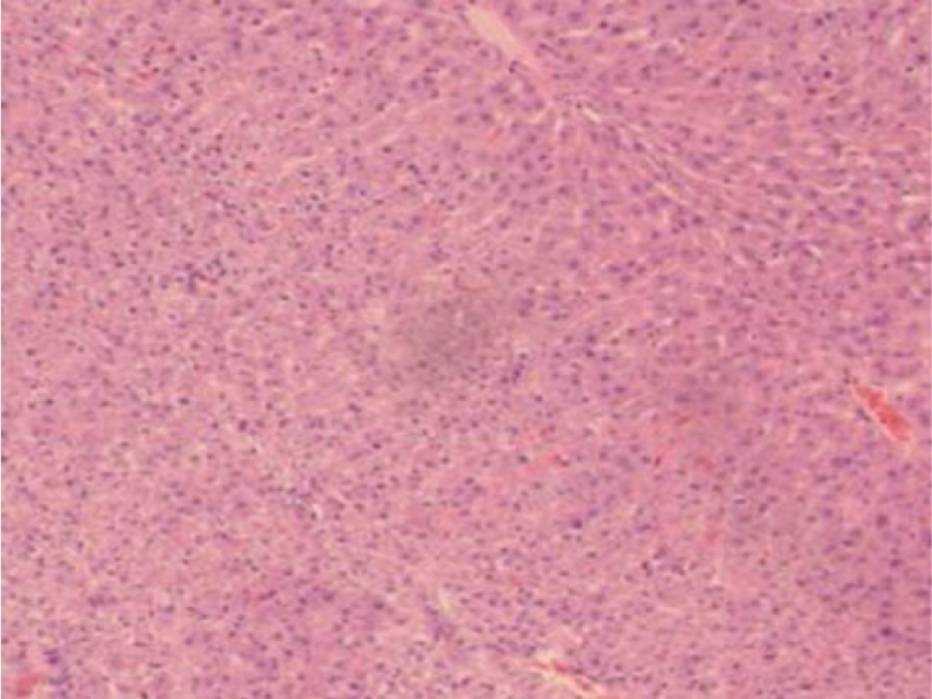
The hepatocytes, portal triads and vasculature were found to be within normal limits during histopathological assessments. There was no pathological modification such as inflammation, necrosis and/or fibrosis in the liver of curcumin-treated animals (Figure 1). A massive fatty change along with centrilobular necrotic shift was observed in the liver of TAA-treated rats. Apoptosis (single hepatic cell necrosis) was also very clearly observed, as well as mononuclear cells infiltration, mostly macrophages and lymphocytes, around central veins and in portal areas in TAA-treated animals (green arrow). The liver section showed extensive damage, characterized by severe necrosis, fatty degeneration, sinusoidal dilatation, and the presence of collagen bundles surrounding the lobules leading to thick fibrotic septae that disrupt the cellular architecture (Figure 2). A complete regeneration of hepatocytes and hepatic tissues appeared in most of the cases in curcumin + TAA-treated animals (Figure 3).
Histological observation of liver tissue from the curcumin-treated group.

Histological observation of liver tissue from the TAA-treated group.

Histological observation of liver tissue from the curcumin+TAA-treated group.
The levels of AST, ALT and ALP were estimated in serum to detect any possible deviations from normal in liver functions (Table 1). The liver-specific enzymes were found to be markedly affected in the group treated with TAA. Significantly higher (p<0.05) levels of serum AST, ALT and ALP in TAA-treated rats suggested elevations in the release of the enzymes due to serious damage in the liver. However, a significantly lower (p<0.05) level was observed for these enzymes in the study group treated with TAA+curcumin compared with the TAA-treated group.
Serum liver enzymes in the study groups.
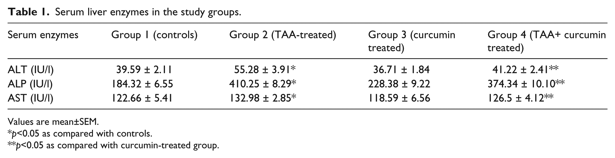
Values are mean±SEM.
p<0.05 as compared with controls.
p<0.05 as compared with curcumin-treated group.
Activity of the anti-oxidant enzyme SOD activity was significantly lower (p<0.05) in TAA-treated cirrhotic rats, whereas no significant difference was observed in curcumin-treated cirrhotic rats as compared with controls. CAT activity was found to be significantly lower (p<0.05) in TAA-treated cirrhotic rats. No significant change was observed in the level of CAT in curcumin-treated and TAA-treated cirrhotic rats as compared with controls (Table 2).
Anti-oxidant and lipid peroxidation status in study groups.

Values are mean±SEM.
p<0.05 as compared with controls.
p<0.05 as compared with curcumin-treated group.
Tissue MDA as a measure of tissue lipid peroxidation was found to be significantly higher (p<0.05) in TAA-treated cirrhotic and curcumin-treated cirrhotic rats as compared with controls (Table 2). ACE gene expression in the study groups is shown in Figure 4, which was significantly higher (p<0.05) after 8 weeks of TAA treatment in cirrhotic rats as compared with the control group. This gene expression was found to be reversed in the curcumin-treated cirrhotic rats (Figure 4).

ACE gene expression in study groups.
Discussion
In order to understand the underlying mechanisms involved in the development of cirrhosis, the present study inspected the role of oxidative stress in the mediation of hepatic injury. The TAA-induced cirrhosis resulted in oxidative stress in the liver, as observed by a high level of lipid peroxidation, accompanied by distorted status of antioxidants. In end-stage liver cirrhosis, organ failure has been observed due to severe renal vasoconstriction, which frequently develops in patients with cirrhosis.20,21
Even though the exact molecular mechanisms by which TAA induces toxic effects in different organs is not understood, it is well known that TAA interferes with ribosomal activity, thereby interfering with protein synthesis and stimulating DNA synthesis. 22
The present study aimed to determine the expression of the ACE gene in rats suffering from TAA toxicity and their treatment with curcumin, and compare their levels with control groups by using the quantitative RT-PCR method. ACE gene expression was significantly raised in the TAA group as compared with the control group, indicating that TAA induced changes in the ACE gene, producing more ACE protein than would the normal gene. Furthermore, high activity of the RAAS causes hepatic scarring and decreases the number of functioning hepatocytes. 23 This increase in ACE gene expression was overcome in the experimental animals who received curcumin after TAA; hence, they seemed to be protected against abnormally increased levels of ACE in the liver caused by TAA.24,25
The boost in lipid peroxidation and generation of ROS may diminish cell viability. TAA-induced lipid peroxidation is reported to disturb the reliability of the membrane, leading to inhibition of membrane-bound enzymes. Higher levels of lipid peroxidation have also been observed in rat liver exposed to TAA. A significant decline in lipid peroxidation caused by administration of antioxidants was found after the treatment with curcumin; therefore, it can be said that the curcumin restrained the production of ROS, which has been shown to decrease lipid peroxidation possibly by its anti-oxidant mechanism. 26
SOD is a metalloprotein widely distributed in all cells and plays a significant role in protecting against oxidative damage induced by ROS. Reduced SOD levels may possibly be due to the generation of more superoxide anions. Higher levels of production of H2O2 lowered the activity of CAT. 27 The activity of CAT was found to be significantly lower (p<0.005) in TAA-treated rats as compared with the control group, and after treatment with curcumin CAT activity was found to be significantly reducing the effects of TAA in liver.
The non-enzymatic anti-oxidant GSH is thought to be a key player in the process of detoxification, which actively participates in reactions which lead to the destruction of H2O2, free radicals, and certain foreign compounds.28,29 The higher consumption of GSH to scavenge the toxic intermediates formed by TAA led to significantly lower levels of GSH in liver tissue of the TAA group. On the other hand, a significant rise in GSH level was observed following the administration of curcumin to TAA-induced liver.
Significantly higher ACE gene expression was observed in the TAA-induced group when compared with controls, indicating that more ACE proteins are being formed. It seems that treatment with curcumin increases ACE gene expression in liver as compared with the TAA-induced tissue, approaching normal levels.
Conclusion
It may be concluded from the findings of this study that curcumin administration may produce a defensive effect against TAA-induced liver injury and oxidative stress in rats, which otherwise leads to liver cirrhosis. Further long-term follow-up studies may be of great benefit to understand and to establish the hepatoprotective nature of curcumin administration in the animals under observation.
Footnotes
Conflict of interest
None declared.
Funding
Research grant from Dean, Faculty of Science, University of Karachi is gratefully acknowledged for this study.