
Abstract
Background and aims:
Adenosine is a widely used potent cardioprotective drug, but the effect of an adenosine bolus in initial cardioplegia on cardioprotection in aortic valve replacement (AVR) patients has not been demonstrated. The aim of this double-blind randomized clinical trial was to compare intra-aortic adenosine bolus with saline on the postoperative myocardial function in patients undergoing AVR.
Methods:
Aortic valve stenosis patients scheduled for elective or urgent AVR surgery were randomized to receive either a 20 mg (4 mL) single dose of adenosine or a saline into the ascending aorta during the first cardioplegia infusion. The primary outcome was cardiac index (CI (L/min/m2) at four timepoints (before incision, after weaning from cardiopulmonary bypass (CPB), at 7 p.m. on the operation day, and at 6 a.m. the next morning). Secondary outcomes included left ventricular stroke work index, right ventricular stroke work index, and myocardial biomarkers at the same timepoints.
Results:
Between November 2015 and March 2018, 45 patients were recruited, 23 in the adenosine group and 22 in the placebo group. The last follow-up date was 17 March 2018. There were no statistically significant differences in CI (mean differences with 95% confidence interval (95% CI): 0.09 L/min/m2 at baseline (−0.20 to 0.38), −1.39 L/min/m2 (−3.47 to 0.70) at post-CPB, −0.39 L/min/m2 (−0.78 to 0.004) at 7 p.m., and −0.32 L/min/m2 (−0.68 to 0.05) at 6 a.m., (p = 0.066)), right ventricular stroke work index, (p = 0.24), or cardiac biomarkers between the groups. Left ventricular stroke work index was lower in the adenosine group (−3.66 gm/m2 (−11.13 to 3.81) at baseline, −17.42 gm/m2 (−37.81 to 2.98) at post-CPB, −3.36 gm/m2 (−11.10 to 4.38) at 7 p.m., and −3.77 gm/m2 (−10.19 to 2.66) at 6 a.m. (p = 0.021)).
Conclusions:
There were no differences between 20 mg adenosine bolus and saline in the first cardioplegia infusion in CI improvement in AVR surgery for aortic valve stenosis.
EudraCT number: 2014-001382-26.
Keywords
Context and Relevance
Endogenous adenosine regulates many physiological functions in human organ systems. Adenosine’s cardioprotective effects create interest to investigate its use in myocardial protection during cardiac surgery. Exogenous adenosine has been shown to improve certain outcomes in cardiac surgery, but only in specific patient groups and research settings. Hypertrophic left ventricular myocardium is a challenge for myocardial protection during aortic valve replacement (AVR) surgery for aortic valve stenosis. To our knowledge, no previous study has assessed the effect of adenosine-induced fast cardioplegic arrest in providing better postoperative myocardial recovery compared with conventional hyperkalemic depolarizing cardioplegia in AVR. In this randomized clinical trial, we found that a 20 mg adenosine bolus in the first cardioplegia did not provide better myocardial function recovery postoperatively when compared to placebo. Adenosine bolus in the first cardioplegia did not add to cardioprotection in AVR surgery.
Introduction
Hypoxia, inflammation, and cellular stress stimulate the generation of endogenous adenosine, a nucleoside that provides protection against tissue damage. Adenosine’s cardioprotective effects include coronary artery vasodilatation, reduced myocardial oxygen demand, and antiplatelet activation. 1 Contractile cells, pericytes, located on the abluminal surface of capillary endothelium likely regulate regional capillary flow by decreasing or increasing capillary diameter. Cardiac pericytes relax in response to nitric oxide and adenosine, causing capillary diameter to increase, leading to elevation in red blood cell density, hematocrit, and transit time thus facilitating oxygen delivery to myocardium. 2 O’Farrell et al. managed to increase coronary capillary diameter by 21% and perfusion volume by 57% in ischemic rat hearts by intravenous adenosine infusion in areas where pericytes were present in the myocardium. 3 Adenosine infusion has been shown to reduce infarct size in anterior myocardial infarction patients when given together with reperfusion therapy. 4 Adenosine has also been used as a component in new polarizing cardioplegia solutions, such as in adenosine-lidocaine-magnesium polarizing cardioplegia. Lower postoperative high-sensitivity troponin I levels have been measured with adenosine-lidocaine-magnesium polarizing cardioplegia use in comparison with hyperkalemic depolarizing cardioplegia in retrospectively analyzed propensity score matched pairs. 5 The wide-spread traditional hyperkalemic depolarizing cardioplegia has still remained as a routine in myocardial protection.
Theoretically, faster cessation of myocardial work with the help of adenosine bolus could provide better delivery of initial cardioplegia into the myocardium in diastolic arrest. Intra-aortic adenosine bolus has been shown to shorten the time to asystole when administered at cardioplegic induction in coronary artery bypass grafting (CABG) surgery and in rheumatic valve surgery.6 –12 When adenosine has been used to induce asystole with the first cardioplegia, better postoperative cardiac performance with higher cardiac index (CI), higher cardiac output, higher stroke volume and stroke index, better right ventricular stroke work index (RVSWI), lower filling pressure (pulmonary artery wedge pressure), lower mean pulmonary artery pressure, and reduced need for inotropes are reported after surgery.6 –9,12 Also, lower levels of troponin I and creatine kinase myocardial band isoenzyme (CK-MB) were found after surgery when adenosine was used in cardioplegic induction.7 –9 Adenosine demonstrates cardioprotective potential in cardiac surgery, but it has not previously been investigated in AVR surgery for aortic valve stenosis as a single high-dose bolus injection. Left ventricular myocardium provides a challenge for prevention of ischemic injury in surgery for aortic valve stenosis because of the often-associated muscular hypertrophy, stiffness, and high intracavitary pressure.
In the present randomized placebo-controlled trial, we wanted to assess whether a 20 mg adenosine bolus into the ascending aorta at the beginning of the first cardioplegia infusion could provide better postoperative CI during AVR surgery when compared to placebo.
Methods
Hypothesis
Based on our previous findings,13,14 we hypothesized that a fast induction of asystole could be achieved with a 20 mg intra-aortic adenosine bolus in the beginning of the first cardioplegia infusion. This could minimize the duration of myocardial activity after the aortic cross-clamping and possibly lead to better cardioplegia delivery in a relaxed and arrested myocardium. We presumed that this faster cardiac arrest added with the vasodilative properties of adenosine could improve myocardial protection and postoperative performance.
Trial design
We carried out a randomized, double-blind, placebo-controlled superiority clinical feasibility pilot trial in Tampere University Heart Hospital, Tampere, Finland, a tertiary referral center. The patients were enrolled between November 2015 and March 2018. This study was approved by the Tampere University Hospital Ethics Committee and the Finnish Medicines Agency (registration number ETLR14065M). Before initiation and patient enrollment, the study was registered in the European Union Drug Regulating Authorities Clinical Trials Database (EudraCT number 2014-001382-26). All patients provided written informed consent and conducted according to the Declaration of Helsinki.
Patients
The inclusion criterion was elective or urgent AVR for severe aortic valve stenosis. Only minor additional procedures for secondary indications were allowed at the discretion of the operating surgeon. Elective operations were scheduled to start at approximately 8:45 a.m., and the patients were given the first available elective study operation date. The exclusion criteria were previous cardiac surgery, preoperative ejection fraction less than 40%, severe renal insufficiency, severe aortic regurgitation, and critical medical condition.
Primary, secondary, and post hoc outcomes
The primary outcome was cardiac ventricular performance measured with CI. CI was measured in four timepoints using a Swan-Ganz thermodilution catheter. The secondary predefined outcomes were hemodynamic indices of CI describing work of individual ventricles of the heart, that is, left ventricular stroke work index (LVSWI), RVSWI, and the plasma concentrations of cardiac biomarkers’ creatine kinase (CK), CK-MB, and cardiac troponin T (TnT). The plasma concentrations of cardiac biomarkers were collected at all four timepoints. Clinical outcomes, time to asystole and other intraoperative ECG data, additional hemodynamic measurements, and adverse events were compared as post hoc outcomes.
Randomization
The research group surgeons enrolled the patients after which they were randomized in the adenosine or placebo arm in 1:1 allocation 1 day before surgery. Randomization was unstratified, and an unblocked randomization scheme based on computer-generated digits for 45 patients was used. The research administrator, who was not involved in the patient selection or the operation, produced sealed non-transparent envelopes containing the study group information. The study group information was only viewed by a surgical nurse, who accordingly prepared the study medicine bolus in an unlabeled syringe and delivered it to the scrub nurse. The surgical and anesthesia staff were blinded to the study group information.
Interventions
A standard midline sternotomy and cardiopulmonary bypass (CPB) were used. The anesthesia and CPB protocols and the characteristics and delivery of blood cardioplegia are described in detail in Supplement materials 1 and 2. In the adenosine group, the operating surgeon injected 4 mL (20 mg) of adenosine (Adenosin Life Medical 5 mg/mL, Life Medical Sweden AB, Stockholm, Sweden) into the ascending aorta after cross-clamping the ascending aorta at the beginning of the first cardioplegia infusion. The dose was based on our previous studies.13,14 In the control group, 4 mL of saline (Natriumchlorid Braun 9 mg/mL, B. Braun Melsungen AG, Melsungen, Germany) was injected at the same time point. Blood cardioplegia infusion started immediately after aortic cross-clamping to fill the aortic root, and the study medicine bolus was injected into the ascending aorta cardioplegia cannula 5 s after the start of the first cardioplegia infusion. Blood samples were collected from the radial artery before the surgical incision, 15 min after the de-clamping of the aorta, at 7 p.m. on the day of the operation and at 6 a.m. on the first postoperative morning in the intensive care unit to measure the plasma concentrations of CK, CK-MB, and TnT. A Swan-Ganz thermodilution catheter inserted into the pulmonary artery was used for hemodynamic measurements before incision, immediately after weaning from CPB in a stable phase, at 7 p.m. and at 6 a.m. on the first postoperative morning in the intensive care unit.
Datex Ohmeda S5 Collect software (Datex Ohmeda, Inc., Madison, WI, USA) installed on a laptop computer was used to record the patients’ electrocardiogram (ECG) during the operation. The ECG outcomes were manually analyzed from these recordings according to following definitions. Because of the possibility of a restart of cardiac activity soon after the cardioplegia-induced arrest, we differentiated the concepts of first asystole and asystole. The time to the first asystole was measured from the study medication injection to a visual and electrical cardiac standstill. The surgeon affirmed the cardiac arrest visually in the operating field and electrically from the ECG. The first asystole was defined as a cessation of the electrical and mechanical activity of the heart for 10 s or more. A return of myocardial electrical and mechanical activity within 1 min of cessation was defined as a restart of cardiac activity. If the first asystole remained for more than 1 min or, in the case of a restart, the second asystole remained for more than 30 s, the state was defined as asystole. Cardiac rhythms after aortic de-clamping were classified as (1) supraventricular (sinus rhythm, atrial fibrillation, and junctional rhythm with narrow QRS complexes (QRS deflections in the ECG), (2) ventricular (broad QRS with no atrial activity), and (3) bradyarrhythmia (pacemaker rhythm and grade III atrioventricular block).
Statistical methods
Based on our previous experience, sample size was evaluated with the expected superiority difference in CI of 0.25 L/min/m2 (with two-sided standard deviations (SD) of 0.25) at the earliest time point after baseline (i.e., post-CPB CI). Calculations resulted in 16 patients required for each study group, that is, taking into possible drop-outs, a minimum of 20 patients were planned for randomization to each group. Type I error level was 0.05, and the desired power was 0.80. All randomized patients were included in the analysis until the end of the trial time on the first postoperative morning. The trial patient inclusion ended when all the intended patients had been operated and the data had been obtained.
Statistical analyses were performed comparing normally distributed continuous variables with the independent samples t-test, and the Mann–Whitney U test was applied for non-normally distributed continuous variables. Categorical variables were compared using Fisher’s exact test or Pearson’s chi-square test when appropriate. Two-way repeated measures analysis of variance (ANOVA) was used for comparing continuous variables of hemodynamic parameters and serial blood samples between the groups at multiple time points. Greenhouse–Geisser correction was used for non-sphericity, and Bonferroni correction was used for multiple testing. A p value of less than 0.05 was considered significant. The 95% confidence interval (95% CI) was used as an indicator of parameter precision. Statistical analysis was performed using SPSS software (IBM Corp. Released 2017. IBM Statistics for Macintosh, Version 25.0. Armonk, NY: IBM Corp.). Statistical analysis plan is attached as Supplement material 3.
Results
During the time of the study, altogether 269 elective or urgent patients were operated for aortic valve stenosis at the Heart Hospital, including the patients with concomitant CABG for maximum of two coronary target vessels. Since only one operating room could be used for the direct electronic study data recording, 45 applicable patients were recruited to the study on predetermined study days and all study patients were operated as scheduled elective surgery. The study flow chart is demonstrated in Fig. 1, and patient baseline characteristics are reported in Table 1. Aortic valve stenosis was graded as severe in the echocardiographic evaluation both in the adenosine and in the control groups. Interventricular septum and posterior wall thickness were above normal indicating left ventricular hypertrophy in both groups. Trace to mild mitral regurgitation occurred significantly more often in the adenosine group compared with the control group (difference in proportions 0.33, 95% CI = 0.04–0.56), but otherwise the preoperative structural echo-derived parameters of the hearts were similar between the groups (Table 2).
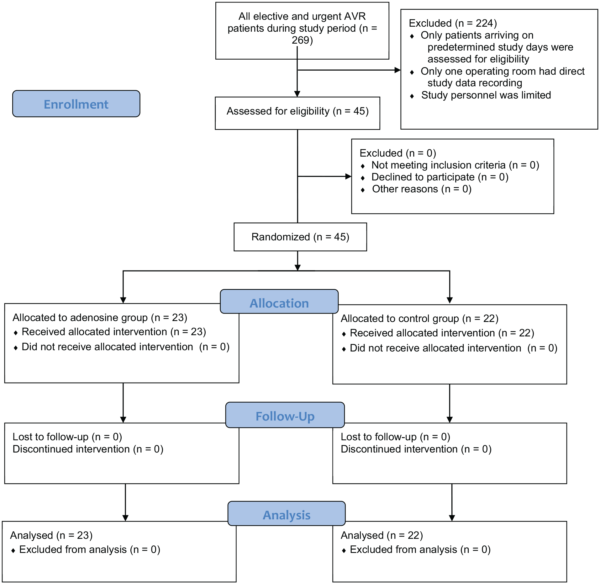
CONSORT flow diagram.
Patient baseline characteristics.
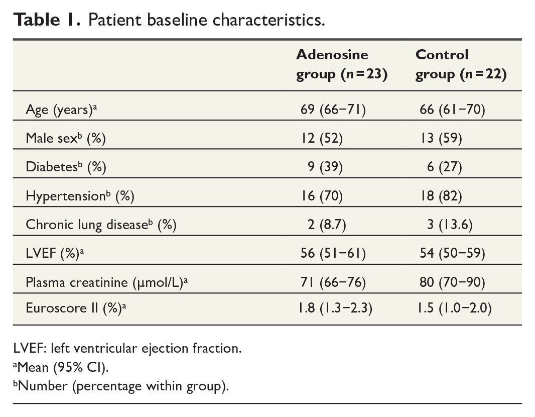
LVEF: left ventricular ejection fraction.
Mean (95% CI).
Number (percentage within group).
Preoperative baseline structural heart parameters.
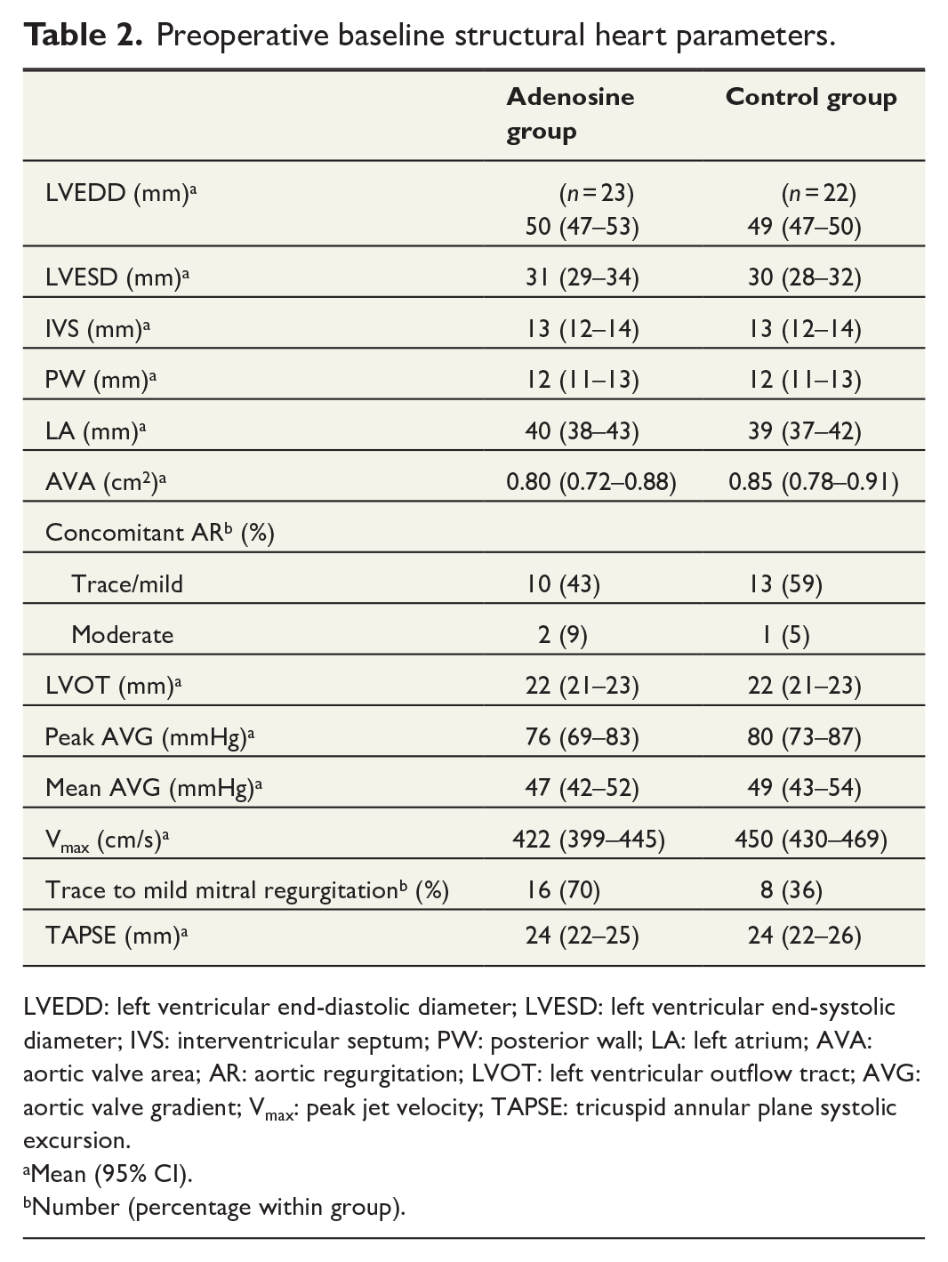
LVEDD: left ventricular end-diastolic diameter; LVESD: left ventricular end-systolic diameter; IVS: interventricular septum; PW: posterior wall; LA: left atrium; AVA: aortic valve area; AR: aortic regurgitation; LVOT: left ventricular outflow tract; AVG: aortic valve gradient; Vmax: peak jet velocity; TAPSE: tricuspid annular plane systolic excursion.
Mean (95% CI).
Number (percentage within group).
Primary outcome
Adenosine bolus had no statistically significant effect on CI. In individual timepoint comparisons, the mean differences and associated 95% CIs were 0.09 L/min/m2 (−0.20 to 0.38) at baseline, −1.39 L/min/m2 (−3.47 to 0.70) at post-CPB, −0.39 L/min/m2 (−0.78 to 0.004) at 7 p.m., and −0.32 L/min/m2 (−0.68 to 0.05) at 6 a.m. The primary hemodynamic outcome parameter results of CI and secondary hemodynamic outcome parameter results of LVSWI and RVSWI are presented in Table 3.
Primary and secondary outcome hemodynamic measurements.
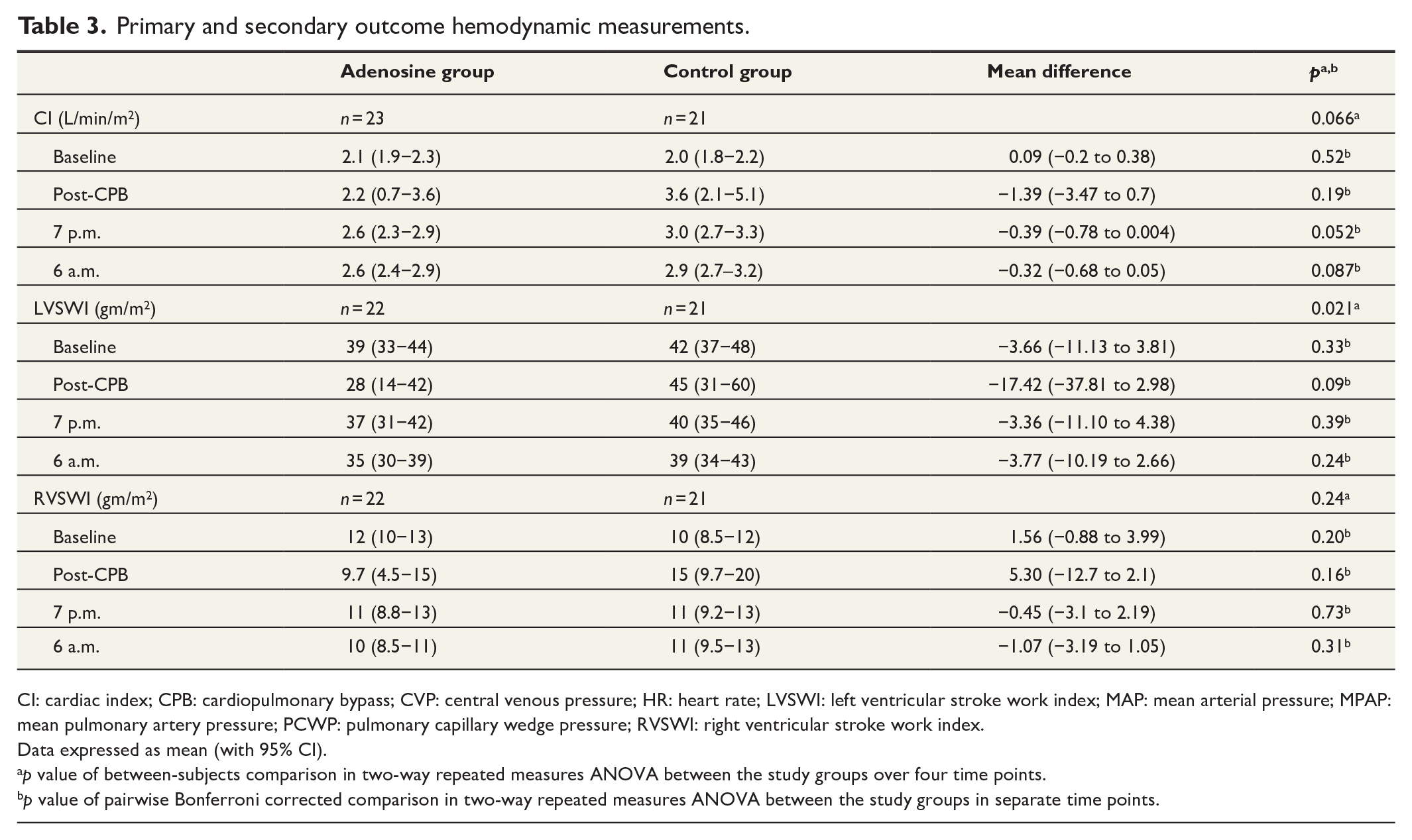
CI: cardiac index; CPB: cardiopulmonary bypass; CVP: central venous pressure; HR: heart rate; LVSWI: left ventricular stroke work index; MAP: mean arterial pressure; MPAP: mean pulmonary artery pressure; PCWP: pulmonary capillary wedge pressure; RVSWI: right ventricular stroke work index.
Data expressed as mean (with 95% CI).
p value of between-subjects comparison in two-way repeated measures ANOVA between the study groups over four time points.
p value of pairwise Bonferroni corrected comparison in two-way repeated measures ANOVA between the study groups in separate time points.
Secondary outcomes
LVSWI was significantly reduced in the adenosine group. In individual timepoint comparisons, the mean differences and associated 95% CIs were −3.66 gm/m2 (−11.13 to 3.81) at baseline, −17.42 gm/m2 (−37.81 to 2.98) at post-CPB, −3.36 gm/m2 (−11.10 to 4.38) at 7 p.m., and −3.77 gm/m2 (−10.19 to 2.66) at 6 a.m. Adenosine bolus did not affect RVSWI. In individual timepoint comparisons, the mean differences and associated 95% CIs were 1.56 gm/m2 (−0.88 to 3.99) at baseline, −5.30 gm/m2 (−12.70 to 2.10) at post-CPB, −0.45 gm/m2 (−3.10 to 2.19) at 7 p.m., and −1.07 gm/m2 (−3.19 to 1.05) at 6 a.m.
The secondary outcome, cardiac biomarker values remained low and there were no significant differences between the groups in the consecutive plasma concentrations of CK, CK-MB, or TnT throughout the study (Table 4). In individual timepoint comparisons, the mean differences and associated 95% CIs were 1.63 U/L (−26.79 to 30.06) for CK, −5.83 U/L (−14.99 to 3.31) for CK-MB, and −0.38 ng/L (−7.43 to 6.67) for TnT at baseline. Post-CPB mean differences were 0.95 U/L (−36.45 to 38.33) for CK, −4.26 U/L (−12.61 to 4.10) for CK-MB, and 8.38 ng/L (−183.70 to 200.47) for TnT. At 7 p.m., the mean differences were −90.01 U/L (−216.69 to 36.67) for CK, −6.10 U/L (−17.69 to 5.50) for CK-MB, and 37.86 ng/L (−191.51 to 267.22) for TnT. At 6 a.m., the mean differences were −121.0 U/L (−333.51 to 91.51) for CK, −6.32 U/L (−17.77 to 5.13) for CK-MB, and −128.86 ng/L (−294.52 to 36.81) for TnT.
Cardiac biomarkers.
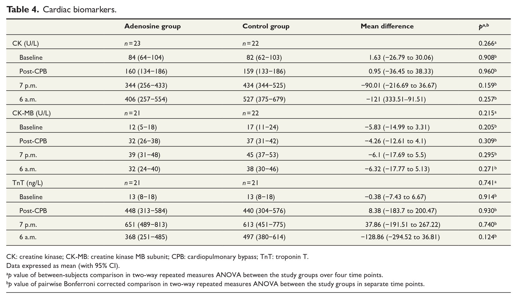
CK: creatine kinase; CK-MB: creatine kinase MB subunit; CPB: cardiopulmonary bypass; TnT: troponin T.
Data expressed as mean (with 95% CI).
p value of between-subjects comparison in two-way repeated measures ANOVA between the study groups over four time points.
p value of pairwise Bonferroni corrected comparison in two-way repeated measures ANOVA between the study groups in separate time points.
Post hoc outcomes
Adenosine did not decrease time to asystole (median difference = 5.5 s, 95% CI = (−59 to 62). In detailed analysis, adenosine did decrease the time to the first asystole (median difference = −89 s, 95% CI = (−116 to −69), but there was a higher restart rate (difference in proportions = 0.47, 95% CI = (0.18–0.66) in the adenosine group compared with the control group. Ventilation time was slightly longer in the adenosine group (mean difference = 1.4 h, 95% CI = (0.05–2.80). No other statistically significant differences were found in clinical outcomes between the groups. Minor additional procedures were made in addition to the AVR operation in both groups at the discretion of the operating surgeon (Table 5). The adenosine group received more concomitant single coronary vessel bypass procedures, but the difference in proportions was not significantly different (0.21, 95% CI = (−0.03 to 0.42). Concomitant coronary artery bypass operation had no influence on the primary or secondary outcome hemodynamic variables in between-groups comparison (Supplemental Table 1). There was significantly more often a need for temporary pacing immediately after cross-clamp removal in the adenosine group (difference in proportions = 0.38, 95% CI = (0.1–0.59). Besides the need for temporary pacing, the time to the first asystole, and the restart of cardiac activity after the first asystole, no substantial other differences in the intraoperative ECG data were found between the adenosine and the control groups (Table 6). The mean arterial pressure (MAP) was lower in the adenosine group immediately after CPB (mean difference = −5.3 mmHg, 95% CI = (−10.39 to −0.16). Otherwise, no significant differences were found in post hoc hemodynamic measurements (Supplemental Table 2). There were no significant differences in the adverse events during hospital stay between the groups (Supplemental Table 3). Trace to mild mitral insufficiency had no influence on the primary or secondary outcome hemodynamic variables in between-groups comparison (Supplemental Table 4).
Clinical outcomes.
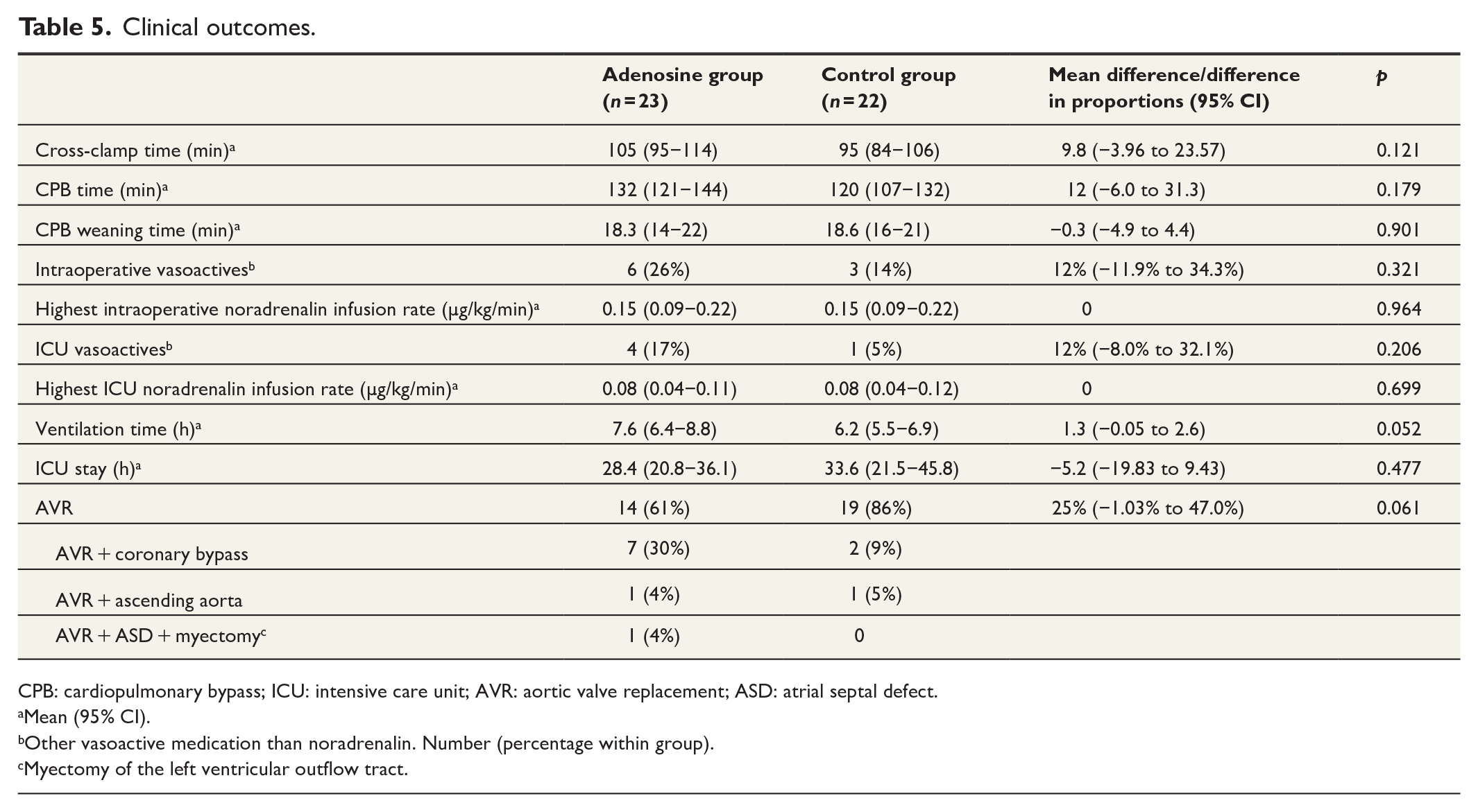
CPB: cardiopulmonary bypass; ICU: intensive care unit; AVR: aortic valve replacement; ASD: atrial septal defect.
Mean (95% CI).
Other vasoactive medication than noradrenalin. Number (percentage within group).
Myectomy of the left ventricular outflow tract.
Intraoperative ECG data.
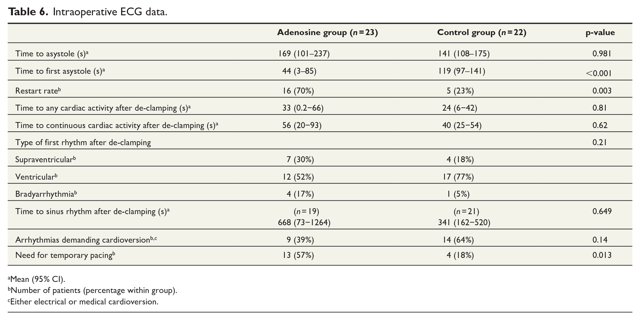
Mean (95% CI).
Number of patients (percentage within group).
Either electrical or medical cardioversion.
Representativeness of study patients
We compared the randomized study population’s characteristics and outcomes to the excluded source population of 224 elective and urgent AVR patients that were operated in Heart Hospital during the study period to evaluate the generalizability of the randomized controlled trial (RCT) results (Supplemental Table 5). The randomized population had more often diabetes (difference in proportions = 0.17, 95% CI = (0.04–0.33), lower left ventricular ejection fraction (LVEF) (mean difference = −3.5%, 95% CI = (−7.19 to 0.25), and lower plasma creatinine (median difference = −9 µmol/L, 95% CI = (−14 to −3) compared with the excluded source population.
Discussion
To our knowledge, this is the first randomized clinical pilot trial to investigate the effects of intra-aortic high-dose adenosine bolus combined with first blood cardioplegia on postoperative ventricular function in aortic valve stenosis in a limited number of patients undergoing AVR. In this study, adenosine did not improve CI compared with placebo. The other main findings included that adenosine decreased LVSWI, but did not affect RVSWI significantly and there were no differences in cardiac biomarker values indicating no difference in the level of myocardial injury between the groups. Adenosine bolus also did not lead to faster asystole. Although it reduced the time to the first asystole, there was a high restart rate. Moreover, the adenosine group demonstrated a significantly higher initial need for temporary cardiac pacing after the heart started.
Our findings in AVR patients differ from previous positive reports about adenosine-induced cardioplegic arrest in valve operations and CABG patients. Ahlsson et al. compared adenosine to mannitol in a RCT in aortic valve stenosis patients, but adenosine was given as mixed in continuous cardioplegia infusion through the operation. They found no enhancement of myocardial protection as measured by myocardial enzyme release, adenine nucleotide and oxygen/lactate metabolism, or hemodynamic performance. 15 Liu et al. had only 5 AVR patients in their trial on 30 rheumatic valve patients, 11 and Abdelwahab et al. had 16 AVR patients in their series of 45 rheumatic valve patients. 7 The previous positive findings with adenosine-induced cardioplegic arrest are from CABG and rheumatic valve disease patient populations, which are different cardiac pathologies compared with aortic valve stenosis. In aortic valve stenosis, the left ventricular myocardium is often hypertrophied creating a challenge for cardioprotection. The hypertrophied left ventricular myocardium probably is more prone to injury during cardiac surgery due to larger myocardial mass to protect. In CABG, adenosine may offer a protection benefit because of its vasodilative properties in stenotic coronary arteries and shorter time to asystole. We think that the increased left myocardial mass may have resulted in an insufficient adenosine dose not being able to improve cardioprotection or shorten the time to asystole in the hypertrophied left ventricles. Also, contrary to CABG patients, the myocardium in AVR patients might not be ischemic and the addition of adenosine to cardioplegia may not offer extra protection with a single dose. The majority of stenotic aortic valves in this trial were associated with trace to moderate aortic regurgitation leading to possible leakage of adenosine from the aortic root into the left ventricle (Table 2). The half-time of adenosine being only about 10 s, and the whole dose may never have reached the myocardium. Only the patients with severe regurgitation were excluded from the study. Considering the similarly low cardiac biomarker values between the groups, we hypothesize that the higher LVSWI in the control group may not have been due to higher myocardial injury in the adenosine group. MAP is a factor in the equation of LVSWI, so the higher LVSWI in the control group might be attributed to the higher MAP values in the control group measured right after weaning from CPB. Since postoperative MAP in cardiac patients is also dependent on the peripheral resistance, we wanted to control, if vasoactive medication was part of this finding. However, there was no difference in vasoactive medication use between the groups to affect MAP (Table 5) and adenosine may have lowered the MAP in the adenosine group.
Limitations
The main limitation of the study is the pilot nature of the RCT with a very low number of patients based on both limited study personnel within the acute care surgery setting and limited resources of a single operating room as assessment of eligibility and study recruitment was only possible for patients arriving on predetermined study days. These circumstances also markedly prolonged the study time. The second limitation is the limited generalizability of the results as compared to the excluded source population, the study population had statistically significantly higher prevalence of diabetes and lower preoperative LVEF and plasma creatinine values compared with the excluded source population although both parameter means were within the normal range in both populations. However, otherwise the populations were comparable. Third, despite the double-blinding, the shorter time to first asystole might have been obvious to the surgical team in the adenosine group; however, we have no reason to believe that this would have affected the decision-making during the operation or determining the main study outcomes.
Conclusion
Contrary to our previous findings in CABG patients, a single 20 mg adenosine bolus in the first cardioplegia infusion did not improve the primary outcome CI or the secondary outcomes LVSWI and RVSWI. Due to the decreased LVSWI in the adenosine group and only a small observed difference in the primary outcome, a larger trial or a higher dose adenosine bolus is required to study the effects of adenosine-induced cardioplegic arrest in AVR patients.
Supplemental Material
sj-docx-1-sjs-10.1177_14574969241266716 – Supplemental material for The cardioprotective effects of adenosine-induced cardioplegic arrest versus saline in aortic valve replacement patients: A randomized controlled trial
Supplemental material, sj-docx-1-sjs-10.1177_14574969241266716 for The cardioprotective effects of adenosine-induced cardioplegic arrest versus saline in aortic valve replacement patients: A randomized controlled trial by Mikko S. Mattila, Kati M. Järvelä, Markku J. Rantanen, Suvi Tuohinen, Kjell C. Nikus and Jari O. Laurikka in Scandinavian Journal of Surgery
Supplemental Material
sj-docx-2-sjs-10.1177_14574969241266716 – Supplemental material for The cardioprotective effects of adenosine-induced cardioplegic arrest versus saline in aortic valve replacement patients: A randomized controlled trial
Supplemental material, sj-docx-2-sjs-10.1177_14574969241266716 for The cardioprotective effects of adenosine-induced cardioplegic arrest versus saline in aortic valve replacement patients: A randomized controlled trial by Mikko S. Mattila, Kati M. Järvelä, Markku J. Rantanen, Suvi Tuohinen, Kjell C. Nikus and Jari O. Laurikka in Scandinavian Journal of Surgery
Supplemental Material
sj-docx-3-sjs-10.1177_14574969241266716 – Supplemental material for The cardioprotective effects of adenosine-induced cardioplegic arrest versus saline in aortic valve replacement patients: A randomized controlled trial
Supplemental material, sj-docx-3-sjs-10.1177_14574969241266716 for The cardioprotective effects of adenosine-induced cardioplegic arrest versus saline in aortic valve replacement patients: A randomized controlled trial by Mikko S. Mattila, Kati M. Järvelä, Markku J. Rantanen, Suvi Tuohinen, Kjell C. Nikus and Jari O. Laurikka in Scandinavian Journal of Surgery
Supplemental Material
sj-docx-4-sjs-10.1177_14574969241266716 – Supplemental material for The cardioprotective effects of adenosine-induced cardioplegic arrest versus saline in aortic valve replacement patients: A randomized controlled trial
Supplemental material, sj-docx-4-sjs-10.1177_14574969241266716 for The cardioprotective effects of adenosine-induced cardioplegic arrest versus saline in aortic valve replacement patients: A randomized controlled trial by Mikko S. Mattila, Kati M. Järvelä, Markku J. Rantanen, Suvi Tuohinen, Kjell C. Nikus and Jari O. Laurikka in Scandinavian Journal of Surgery
Supplemental Material
sj-docx-5-sjs-10.1177_14574969241266716 – Supplemental material for The cardioprotective effects of adenosine-induced cardioplegic arrest versus saline in aortic valve replacement patients: A randomized controlled trial
Supplemental material, sj-docx-5-sjs-10.1177_14574969241266716 for The cardioprotective effects of adenosine-induced cardioplegic arrest versus saline in aortic valve replacement patients: A randomized controlled trial by Mikko S. Mattila, Kati M. Järvelä, Markku J. Rantanen, Suvi Tuohinen, Kjell C. Nikus and Jari O. Laurikka in Scandinavian Journal of Surgery
Supplemental Material
sj-docx-6-sjs-10.1177_14574969241266716 – Supplemental material for The cardioprotective effects of adenosine-induced cardioplegic arrest versus saline in aortic valve replacement patients: A randomized controlled trial
Supplemental material, sj-docx-6-sjs-10.1177_14574969241266716 for The cardioprotective effects of adenosine-induced cardioplegic arrest versus saline in aortic valve replacement patients: A randomized controlled trial by Mikko S. Mattila, Kati M. Järvelä, Markku J. Rantanen, Suvi Tuohinen, Kjell C. Nikus and Jari O. Laurikka in Scandinavian Journal of Surgery
Supplemental Material
sj-docx-7-sjs-10.1177_14574969241266716 – Supplemental material for The cardioprotective effects of adenosine-induced cardioplegic arrest versus saline in aortic valve replacement patients: A randomized controlled trial
Supplemental material, sj-docx-7-sjs-10.1177_14574969241266716 for The cardioprotective effects of adenosine-induced cardioplegic arrest versus saline in aortic valve replacement patients: A randomized controlled trial by Mikko S. Mattila, Kati M. Järvelä, Markku J. Rantanen, Suvi Tuohinen, Kjell C. Nikus and Jari O. Laurikka in Scandinavian Journal of Surgery
Supplemental Material
sj-docx-8-sjs-10.1177_14574969241266716 – Supplemental material for The cardioprotective effects of adenosine-induced cardioplegic arrest versus saline in aortic valve replacement patients: A randomized controlled trial
Supplemental material, sj-docx-8-sjs-10.1177_14574969241266716 for The cardioprotective effects of adenosine-induced cardioplegic arrest versus saline in aortic valve replacement patients: A randomized controlled trial by Mikko S. Mattila, Kati M. Järvelä, Markku J. Rantanen, Suvi Tuohinen, Kjell C. Nikus and Jari O. Laurikka in Scandinavian Journal of Surgery
Footnotes
Author contributions
M.S.M.: Data curation; Formal analysis; Funding acquisition; Investigation; Project administration; Writing—Original draft. K.M.J.: Validation; Writing—Review & editing. M.J.R.: Resources; Methodology. S.T.: Methodology; Formal analysis; Investigation; Writing—Review & Editing. K.C.N.: Formal analysis; Validation; Writing—Review & editing. J.O.L.: Conceptualization; Data curation; Formal analysis; Funding acquisition; Investigation; Methodology; Project administration; Supervision; Validation; Writing—Review & editing.
Declaration of conflicting interests
The author(s) declared no potential conflicts of interest with respect to the research, authorship, and/or publication of this article.
Funding
The author(s) disclosed receipt of the following financial support for the research, authorship, and/or publication of this article: This work was supported by the Heart Hospital research grant and the Finnish Cultural Foundation research grant.
Supplemental material
Supplemental material for this article is available online.
References
Supplementary Material
Please find the following supplemental material available below.
For Open Access articles published under a Creative Commons License, all supplemental material carries the same license as the article it is associated with.
For non-Open Access articles published, all supplemental material carries a non-exclusive license, and permission requests for re-use of supplemental material or any part of supplemental material shall be sent directly to the copyright owner as specified in the copyright notice associated with the article.