
Abstract
Background:
The saphenous vein is the most frequently used bypass conduit for vascular reconstructions, which may need to be stored for a prolonged time. The aim of this study was to compare the effect of different preservation solutions on the morphology of saphenous veins during the long-term cold storage.
Design:
An in vitro study
Material and Methods:
Saphenous vein samples, collected from 29 patients undergoing varicose vein surgery, were stored at +4°C in (1) 10% formalin, (2) isotonic saline with heparin and antibiotic, (3) phosphate-buffered saline, (4) 2.5% glutaraldehyde + phosphate-buffered saline, and (5) Custodiol (histidine–tryptophan–ketoglutarate). Changes in the vein wall were histologically investigated up to day 35. Possible retention of the capacity of endothelial function was evaluated by immunohistochemical detection of endothelial nitric oxide synthase.
Results:
Formalin as the control medium well preserved the vein wall morphology, but endothelial nitric oxide synthase immunostaining was very weak. Phosphate-buffered saline and isotonic saline with heparin and antibiotic poorly preserved vein wall morphology. Phosphate-buffered saline endothelial nitric oxide synthase staining decreased dramatically throughout the study period. Compared to phosphate-buffered saline, stronger isotonic saline with heparin and antibiotic endothelial nitric oxide synthase staining was noted at day 35 (p < 0.001). Custodiol and glutaraldehyde better preserved vein morphology compared to ISHA and PBS at day 5 and later (p < 0.001), but compared to stronger isotonic saline with heparin and antibiotic their endothelial nitric oxide synthase staining was weaker.
Conclusion:
In terms of preserving the morphology of saphenous veins, phosphate-buffered saline and isotonic saline with heparin and antibiotic were the poorest, while Custodiol and glutaraldehyde were the best. Demonstrating good retention of endothelial nitric oxide synthase staining throughout the study period, isotonic saline with heparin and antibiotic seems to have the best potential to retain vein wall functionality, despite relatively poor morphological preservation.
Introduction
The optimal graft material for infrainguinal bypass is autologous great saphenous vein. However, this vein is unavailable or inadequate in about one-third of the patients (1). The use of prosthetic grafts has consistently failed to meet the standards of autologous vein graft patency, not only for infrapopliteal reconstructions (2, 3) but also for above-knee femoropopliteal bypass (4, 5). Thus, the use of a vascular allograft is an attractive alternative for patients without suitable autologous vein. Despite the availability of several preservation methods, for example, cryopreservation, cold storage in saline, or use of various preservation solutions, search for an optimal solution for vessel storage is continuing. Preserving the structural integrity of vessel wall, in particular, the endothelial lining, is very important for the quality and function of preserved vein grafts. It has been reported that even short-term storage of saphenous vein grafts in the physiological saline solution impairs endothelial function (6, 7). In order to improve long-term outcome, extra constituents are added. In our clinic, venous allografts are preserved in isotonic saline, with the addition of heparin and antibiotics at +4°C for up to 10–15 days (2, 8). Also, more advanced solutions are suggested, such as the transplantation media Custodiol or HTK (histidine–tryptophane–ketoglutarate) solutions, which have shown good results in preventing endothelial damage of vein grafts preserved over significant time (9).
Preserving allografts at 4°C causes exfoliation of the endothelial cells and reduces the amount of smooth muscle cells in the medium. The low incidence of graft rejection with the use of venous allografts may be due to the lack of HLA-II antigen expression in the wall of large vessels. The rejection process involves cell-mediated immunity to venous allografts and is not eliminated with low-dose azathioprine. There is also indirect evidence of participation of humoral immunity in venous allograft rejection (8). Freshly harvested vein with preserved intimal functions is preferred (stored in non-denaturing solutions, for example, isotonic saline with heparin and antibiotic (ISHA), phosphate-buffered saline (PBS), Custodiol) to avoid any toxic reactions in patients caused by denaturing preserving solutions (glutaraldehyde and formalin).
In this study, the effect of different solutions on preservation of the structure of saphenous vein samples from 1 day up to 35 days was compared. In addition, the expression of endothelial nitric oxide synthase (eNOS) was determined by immunohistochemistry at days 1, 14, and 35 to estimate the possible loss of endothelial functionality. Detection of constitutive eNOS has been commonly used to follow possible endothelium-driven functional changes in vein wall as nitric oxide (NO) generated by eNOS is an important marker molecule involved in smooth muscle relaxation (10, 11).
Material and Methods
Saphenous vein samples were collected from the Department of Vascular Surgery at Tartu University Hospital from 29 patients undergoing saphenectomy with a vein stripper during the period January 2011–March 2012. All patients gave their informed consent. The study was approved by the Ethics Review Committee on Human Research of the University of Tartu.
A visually healthy, manually dissected proximal part of saphenous vein segments, removed from retained fat, were taken for histological analysis before inserting the vein stripper.
Histology
A vein sample from each patient was divided into five parts, which were randomly immersed into one of the following solutions: (1) 10% buffered formalin (Sigma-Aldrich, Germany); (2) isotonic saline solution (0.9% NaCl (B. Braun Melsungen AG, Germany) + 5000 IU heparin (Rotexmedica, Germany) + antibiotic (penicillin 1 g per 500 mL; Sandoz, Germany)); (3) PBS (pH 7.4; Gibco Invitrogen, USA); (4) 2.5% glutaraldehyde (Sigma-Aldrich) + PBS; and (5) Custodiol (HTK; Dr. Franz Köhler Chemie GmbH, Germany). The samples were stored in the above-mentioned solutions at +4°C and 5–6 mm consecutive transverse sections were cut after 1, 5, 9, 14, 21, 28, and 35 days. The specimens were subsequently embedded in paraffin according to standard methods. The 4-µm-thick sections were stained with hematoxylin–eosin and van Gieson for light microscopic examinations. Pathomorphological changes in vein wall were estimated by a licensed pathologist. For a single patient at every time point, eight slides were analyzed. To evaluate the extent of changes, a scale ranging from 0 to 4 (0—no change, 1—weak, 2—moderate, 3—marked, and 4—very marked change) was applied. Separate estimations were performed for the intima, the media, and the adventitia. In the intima, the integrity of the endothelial lining and of the structure of the subendothelial basement membrane (BM) was evaluated (0—complete or almost complete preservation of the endothelium and BM, 1—preservation of the endothelium and BM more than 50%, 2—preservation of the endothelium and BM more than 25%, 3—preservation of the endothelium and BM less than 25%, and 4—lack of the endothelium and marked BM degenerative changes). In the media and the adventitia, degenerative changes in the connective and muscle tissues were graded from 0 to 4 (0—no changes, 1—weak, 2—moderate, 3—marked, and 4—very marked changes). Evaluation was performed in a blinded fashion, that is, the pathologist conducted the grading being unaware of the coding of the specimens. The obtained scores were summed and used for statistical analysis.
Immunohistochemistry
The 3-µm-thick paraffin sections mounted on poly-
Statistical analysis was carried out using the commercial program InStat (GraphPad Software Inc). The Kruskal–Wallis Test (nonparametric analysis of variance (ANOVA)) was applied followed by Dunn’s multiple comparisons test.
Results
Structural Changes in the Vein Wall
Among the five tested preservation solutions, formalin is known to well infiltrate and fix large specimens; therefore, it was used in this study as the reference solution. For all tested vein preservation times, formalin yielded score 0, that is, there were no significant patho-morphological changes in the structure of vein wall regardless of the preservation time (Fig. 1). Out of the four other preservation solutions, ISHA and PBS showed similar high scores, indicating significant morphological changes, which increased over the preservation time. On the contrary, the structure of the veins preserved with glutaraldehyde and Custodiol underwent less change and the samples stored in these solutions demonstrated low scores throughout the study period. The representative micrographs of vein wall at days 5 and 35 are presented in Figs 2 to 4.
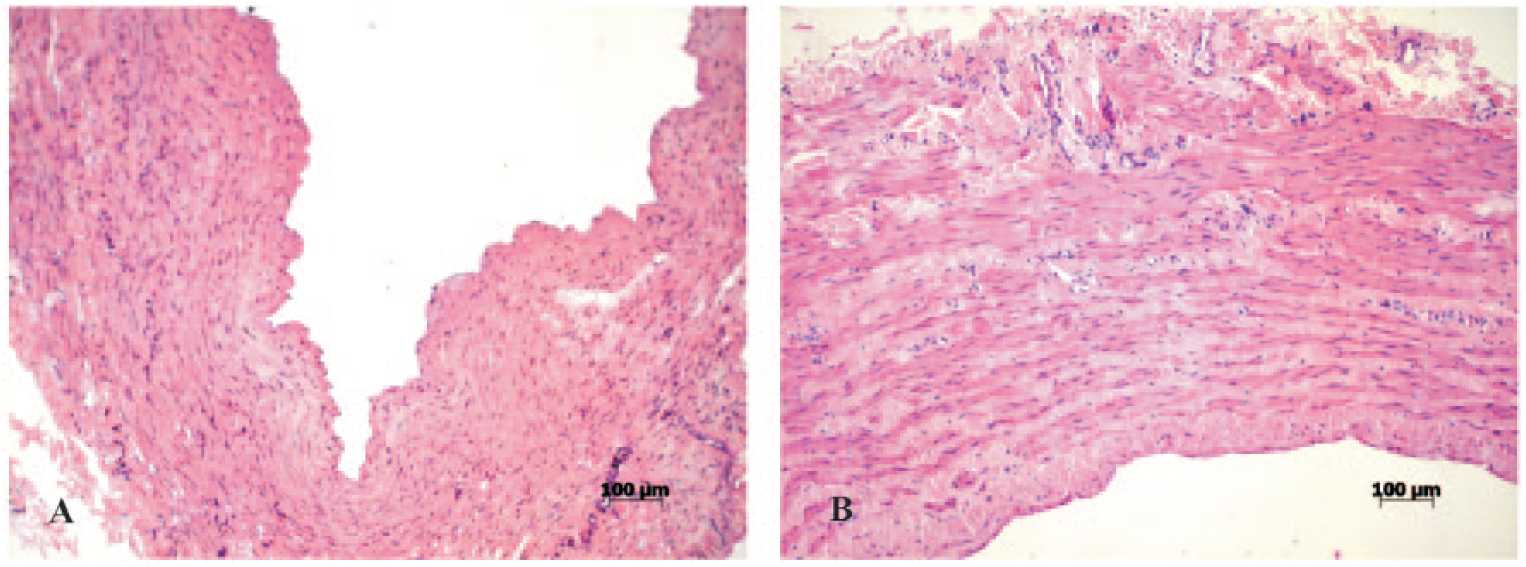
Vein specimens stored in 10% formalin forA) 5 days and B) 35 days. Regular structure of the vein wall without changes in layers (pathological changes scores in the intima, media, and adventitia were estimated as 0). Hematoxylin–eosin staining.
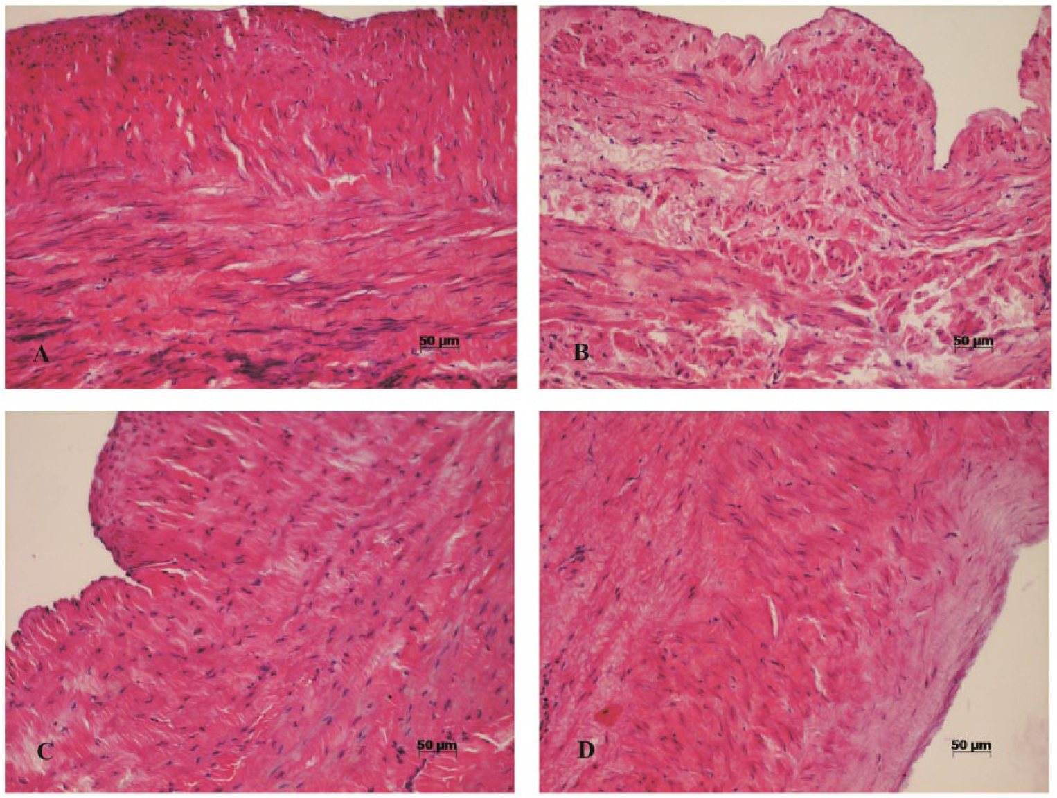
Vein specimen storage at 5 days in A) Custodiol, B) glutaraldehyde, C) PBS, and D) ISHA resulted in minimal vein wall structure changes. For the vein specimens shown in the micrographs of this figure, the estimated pathological changes score was 0 for all vein wall layers; only the veins stored in D) ISHA had the score for changes in the intima estimated as 1. Hematoxylin–eosin staining.
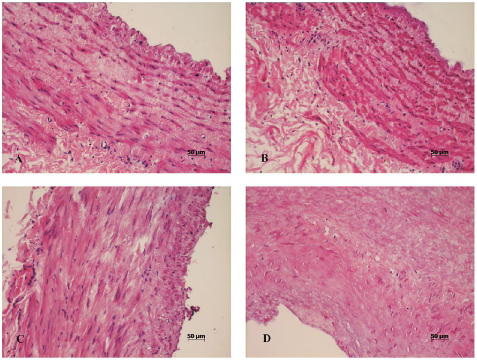
Vein specimen storage at 35 days in A) Custodiol, B) glutaraldehyde, C) PBS, and D) ISHA resulted in significant vein wall structure changes of varying grade. Moderate grade changes were noted in the veins stored in Custodiol or glutaraldehyde. In the vein specimens demonstrated in the micrographs of this figure, the pathological changes score was estimated as 1 for all layers of the vein stored in A) Custodiol and as 2 for the specimen stored in B) glutaraldehyde. Marked changes, in particular, detachment of the endothelium, were seen in the veins stored in PBS and ISHA. In the vein specimens demonstrated in the micrographs of this figure, the changes score was estimated as 3 for all vein layers stored in C) PBS and as 4 for all vein layers stored in D) ISHA. Hematoxylin–eosin staining.
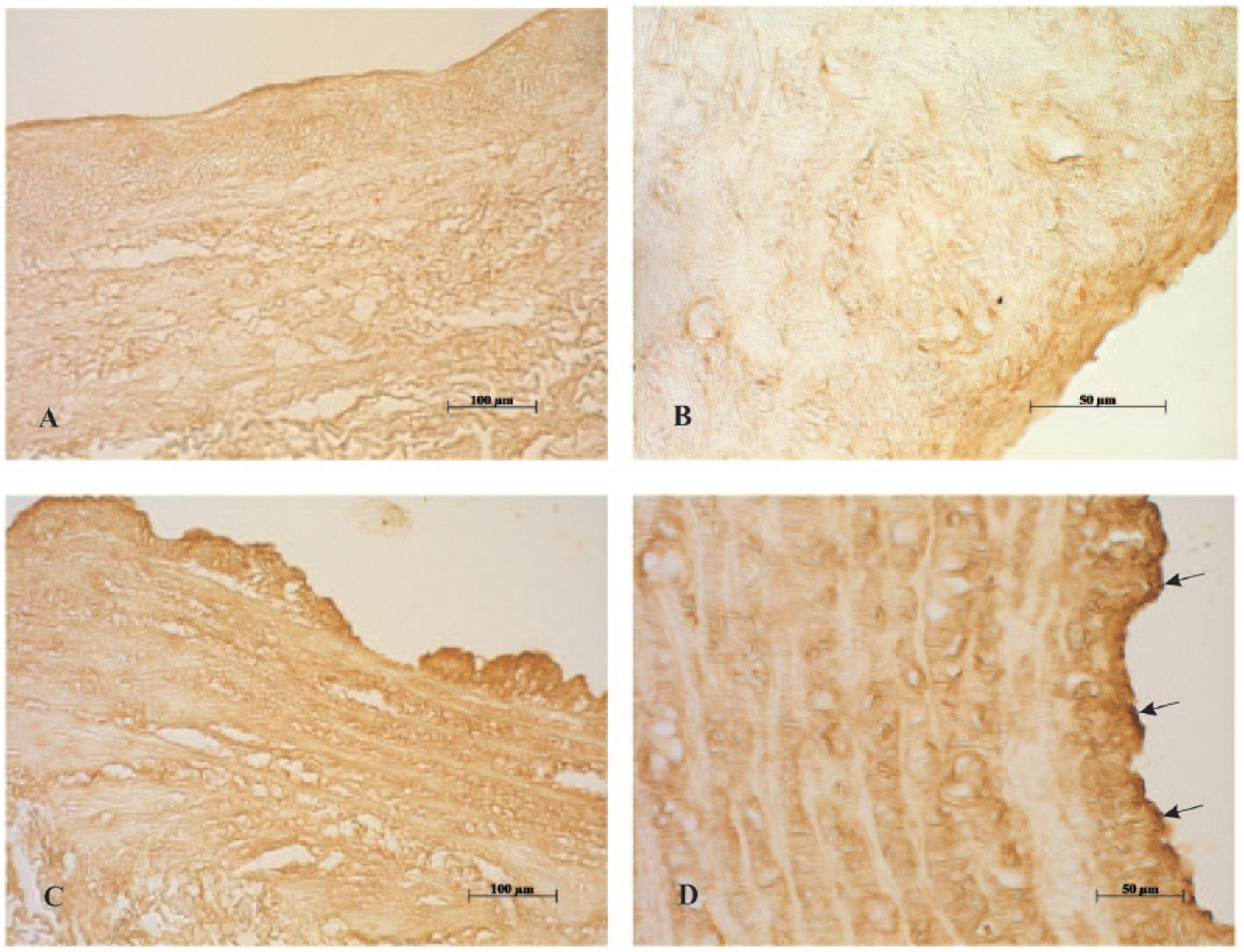
Vein specimens stored for 35 days in A) Custodiol showed weak eNOS staining (brown color) and in B) PBS showed very weak eNOS staining. Despite poor structural preservation at day 35 in the C) ISHA group, there are still areas of relatively well-preserved intimal regions with (D, arrows) moderate eNOS staining. DAB.
Pathomorphological alterations in the veins, estimated separately in the three vessel tunics, are presented in Figs 5 to 7.
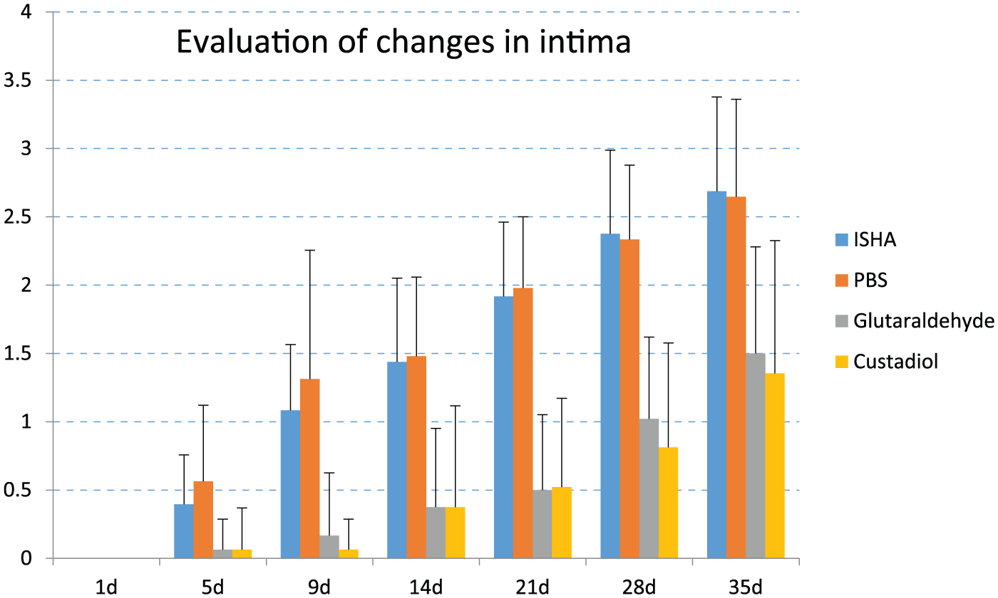
Evaluation of changes in the structure of the intima of veins. The extent of changes was estimated from 0 (no changes) to 4 (strong changes). Values are expressed as mean ± SD. Statistically significant differences: ISHA vs glutaraldehyde, ISHA vs Custodiol, PBS vs Custodiol, and PBS vs glutaraldehyde p < 0.001 at days 5, 9, 14, 21, 28 and 35; ISHA vs PBS p < 0.05 at day 5.

Evaluation of changes in the structure of the media of veins. The extent of changes was estimated from 0 (no changes) to 4 (strong changes). Values are expressed as mean ± SD. Statistically significant differences: ISHA vs glutaraldehyde, ISHA vs Custodiol, PBS vs Custodiol, and PBS vs glutaraldehyde p < 0.001 at days 5, 9, 14, 21, 28, and 35; ISHA vs PBS p < 0.05 at day 5.
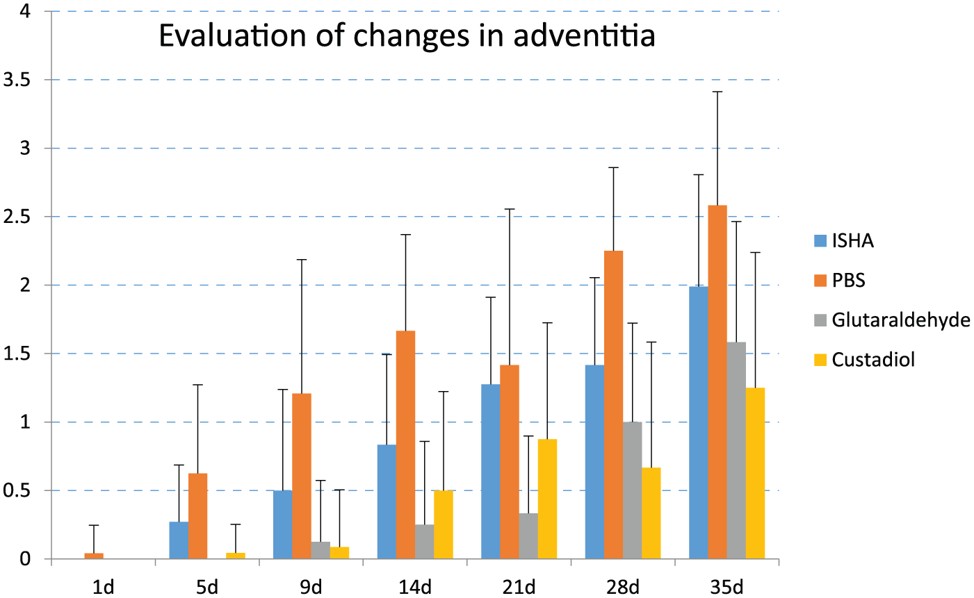
Evaluation of changes in the structure of the adventitia of veins. The extent of changes was estimated from 0 (no changes) to 4 (strong changes). Values are expressed as mean ± SD. Statistically significant differences: ISHA vs glutaraldehyde, ISHA vs Custodiol, PBS vs Custodiol, and PBS vs glutaraldehyde p < 0.001 at days 5, 9, 14, 21, 28, and 35; ISHA vs PBS p < 0.05 at days 5, 9, and 14.
Evaluation of Changes in the Intima
As seen in Fig. 5, no morphological alterations occurred in the endothelium or in the intima at day 1 in any of the tested preservation solutions. From day 5 to day 35, the score of morphological changes showed a gradual increase. For glutaraldehyde and Custodiol, the score increased relatively slowly from day 5 to day 35. In ISHA and PBS, morphological alterations in the intima occurred much faster and the corresponding scores were high compared to glutaraldehyde and Custodiol (Fig. 5). No significant differences in the scores were noted when comparing ISHA vs PBS and glutaraldehyde vs Custodiol (Fig. 5).
Evaluation of Changes in the Media
As seen in Fig. 6, no structural changes in the media were found at day 1 in any of the tested solutions. At days 5 and 9, very slight changes were seen in the media of the veins in glutaraldehyde and Custodiol, while changes became more pronounced only from day 14 to day 35. ISHA and PBS showed clear changes at day 5, and the scores increased gradually at the later time points throughout the study period. The corresponding scores were high compared to glutaraldehyde and Custodiol (Fig. 6). When comparing ISHA and PBS, the former showed low scores at day 5, but no significant differences were noted at the later time points. No significant differences in the average scores were found between glutaraldehyde and Custodiol.
Evaluation of Changes in the Adventitia
The veins preserved in PBS showed the highest scores throughout the study; some slight changes in the adventitia were noted already at day 1. No changes were found in the case of ISHA, glutaraldehyde or Custodiol at day 1. In the case of ISHA, the scores were low compared to PBS throughout the study; statistically significant differences were recorded at days 5, 9 and 14 (Fig. 7). The lowest scores, which were slowly decreasing throughout the study period, were seen for glutaraldehyde and Custodiol. Differences from ISHA and PBS were statistically significant at days 5, 9, 14, 21, 28, and 35 (Fig. 7). No significant differences in the average scores were found between glutaraldehyde and Custodiol.
Evaluation of Immunohistochemical Studies of Enos
Immunohistochemical studies of eNOS performed at days 1, 14, and 35 demonstrated very weak immunostaining of endothelial cells in formalin with no detectable staining at day 35 (Fig. 8). At day 1, the strongest eNOS staining was seen in PBS and ISHA, while staining in glutaraldehyde and Custodiol was moderate (Fig. 8). At day 14, eNOS staining declined in all groups. The most dramatic decrease was seen in PBS, while changes in the other groups were less marked. Immunostaining was the most expressed in ISHA and glutaraldehyde compared to formalin and PBS (Fig. 8). At day 35, eNOS staining was further decreased in all groups except ISHA (Figs. 4 and 8). In this group, eNOS staining did not show significant changes in comparison with the situation at day 14. The low scores of eNOS staining at day 35 were recorded for PBS and Glutaraldehyde (Fig. 8).
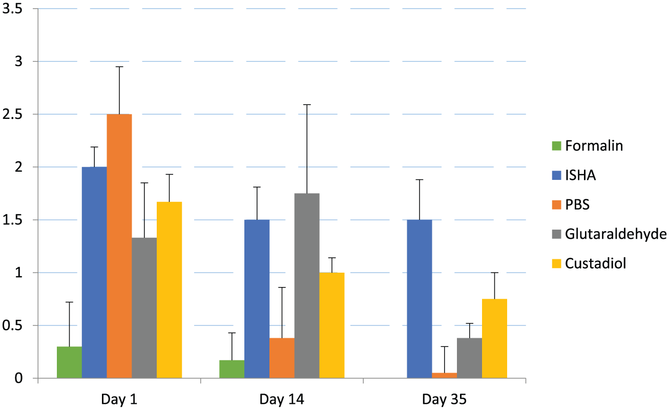
Evaluation of eNOS staining in the intima of the veins preserved in different solutions at days 1, 14, and 35. The eNOS staining was evaluated by a scale ranging from 0 to 3 (0—no staining, 1—weak staining, 2—moderate staining, and 3—strong staining). Values are expressed as mean ± SD. Statistically significant differences: formalin vs ISHA, and formalin vs PBS p < 0.001 at days 1, 14, and 35; ISHA vs PBS p < 0.001 at day 35; formalin vs glutaraldehyde, and formalin vs Custodiol p < 0.005 at days 14 and 35.
Discussion
As expected, 10% formalin as a widely known fixative preserved well all structures of the vein specimens with no evident morphological changes throughout the testing period. However, it has to be emphasized that the use of formalin in the clinical situation is complicated since it is recognized as a human carcinogen (12). Furthermore, eNOS expression in the formalin-preserved samples was very weak at days 1 and 14 and absent at day 35, indicating that under the current experimental conditions formalin may not necessarily retain the functional capacity of the endothelium. Also, formaldehyde may mask the epitopes to which eNOS antibodies bind, resulting in very weak immunostaining.
PBS turned out to be the poorest of the tested solutions in terms of preserving the morphology of vein wall structure. The structure of vein wall stored in PBS was almost intact at day 1; however, starting from day 5, obvious alterations in the structure were seen in all vein wall tunics. Also, eNOS immunostaining underwent clear changes from moderate staining at day 1 to very weak staining at days 14 and 35, which is supportive of significant loss of endothelial functionality.
In a recent literature survey of vein graft preservation solutions (13), it was shown that 0.9% saline solution can be harmful for the endothelium. Therefore, saline solution may not be the best even for intraoperational graft storage (14). At the same time, several clinical studies have reported reasonable outcome of bypass operations where venous allografts were stored at +4°C in 0.9% saline solution containing antibiotics (2, 15–17). In our study, the storage of the vein specimens in ISHA which also contained, in addition to 0.9% saline solution, heparin and antibiotics yielded results similar to those obtained with PBS, especially at the later time points. Although the average scores of vein wall changes for ISHA were slightly lower compared to the scores for PBS at days 5 and 9, these differences more or less leveled out at later time points. Thus, it seems that ISHA has a slight advantage over PBS in preserving morphology during short-term storage of vein specimens. However, the most striking difference was seen in the immunostaining of eNOS: when for PBS it was very weak at days 14 and 35, then for ISHA it was quite similar at days 14 and 35 to that recorded at day 1, which may reflect the possible retention of vein wall functionality.
Both glutaraldehyde and Custodiol preserved the structure of veins significantly better than ISHA or PBS. For glutaraldehyde, good preservation of vein morphology can be expected as this solution is widely used for sterilization and preservation. Glutaraldehyde-stabilized umbilical veins have been used as a biological alternative to autologous vein for lower limb revascularization for decades with favorable results (18–20). It is suggested that glutaraldehyde diminishes allograft rejection, masking the histocompatibility of the antigen sites, which results in low rate of biodegeneration of glutaraldehyde-stabilized veins (20). Hence, in our study, the relatively weaker expression of eNOS in the vein samples stored in glutaraldehyde, compared to PBS and ISHA, can be also explained by the effect of glutaraldehyde to mask the epitopes for binding antibodies.
Custodiol, or HTK, or Bretschneider’s solution was primarily designed for preserving donor organs in hypothermic conditions prior to transplantation. Custodiol cardioplegia has been particularly attractive for cardiac surgery in providing myocardial protection (21). As Custodiol is aimed for organ function inactivation, it is not surprising that this solution preserved well the vein sample morphology in our study. For all vein wall layers, the average grades of morphological changes were similar to the corresponding grades of glutaraldehyde. Custodiol has been shown to preserve vein endothelial integrity for up to 5 days, differently from 0.9% saline solution and the formalin control groups (9). Also in our study, very low grades of endothelial (intimal) damage were seen, when vein samples were stored up to day 9; thereafter, the grades started slowly to increase up to the end of the study. The expression of eNOS in samples preserved in Custodiol was slightly stronger in comparison with glutaraldehyde, but compared with ISHA relatively weaker eNOS staining was seen. Thus, it can be speculated that ISHA may have an advantage over Custodiol in retaining vein wall functionality despite poorer morphology preservation. However, as a limitation of the given experiment, it has to be emphasized that it is an in vitro study which does not mimic in vivo conditions. As a next step, further animal experiments are required for proper in vivo testing of the behavior of these long-term preserved vein grafts.
In conclusion, our morphological study comparing different solutions for prolonged cold storage of saphenous veins demonstrated that PBS was the poorest in preserving vein wall structure. ISHA had only a slight advantage over PBS in preserving vein wall morphology in short-term storage, but was the best of the tested solutions in possible vein wall functionality retention, as indicated by the highest scores of eNOS staining at day 35. Although Custodiol and glutaraldehyde were superior to PBS and ISHA in preserving the morphology of vein wall, ISHA retained eNOS expression better. Thus, despite the fact that ISHA may not be the best to preserve vein wall structure, it seems to have the best potential in maintaining the functional capacity of vein wall. Based on a previous study, the authors recommend ISHA for preserving venous allografts, with a maximum storage time of up to 3 weeks. However, more extensive functionality studies are required to verify this speculation.
Footnotes
Acknowledgements
The authors appreciate the work performed by Dr Hannes Tamm.
Declaration of Conflicting Interests
The author(s) declared no potential conflicts of interest with respect to the research, authorship, and/or publication of this article.
Funding
The author(s) disclosed receipt of the following financial support for the research, authorship, and/or publication of this article: This study was supported by target financing (project no 0180012s11) from the Ministry of Education and Research of Estonia.