
Abstract
Background
Porcine reproductive and respiratory syndrome virus (PRRSV) is a pathogen that affects swine and causes substantial economic losses in the global pig industry. Despite the availability of vaccines, it remains crucial to explore innovative therapeutic strategies to control PRRSV infections. Magnolol, a bioactive compound extracted from the root and bark of Magnolia officinalis, has demonstrated broad-spectrum antiviral activity in previous studies.
Methods
The cytotoxicity of magnolol was determined by the CCK-8 method. RT-qPCR, western blot, and immunofluorescence were used to study the inhibitory effect of magnolol on PRRSV N gene and protein expression through antiviral assay and viral attachment, internalization, replication and release assays. The effect of magnolol on immune-related gene expression was analysed by RT-qPCR.
Results
We found magnolol hinders multiple facets of the PRRSV lifecycle, encompassing the stages of viral attachment and replication. Furthermore, magnolol enhances the expression of pivotal cytokines, including interleukin-6 (IL-6), interleukin-8 (IL-8), and tumour necrosis factor-α (TNF-α), during PRRSV infection, thereby reinforcing the host cells’ capacity to mount an effective antiviral defence. Additionally, it exerts inhibitory effects on PRRSV replication by upregulating the expression of a disintegrin and metalloprotease 17 (ADAM17) at both the protein and mRNA levels.
Conclusions
In this study, we provide evidence demonstrating the potent efficacy of magnolol in inhibiting PRRSV replication within Marc-145 cells. Our findings underscore the potential of magnolol as a novel antiviral agent for the PRRSV control.

Keywords
Introduction
Porcine reproductive and respiratory syndrome (PRRS), caused by the PRRS virus (PRRSV), is characterized by respiratory disorder, reproductive failure in pregnant sows, and high mortality rates among piglets.1,2 This disease has emerged as a predominant pathogen responsible for significant economic losses in the global swine industry.3,4 Currently, there are no effective treatments for PRRS, with management strategies primarily focused on prevention through vaccination.5,6 Given the virus’s extensive genetic diversity and high mutation rates of PRRSV, the protection offered by existing commercially available vaccines is limited.6,7 Consequently, there is an urgent need to develop novel anti-PRRSV strategies.
In recent years, numerous antiviral agents, including small-molecule inhibitors and natural compounds, have been investigated to combat viral infections. Notable examples include ensitrelvir, 8 simnotrelvir, 9 cordycepin, 10 riboprine, 11 molnupiravir, 12 remdesivir, 13 and natural extracts (such as strychnine bush, pineapple, and ginger ingredients), 14 which have shown efficacy against various viral pathogens such as SARS-CoV-2. These compounds primarily target viral entry, replication, anti- RNA-dependent RNA polymerase (RdRp), or immune evasion mechanisms, offering valuable insights into antiviral strategies. Despite these advancements, the application of synthetic antivirals often faces challenges related to drug resistance, adverse effects, and high production costs.
In China, approximately 10% of prescriptions incorporate Traditional Chinese herbs. Valued for their efficacy, cost-effectiveness, and safety, these herbs have become a key focus for developing novel antiviral therapy.15,16 Numerous natural compounds and herbal components have demonstrated antiviral properties.17,18 For example, compounds such as sanggenon C, 19 ginsenoside Rg1, 20 andrographolide, 21 and baicalin 22 have exhibited promising antiviral effects against PRRSV and other RNA viruses, mainly through the modulation of host immune responses and inhibition of viral replication pathways. Magnolol, a bioactive constituent extracted from the roots and bark of Magnolia officinalis, a renowned Chinese herb, 23 has garnered attention in previous studies due to its antiviral potential. 24 Specifically, magnolol has exhibited inhibitory effects against H1N1 influenza virus by stimulating the secretion of interferon-β (IFN-β). 25 Simultaneously, it has bolstered the resilience of grass carp against grass carp reovirus (GCRV) infection. 26 Moreover, magnolol’s unique structural characteristics, featuring hydroxylated phenyl groups, contribute to its antioxidant, anti-inflammatory, and antiviral properties, making it a promising candidate for combating PRRSV infection. 27 Nevertheless, whether magnolol exerts inhibitory effects on PRRSV infection and replication remains uncertain.
In this study, we demonstrated that rifampicin effectively inhibits PRRSV nucleocapsid protein levels in Marc-145 cells in a dose-dependent manner, effectively targeting both the attachment and replication phases of the PRRSV life cycle. Remarkably, magnolol also amplifies the Marc-145 cell’s antiviral immune response, elevating the transcription levels of interleukin-6 (IL-6), IL-8, and tumour necrosis factor-α (TNF-α) during PRRSV infection. Remarkably, we extended the investigation to assess the impact of magnolol on ADAM17 (a disintegrin and metalloprotease 17). In conclusion, the potential of magnolol as an innovative antiviral agent, opening new avenues for the controlling and preventing PRRSV infection.
Materials and methods
Cells and viruses
Marc-145 cells, a widely used cell line for the propagation of PRRSV, were maintained in Dulbecco’s modified Eagle’s medium (DMEM, Gibco) supplemented with 10% fetal bovine serum (FBS, Gibco), 100 U/mL penicillin, and 100 µg/mL streptomycin at 37°C with 5% CO2. The PRRSV strain CHR6, classified as a classical North American type PRRSV strain, 28 was kindly provided by Dr Yaosheng Chen (Sun Yat-sen University). The virus was propagated in Marc-145 cells using DMEM containing 2% FBS. Virus preparations were stored at −80°C and titrated using the 50% tissue culture infective dose (TCID50) method.
Antibody
The primary antibodies used in this study included anti-PRRSV N protein (1:5000 dilution, GeneTex, USA), anti-ADAM17 (1:1000 dilution, GeneTex, USA), and anti-β-actin (1:2000 dilution, Cell Signalling Technology, USA). The secondary antibody used was goat anti-rabbit IgG H&L conjugated with HRP (1:1000 dilution, ab205718, Abcam).
Preparation of magnolol
Magnolol (purity ≥99.92%) was obtained from Med Chem Express (MCE, USA) and dissolved in dimethyl sulfoxide (DMSO; Sigma-Aldrich, USA) to prepare stock solutions. The final concentration of DMSO in the cell culture medium was maintained below 0.4% in all experiments to avoid solvent-induced cytotoxicity.
Cytotoxicity assay
The cytotoxicity of magnolol on Marc-145 cells was evaluated using the Cell Counting Kit-8 (CCK-8, GlpBio, USA) assay.
29
Briefly, Marc-145 cells were seeded into 96-well plates and incubated in DMEM containing various concentrations of magnolol (0, 1, 2, 4, 8, 16, and 32 µM) for 24 or 48 h (h) at 37°C. Following incubation, the cells were washed twice with phosphate-buffered saline (PBS), and 90 µL of fresh DMEM along with 10 µL of CCK-8 solution was added to each well. Plates were then incubated for an additional 2 h at 37°C. Absorbance at 450 nm was measured using a microplate reader to determine cell viability, and cytotoxicity was calculated using the following formula:

Antiviral activity assay
To assess the antiviral activity of magnolol against PRRSV, Marc-145 cells were seeded in 6-well plates and infected with PRRSV at a multiplicity of Infection (MOI) of 1 for 24 h in the presence of varying concentrations of magnolol. At designated time points post-infection, both cells and supernatants were collected for analysis. The mRNA expression of the PRRSV N gene was quantified by reverse transcription quantitative PCR (RT-qPCR), and the viral N protein levels were assessed by western blotting. Indirect immunofluorescence assay (IFA) was performed to determine the number of PRRSV-infected cells. To facilitate the maximal release of intracellular virions, cells and supernatants were subjected to three freeze-thaw cycles alternating between −80°C and 4°C. The viral titre in the supernatant was subsequently determined using an endpoint dilution assay in Marc-145 cells and expressed as Log10 TCID50/mL. 30
In the pre-treatment model, Marc-145 cells were mock-treated or treated with different concentrations of magnolol for 4 h, followed by three washes with PBS. The cells were then mock-infected or infected with PRRSV (MOI = 1) and cultured for an additional 24 h.
In the post-treatment model, Marc-145 cells were first mock-infected or infected with PRRSV (MOI = 1) for 4 h, after which the medium was replaced with fresh medium containing different concentrations of magnolol. The cells were then cultured for an additional 24 h.
Viral attachment, internalization, replication, and release assays
To investigate the effects of magnolol on different stages of the PRRSV life cycle, specific assays were conducted as described below: (I) Attachment: Cells were pre-chilled at 4°C for 1 h and subsequently mock-infected or infected with PRRSV (MOI = 4) in the presence or absence of magnolol for 2 h at 4°C. The cells were then covered with fresh medium, incubated for an additional 22 h at 37°C. Viral N protein and mRNA levels were assessed by western blot and RT-qPCR, respectively. (II) Internalization: Cells were pre-chilled at 4°C for 1 h. Then cells were mock-infected or infected with PRRSV (MOI = 4) for 2 h at 4°C to allow virus attachment without internalization. After washing twice with cold PBS to remove unbound virus, magnolol at various concentrations was added, and the cells were incubated at 37°C for 3 h to permit viral internalization. The medium was then replaced with fresh culture medium, and cells were incubated for an additional 19 h at 37°C. (III) Replication: Cells were mock-infected or infected with PRRSV (MOI = 4) for 6 h at 37°C to allow viral entry. Subsequently, cells were treated with magnolol for 4 h and harvested at 24 h post-infection (hpi) for analysis. (IV) Release: Cells were mock-infected or infected with PRRSV (MOI = 4) for 24 h at 37°C. Then the medium was replaced with fresh medium containing various concentrations of magnolol. PRRSV N protein and mRNA levels in both cell lysates and culture supernatants were quantified using western blot and RT-qPCR.
31
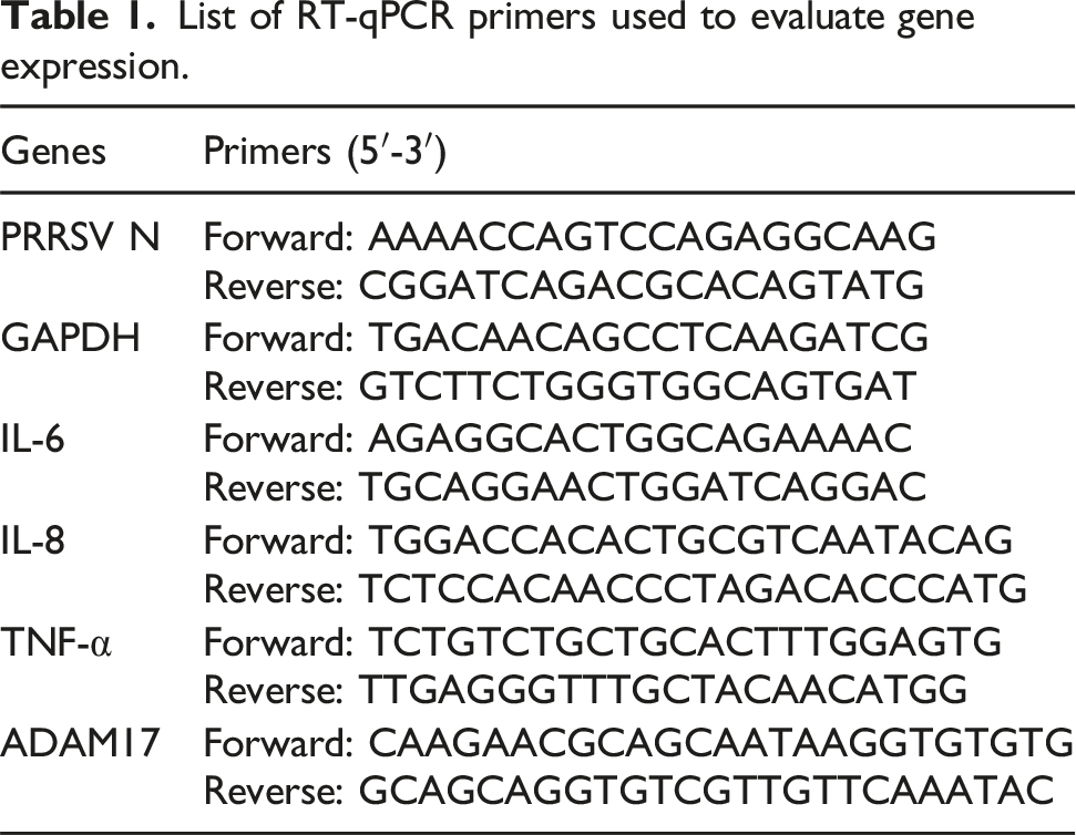
Real-time quantitative reverse transcriptase PCR (RT-qPCR)
List of RT-qPCR primers used to evaluate gene expression.
Western blot
Cells were washed three times with cold PBS and lysed using Radio Immunoprecipitation Assay (RIPA) lysis buffer (Beyotime, China) containing 1% phenylmethylsulfonyl fluoride (PMSF, Beyotime, China). Total protein (20 µg per sample) was subjected to 12% sodium dodecyl sulphate-polyacrylamide gel electrophoresis (SDS-PAGE) gel and transferred to polyvinyl difluoride (PVDF) membrane (Beyotime, China). The PVDF membranes were blocked with 5% bovine serum albumin (BSA, Ruishu, China) in TBST (Tris-buffered saline with 0.1% Tween-20) at room temperature (RT). The PVDF membrane was then incubated overnight at 4°C with the indicated antibodies. Each membrane was washed three times with TBST and incubated with the indicated secondary antibody for 1 h at RT. Finally, the membrane was washed and protein bands were visualized by Gel Imaging System (Tanon, 4200).
Indirect immunofluorescence assay (IFA)
Cells were fixed with 4% paraformaldehyde for 20 min. Then, cells were rinsed twice with PBS and permeated with 0.1% Triton X-100 for 15 min at RT. After permeabilization, cells were blocked with 3% normal goat serum for 2 h at RT and incubated overnight at 4°C with a primary antibody against PRRSV N protein (1:500 dilution, GeneTex, USA). Following primary antibody incubation, cells were washed and incubated in the dark for 2 h with a FITC-conjugated secondary antibody (1:200 dilution, Affinity). DAPI (Solarbio, China) (blue) was used to counterstain nuclei for 5 min. Finally, cells were determined by fluorescence microscopy (Leica DMi8, Germany).
Statistical analysis
All experiments were performed at least three independent replicates. Data are presented as means ± standard deviations (SD). Statistical significance was assessed by Student’s t test when only two groups were compared or by one-way analysis of variance (ANOVA) when more than two groups were compared. P values of <0.05 (*), <0.01 (**), and <0.001 (***) were considered statistically significant. P values of >0.05 (ns) were considered not-significant.
Results
Magnolol inhibits PRRSV replication in Marc-145 cells
To evaluating the antiviral activity of magnolol, the cytotoxicity of magnolol in Marc-145 cells was tested by CCK-8 assay. As shown in Figure 1A, magnolol exhibited no cytotoxic effects at concentrations up to 32 µM in Marc-145 cells. Hence, all subsequent experiments were conducted using magnolol at concentrations not exceeding 32 µM. Subsequently, we evaluated the inhibitory effects of magnolol against PRRSV (MOI = 1) through virus titration, western blot analysis, RT-qPCR, and immunofluorescence assay at 24 hpi. As depicted in Figure 1B, magnolol treatment significantly reduced PRRSV viral titres. Consistently, western blot and RT-qPCR analyses demonstrated a dose-dependent reduction in PRRSV N protein and mRNA levels (Figure 1C and D). Additionally, immunofluorescence analysis revealed a discernible reduction in PRRSV N following magnolol treatment, further confirming the inhibitory effect of magnolol on PRRSV replication (Figure 1E). These results indicate that magnolol effectively suppresses PRRSV replication in vitro. Cytotoxicity and anti-PRRSV activity of magnolol in Marc-145 cells. (A) Marc-145 cells were treated with magnolol at indicated concentrations for 24 or 48 h. (B) Marc-145 cells were mock-infected or infected with PRRSV (MOI = 1) and treated with different concentrations of magnolol (0, 16, and 32 µM) for 24 h. Viral titres in the supernatant were determined using endpoint dilution assays. (C) PRRSV N protein expression was analysed by western blot to evaluate viral protein levels. (D) The mRNA expression of viral ORF7 (N gene) was quantified by RT-qPCR to assess viral gene replication in Marc-145 cells. (E) Cells grown in 24-well plates were infected with PRRSV (MOI = 1) for 24 h in the presence of different concentrations of magnolol (0, 4, 8, 16, and 32 µM). Intracellular viral load was visualized by immunofluorescence staining. ns, not-significant; *P < .05; **P < .01 compared with the virus control (0 μM magnolol). Scale bar, 500 µm.
Magnolol more effective as a post-treatment in inhibiting PRRSV replication
Given magnolol’s demonstrated ability to inhibit PRRSV replication, we further investigated its antiviral efficacy when administered either before (pre-treatment) or after (post-treatment) viral infection (MOI = 1), as illustrated in Figure 2A. In the pre-treatment model, magnolol exhibited minimal impact on PRRSV N protein expression and mRNA levels (Figure 2B and C). In contrast, post-infection treatment with magnolol resulted in a significant reduction in both PRRSV N protein and mRNA expression (Figure 2D and E). These results suggest that magnolol exerts greater antiviral efficacy when applied after viral entry, indicating its predominant role in inhibiting viral replication rather than preventing initial infection. Magnolol more effective as a post-treatment in inhibiting PRRSV replication. (A) Schematic representation of the experimental design. Cell harvest is represented by purple vertical bars, while PRRSV infection is represented by red bars, and magnolol treatment is represented by blue bars. Western blot was used to determine PRRSV N protein expression in pre-treatment model (B) and post-treatment model (D). RT-qPCR was used to determine PRRSV N mRNA expression in pre-treatment model (C) and post-treatment model (E). ns, not-significant; *P < .05; **P < .01 compared with the virus control (0 μM magnolol).
Magnolol blocks viral attachment and replication
The PRRSV infection cycle comprises four principal processes: attachment, internalization, replication, and release.
33
Given the observed inhibitory effect of magnolol on PRRSV replication, we systematically designed experiments targeting these four phases, informed by previous investigations, to determine the specific stage of PRRSV infection affected by magnolol treatment,28,34,35 as illustrated in Figure 3A. Magnolol blocks viral attachment and replication. (A) Schematic diagram of magnolol treatments. Cell harvest is represented by purple vertical bars, while PRRSV infection is represented by red bars and magnolol treatment is represented by blue bars. For the viral attachment (B, C), internalization (D, E), replication (F, G), and release (H, I) assay, western blot and RT-qPCR were used to determine the expression of PRRSV N protein and mRNA. ns, not-significant; *P < .05; **P < .01 compared with the virus control (0 μM magnolol).
As shown in Figure 3B and C, magnolol treatment at 16 µM and 32 µM significantly reduced PRRSV N protein and mRNA expression during the attachment phase, suggesting that magnolol interferes with viral binding to host cells. Conversely, no significant changes in N protein or mRNA levels were observed during the internalization phase (Figure 3D and E), indicating that magnolol does not impact viral entry once attachment has occurred.
Further evaluation during the replication phase revealed a substantial decrease in both N protein and mRNA levels following magnolol treatment (Figure 3F and G), thereby indicating the inhibition of the replication phase of PRRSV.
To assess its influence on viral release, PRRSV N mRNA levels were measured in both cell lysates and culture supernatants. The ratio of PRRSV N mRNA in cells and supernatant provides insight into the virus release rate. As depicted in Figure 3H and I, magnolol does not exert inhibitory effects on the release phase of PRRSV.
Collectively, these findings demonstrate that magnolol primarily exerts its antiviral effects by targeting the attachment and replication stages of PRRSV infection, without affecting internalization or release.
Magnolol enhances the expression of immune-related genes in Marc-145 cells
To evaluate the immunomodulatory effects of magnolol, we assessed the transcriptional levels of key proinflammatory cytokines in Marc-145 cells, such as IL-6, IL-8, and TNF-α. As shown in Figure 4, the mRNA expression of these cytokines was significant promoted in magnolol and PRRSV co-treated Marc-145 cells compared to those treated with PRRSV or magnolol alone. These results suggested that magnolol enhances the host antiviral immune response by promoting the expression of immune-related genes in Marc-145 cells during PRRSV infection. The mRNA expression of IL-6, IL-8, and TNF-α in Marc-145 cells treated with magnolol during PRRSV infection. Marc-145 cells were mock-infected or infected with PRRSV (MOI = 1) in the presence or absence of magnolol (32 µM) for 24 h. (A-C) Relative expressions of IL-6, IL-8, and TNF-α were analysed using RT-qPCR. ns, not-significant; **P < .01; ***P < .01 compared with the control (Mock).
Magnolol upregulates ADAM17 expression
A disintegrin and metalloprotease 17 (ADAM17) has been implicated in the suppression of PRRSV infection through modulation of viral entry and immune signalling pathways. 36 Moreover, cytokines such as IL-8 and TNF-α have been shown to induce ADAM17 expression. 7 To further elucidate the mechanism by which magnolol exerts its antiviral effects, we investigated the potential impact of magnolol on ADAM17 expression.
In Figure 5, both the mRNA and protein levels of ADAM17 were significantly upregulated in Marc-145 cells co-treated with magnolol and PRRSV, relative to cells infected with PRRSV or magnolol alone. These findings suggest that magnolol may suppress PRRSV replication, at least in part, through the upregulation of ADAM17, further supporting its role as an immunomodulatory antiviral agent. Magnolol upregulated the protein and mRNA expression of ADAM17. Marc-145 cells were mock-infected or infected with PRRSV (MOI = 1) in the presence or absence of magnolol (32 µM) for 24 h. (A) The protein expression of ADAM17 was detected by western blot. (B) The mRNA expression of ADAM17 was analysed using RT-qPCR. ns, not-significant; **P < .01.
Discussion
The emergence of PRRSV as a pathogen was first documented in the late 1980s. 37 Since then, it has been widely spread around the world, significantly impacting the global swine industry and causing substantial economic losses.38,39 Over the past decades, researchers spanning various nations have diligently strived to uncover effective strategies for PRRSV management.40,41 However, the task of preventing and curtailing PRRSV is becoming progressively intricate due to its capacity to induce immunosuppression and establish persistent infections.2,3,42 The abuse of vaccines also makes it more and more difficult to control PRRSV. 43 Hence, there is an urgent need to explore novel and effective strategies to combat this widespread and economically damaging pathogen.
In recent years, with the deep research and development on Chinese herbal medicine, it has been found that natural herbal extracts and products have shown notable antiviral effects, such as ginsenoside Rg1, 20 andrographolide, 21 and Magnolia officinalis. 44 Magnolol, a primary active component of Magnolia officinalis, has demonstrated a wide range of pharmacological properties, 44 including antifungal, 45 antimicrobial, 46 and antiviral activities.25,47 Particularly, magnolol has exhibited promising antiviral potential against foodborne viruses like norovirus, making it a candidate for foodborne pathogen control. 47 Given the limited understanding of magnolol’s effects on PRRSV infection and replication, this study aims to provide comprehensive insights into its antiviral potential and underlying mechanisms.
The nucleocapsid protein (N protein) of PRRSV, the most abundant structural proteins, interacts with viral RNA to form nucleocapsids and participates in virion assembly. Additionally, N protein has been proved to regulate the host immune response.48,49 Hence, PRRSV N is a critical target gene and protein for viral detection and replication. Here, we systematically evaluated magnolol’s potential to inhibit PRRSV replication in Marc-145 cells. Our results demonstrated that magnolol effectively reduced PRRSV N mRNA levels and protein expression in a dose-dependent manner, with the best inhibitory effect was at 32 μM, which is consistent with other natural herbal extracts and small-molecule chemical compound, such as glycyrrhizin, 50 rifampicin, 51 baicalin, 22 echinacea, Ilex purpurea Hassk, Ganoderma lucidum Kars, Taraxacum mongolicum, and Ilex rotunda Thunb. 52 Subsequently, we devised distinct temporal treatment models to dissect magnolol impact on PRRSV replication and intriguingly, the results revealed that magnolol exerts inhibitory effect on PRRSV replication detectable in the post-treatment model but not in the pre-treatment model. This indicated that magnolol may directly affect the PRRSV replication, but the specific infection route needs to be further explored.
Given the importance of the timing of magnolol administration, we hypothesized that magnolol may interfere with specific stages of the PRRSV replication cycle, including viral attachment, internalization, replication, and release.3,53 Therefore, we conducted a series of assays to determine which stage(s) of PRRSV infection were affected by magnolol treatment. Notably, we observed that magnolol significantly inhibited viral attachment and replication, while having no apparent effect on internalization or viral release.
Viral invasion can be recognized by host pattern-recognition receptors (PRRs), which initiate effective antiviral responses, including the production of various cytokines and the induction of inflammatory responses.54,55 In this study, we investigated the expression levels specific immune-related genes and found that magnolol upregulated the transcription of IL-6, IL-8, and TNF-α genes during PRRSV infection. This indicates that magnolol enhances the antiviral immune response of Marc-145 cells during PRRSV infection. These proinflammatory cytokines contribute to the establishment of an antiviral state in uninfected neighbouring cells, facilitate the recruitment of lymphocytes to the site of infection, and promote the cytolysis of virus-infected cells. 55 Therefore, the magnolol-induced enhancement of the cellular antiviral immune response may underlie its inhibitory effect during PRRSV infection. Furthermore, the efficacy of magnolol can be compared with other antiviral agents that target RNA viruses. For instance, small-molecule inhibitors such as ensitrelvir 8 and simnotrelvir, 9 developed against SARS-CoV-2, act by targeting viral proteases, while nucleoside analogs like molnupiravir 12 interfere with viral RNA replication. Unlike these compounds, magnolol appears to exert dual antiviral effects by both preventing viral attachment and promoting immune responses through cytokine regulation. This multifaceted mechanism of action could reduce the resistance development, a critical advantage over single-target antivirals such as remdesivir, which primarily inhibits RNA polymerases.
Previous studies have shown that the modulation of ADAM17 impacts PRRSV entry and infection dynamics. Specifically, inhibition of ADAM17 has been shown to enhance PRRSV entry, whereas its overexpression suppresses PRRSV infection. Additionally, activation of ADAM17 may contribute to the stimulation of host immune responses. 36 In the present study, we observed that magnolol treatment significantly increased both the mRNA and protein expression levels of ADAM17, exceeding the levels induced by PRRSV infection alone. Ren et al. reported that TNF-α suppresses PRRSV infection in Marc-145 cells and ADAM17 mediates the production of soluble TNF-α. 56 Given that magnolol upregulates ADAM17 expression, we speculate that the upregulation of TNF-α expression by magnolol may be mediated through ADAM17 activity. The role of magnolol in enhancing cytokine production and ADAM17 expression in Mac-145 cells may provide insight into its efficacy in post-treatment model. This might be attributed to its capacity to rapidly modulate the host cell response, thereby facilitating viral clearance.
Magnolol, a type of bisphenol compound, is characterized by a biphenyl structure with two hydroxyl groups. The structural modification of magnolol can not only increase the structural diversity, but also improve the activities and bioavailability. 57 This underscores the potential of magnolol as a lead compound for drug development. Therefore, studies focussing on the structure-activity relationships of magnolol derivatives are essential to fully realize its therapeutic potential. In practical applications, magnolol offers significant advantages due to its natural origin, low toxicity, and immunomodulatory effects- particularly in livestock management where reducing antibiotic usage is a growing priority. Moreover, the potential antiviral activity of magnolol suggests its utility in combination therapy strategies to enhance efficacy and mitigate drug resistance. While our study provides robust evidence of magnolol’s antiviral activity in Marc-145 cells, it is important to note that these cells do not fully mimic the natural host environment of PRRSV infection in swine. Future studies utilizing porcine alveolar macrophages (PAMs) and animal models will be essential to validate these findings and evaluate the therapeutic efficacy, pharmacokinetic profile, and safety of magnolol. Furthermore, a limitation of this study is the absence of positive control compounds to contextualize magnolol’s antiviral potency. Future investigation will include comparative analyses with clinically relevant antivirals, such as remdesivir or ribavirin, to establish relative efficacy and explore potential synergies.
Conclusions
In summary, our findings demonstrate that magnolol effectively inhibits PRRSV infection in a dose-dependent manner by targeting both the viral attachment and replication stages of the PRRSV life cycle. Moreover, magnolol treatment enhances the antiviral immune response of Marc-145 cells and is associated with increased transcription and protein expression levels of ADAM17. Its multi-target mechanism and potential for combination therapies highlight promise for PRRSV control. Further in vivo and in vitro studies are warranted to validate the mechanistic role of magnolol in antiviral defence and host immune regulation and comparative efficacy with existing antivirals.
Footnotes
Acknowledgements
The authors would like to thank Prof Yaosheng Chen for providing the PRRSV strain utilized in this study.
Author contributions
Conceptualization: S.L. and Q.F.; investigation: M.Z. and J.D.; writing—original draft: S.L.; supervision: Q.F.; writing—review and editing: S.L., Y.L., Y.H., B.G., Q.L., and Q.F. All authors have read and agreed to the published version of the manuscript.
Funding
The authors disclosed receipt of the following financial support for the research, authorship, and/or publication of this article: This research was supported by the National Natural Science Foundation of China (32473024, 31872443); National Natural Science Foundation of Guangdong Province (2022A1515140052); Project of Science and Technology of Guangdong Province (KTP20240768); Project of Department of Education of Guangdong Province (2022ZDJS036); Natural Science Foundation of Foshan (2420001004253).
Declaration of conflicting of interests
The authors declared no potential conflicts of interest with respect to the research, authorship, and/or publication of this article.