
Abstract
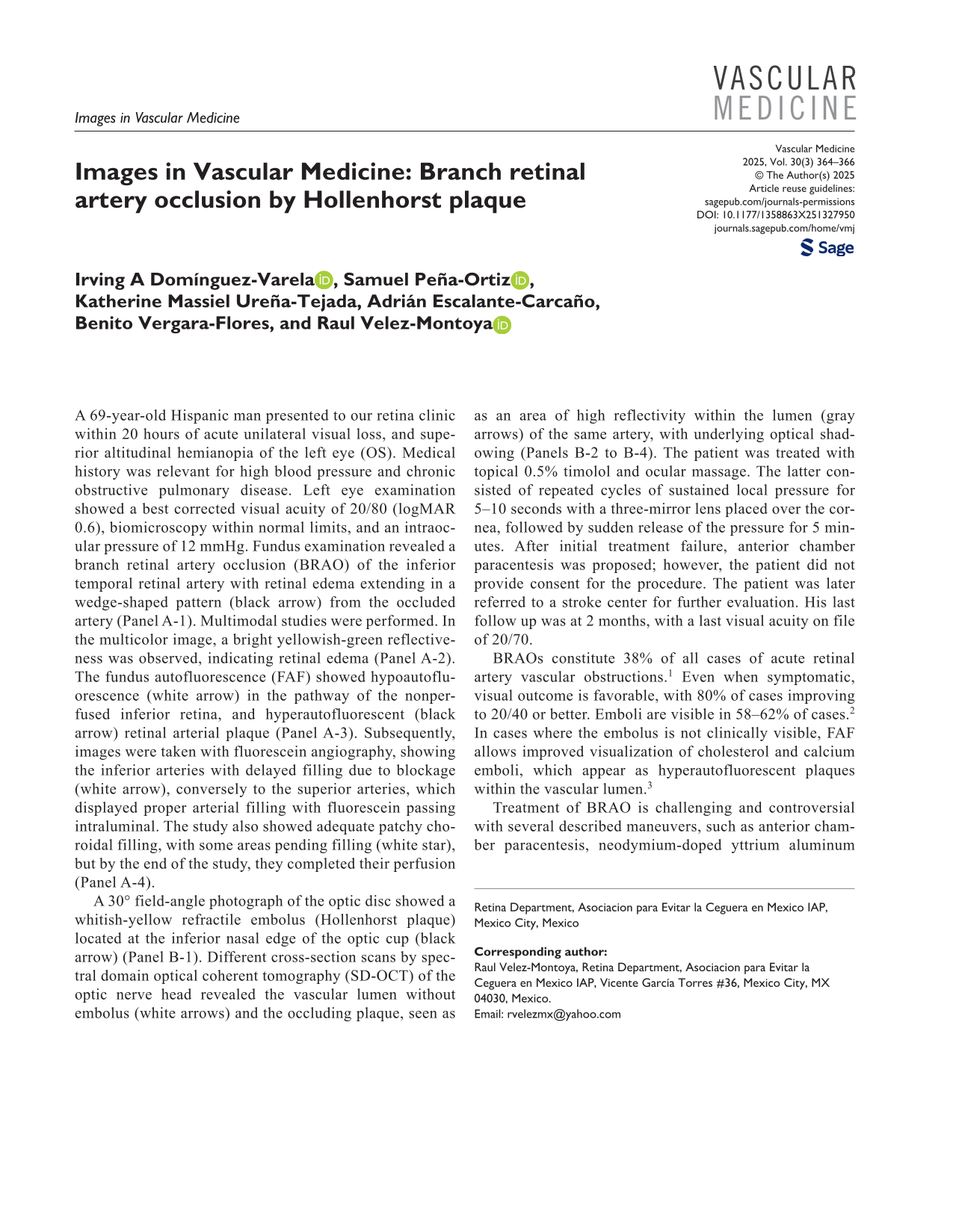
A 69-year-old Hispanic man presented to our retina clinic within 20 hours of acute unilateral visual loss, and superior altitudinal hemianopia of the left eye (OS). Medical history was relevant for high blood pressure and chronic obstructive pulmonary disease. Left eye examination showed a best corrected visual acuity of 20/80 (logMAR 0.6), biomicroscopy within normal limits, and an intraocular pressure of 12 mmHg. Fundus examination revealed a branch retinal artery occlusion (BRAO) of the inferior temporal retinal artery with retinal edema extending in a wedge-shaped pattern (black arrow) from the occluded artery (Panel A-1). Multimodal studies were performed. In the multicolor image, a bright yellowish-green reflectiveness was observed, indicating retinal edema (Panel A-2). The fundus autofluorescence (FAF) showed hypoautofluorescence (white arrow) in the pathway of the nonperfused inferior retina, and hyperautofluorescent (black arrow) retinal arterial plaque (Panel A-3). Subsequently, images were taken with fluorescein angiography, showing the inferior arteries with delayed filling due to blockage (white arrow), conversely to the superior arteries, which displayed proper arterial filling with fluorescein passing intraluminal. The study also showed adequate patchy choroidal filling, with some areas pending filling (white star), but by the end of the study, they completed their perfusion (Panel A-4).

A 30° field-angle photograph of the optic disc showed a whitish-yellow refractile embolus (Hollenhorst plaque) located at the inferior nasal edge of the optic cup (black arrow) (Panel B-1). Different cross-section scans by spectral domain optical coherent tomography (SD-OCT) of the optic nerve head revealed the vascular lumen without embolus (white arrows) and the occluding plaque, seen as as an area of high reflectivity within the lumen (gray arrows) of the same artery, with underlying optical shadowing (Panels B-2 to B-4). The patient was treated with topical 0.5% timolol and ocular massage. The latter consisted of repeated cycles of sustained local pressure for 5–10 seconds with a three-mirror lens placed over the cornea, followed by sudden release of the pressure for 5 minutes. After initial treatment failure, anterior chamber paracentesis was proposed; however, the patient did not provide consent for the procedure. The patient was later referred to a stroke center for further evaluation. His last follow up was at 2 months, with a last visual acuity on file of 20/70.

BRAOs constitute 38% of all cases of acute retinal artery vascular obstructions. 1 Even when symptomatic, visual outcome is favorable, with 80% of cases improving to 20/40 or better. Emboli are visible in 58–62% of cases. 2 In cases where the embolus is not clinically visible, FAF allows improved visualization of cholesterol and calcium emboli, which appear as hyperautofluorescent plaques within the vascular lumen. 3
Treatment of BRAO is challenging and controversial with several described maneuvers, such as anterior chamber paracentesis, neodymium-doped yttrium aluminum garnet (Nd:YAG) laser to disrupt the emboli, hyperbaric oxygen treatment, intra-arterial thrombolysis, and pars plana vitrectomy among others. 4 The objective of the acute treatment (within the first 24 hours from onset) is to attempt to destroy or mobilize the embolus further throughout the vascular stream, to improve retina perfusion. However, the rate of reported success varies widely and is somewhat unpredictable. 4
Footnotes
Declaration of conflicting interests
The authors declared no potential conflicts of interest concerning the research, authorship, and/or publication of this article.
Funding
The authors received no financial support for the research, authorship, and/or publication of this article.