
Abstract
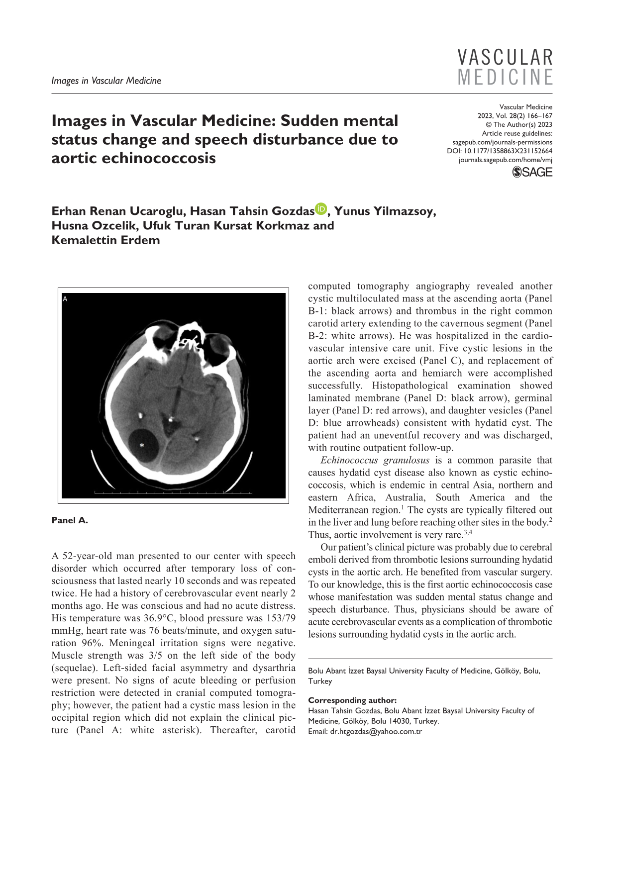
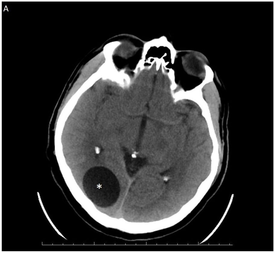
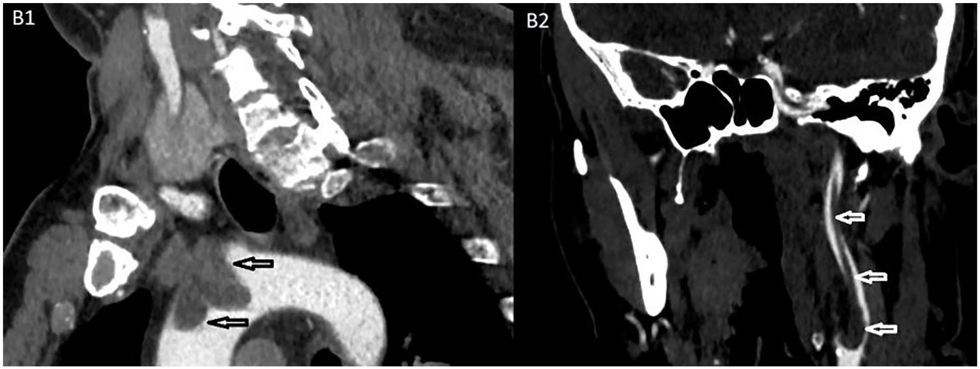
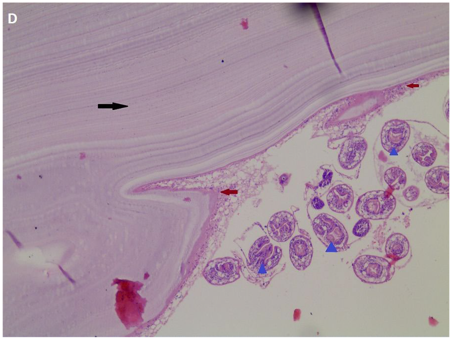
A 52-year-old man presented to our center with speech disorder which occurred after temporary loss of consciousness that lasted nearly 10 seconds and was repeated twice. He had a history of cerebrovascular event nearly 2 months ago. He was conscious and had no acute distress. His temperature was 36.9°C, blood pressure was 153/79 mmHg, heart rate was 76 beats/minute, and oxygen saturation 96%. Meningeal irritation signs were negative. Muscle strength was 3/5 on the left side of the body (sequelae). Left-sided facial asymmetry and dysarthria were present. No signs of acute bleeding or perfusion restriction were detected in cranial computed tomography; however, the patient had a cystic mass lesion in the occipital region which did not explain the clinical picture (Panel A: white asterisk). Thereafter, carotid computed tomography angiography revealed another cystic multiloculated mass at the ascending aorta (Panel B-1: black arrows) and thrombus in the right common carotid artery extending to the cavernous segment (Panel B-2: white arrows). He was hospitalized in the cardiovascular intensive care unit. Five cystic lesions in the aortic arch were excised (Panel C), and replacement of the ascending aorta and hemiarch were accomplished successfully. Histopathological examination showed laminated membrane (Panel D: black arrow), germinal layer (Panel D: red arrows), and daughter vesicles (Panel D: blue arrowheads) consistent with hydatid cyst. The patient had an uneventful recovery and was discharged, with routine outpatient follow-up.
Echinococcus granulosus is a common parasite that causes hydatid cyst disease also known as cystic echinococcosis, which is endemic in central Asia, northern and eastern Africa, Australia, South America and the Mediterranean region. 1 The cysts are typically filtered out in the liver and lung before reaching other sites in the body. 2 Thus, aortic involvement is very rare.3,4
Our patient’s clinical picture was probably due to cerebral emboli derived from thrombotic lesions surrounding hydatid cysts in the aortic arch. He benefited from vascular surgery. To our knowledge, this is the first aortic echinococcosis case whose manifestation was sudden mental status change and speech disturbance. Thus, physicians should be aware of acute cerebrovascular events as a complication of thrombotic lesions surrounding hydatid cysts in the aortic arch.
‘Images in vascular medicine’ is a regular feature of Vascular Medicine. Readers may submit original, unpublished images related to clinical vascular medicine. Submissions may be sent to: Daniella Kadian-Dodov, Images Section Editor, Vascular Medicine, via the web-based submission system at http://mc.manuscriptcentral.com/vascular-medicine
Footnotes
Declaration of conflicting interests
The authors declared no potential conflicts of interest with respect to the research, authorship, and/or publication of this article.
Funding
The authors received no financial support for the research, authorship, and/or publication of this article.