



Abstract

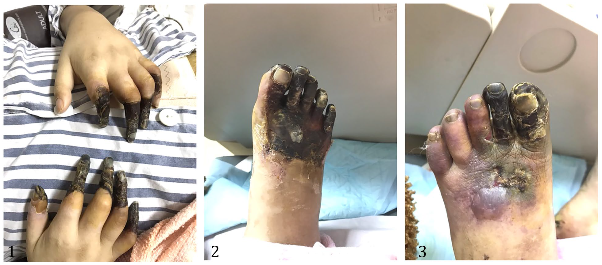
A 30-year-old woman presented to our clinic with a 1-month history of progressive pain in the fingers and toes. She took no regular medication, rarely drank alcohol, and smoked. Physical examination showed black discoloration and dry gangrene of the distal fingers (Panel A-1). Similar changes were visualized on the toes of the right foot (Panel A-2), and purple discoloration of the 3rd to 5th toes of the left foot, with broken skin on the front dorsum (Panel A-3). There was associated edema but no sclerodactyly or telangiectasias. The dorsal pedis pulses were weak. Laboratory tests revealed significantly abnormal cardiac and renal function: brain natriuretic peptide (BNP) of 14,243.3 pg/mL (normal 45–84 μmol/L), creatine kinase (CK) of 869 U/L (normal 26–140 U/L), creatine kinase-MB (CK-MB) of 143.5 ng/mL (normal 0–5 ng/mL), troponin I of 50.988 μg/L (normal 0–0.05 μmol/L), and creatinine of 528 μmol/L (normal 45–84 μmol/L). The test for hepatitis B virus (HBV) was positive; antineutrophil cytoplasmic antibodies, antinuclear antibodies, antiphospholipid antibodies, and cryoglobulin were negative. The cultures of skin purulent secretion indicated the growth of Stenotrophomonas maltophilia. The electrocardiogram showed poor anterior wall R-wave escalation and ST-segment elevation. Vascular computerized tomography examination showed lumen segmental stenosis of the coronary artery (Panel B-1: arrow) and the entire lower limb arteries (Panel B-2: arrow). Skin biopsy revealed a necrotizing vasculitis affecting medium and small arteries in the deep dermis and subcutis (Panel B-3: arrows).

With the diagnosis of polyarteritis nodosa (PAN) combined with infection, she was treated with glucocorticoids (methylprednisolone 1 g/day for 3 days, then 1 mg/kg/day for subsequent therapy) and sensitive antibiotics. The symptoms of pain diminished, and the levels of C-reactive protein (CRP), erythrocyte sedimentation rate (ESR), procalcitonin, creatinine, CK, CK-MB, and BNP declined to almost normal. There was no further deterioration of the remaining limbs. However, 2 weeks after discharge, she died at home suddenly, possibly from a massive heart attack.
PAN is a vasculitic disease characterized by necrotizing vasculitis – inflammatory lesions in blood vessels that lead to vessel wall necrosis. 1 The etiopathogenesis of PAN is still unclear; evidence shows it is associated with HBV infection.2,3 The incidence of cardiac injury is about 36–65%, which is one of the main causes of death. 4 European Alliance of Associations for Rheumatology (EULAR) recommendations suggest the use of glucocorticoids and cyclophosphamide for remission induction. In conclusion, our case illustrates the dangers of polyarteritis nodosa, especially cardiac involvement, which can lead to sudden death, even when clinical symptoms and laboratory indicators are significantly improved.
‘Images in vascular medicine’ is a regular feature of Vascular Medicine. Readers may submit original, unpublished images related to clinical vascular medicine. Submissions may be sent to: Daniella Kadian-Dodov, Images in Vascular Medicine Section Editor, Vascular Medicine, via the web-based submission system at http://mc.manuscriptcentral.com/vascular-medicine
Footnotes
Declaration of conflicting interests
The authors declared no potential conflicts of interest with respect to the research, authorship, and/or publication of this article.
Funding
This research was supported by the Traditional Chinese Medical Science and Technology Project of Zhejiang Province (grant no. 2022ZB130).
