
Abstract

Two patients were referred for pulmonary computer tomographic angiogram (CTA) at our tertiary referral hospital for suspected venous thromboembolic pulmonary emboli (PE).
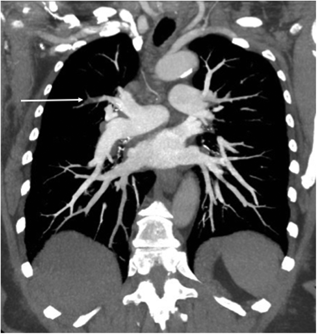
Patient 1, a 69-year-old woman, underwent bilateral total knee arthroplasties for primary osteoarthritis. The 160-minute operation proceeded without complication. Forty hours post-operatively she became acutely dyspnoeic and her peripheral oxygen saturation fell to 67% on room air. The pulmonary CTA demonstrated a 20-mm-long filling defect in the right upper lobe segmental pulmonary artery with a density of −65 Hounsfield units, consistent with fat embolus (Panel A: white arrow). There were no parenchymal lung abnormalities. Compression duplex ultrasound of her lower limb deep veins was normal. A comprehensive neurological and dermatological exam was normal, and the patient was diagnosed with fat embolism and treated with supplemental oxygen, which was successfully weaned over the subsequent 6 days.
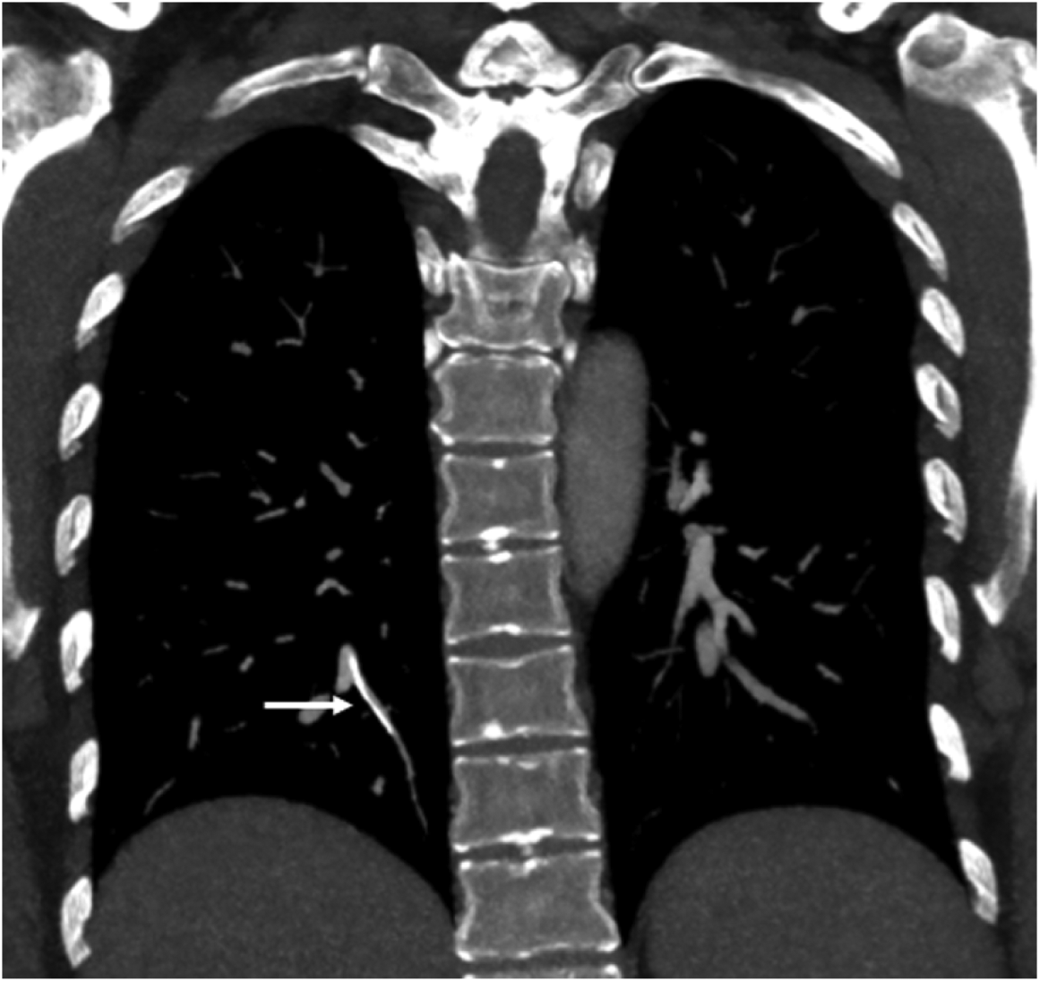
Patient 2, a 42-year-old woman, presented with bilateral leg swelling and pain. She had experienced an iliofemoral deep vein thrombosis (DVT) 5 years prior that was treated with catheter-directed thrombolysis, placement of a Bard Recovery G2X® inferior vena cava (IVC) filter (Tempe, AZ, USA), and 12 months of therapeutic anticoagulation. A CT venogram showed extensive IVC and iliofemoral DVT. The maximum intensity projection sequence of subsequent CTPA revealed three IVC filter struts in the segmental left and right pulmonary arteries without associated thrombotic filling defects on contrast images. The right lower lobe strut is shown lodged in a right posterobasal subsegmental pulmonary artery in Panel B (white arrow). The patient was recommenced on therapeutic anticoagulation, and subsequent removal and inspection of the IVC filter confirmed the fracture of three struts.
Macroscopic non-thrombotic emboli are rarely visualised but are important to consider in the differential diagnosis when pulmonary artery filling defects are visualised on CTA. The correct diagnosis can usually be made with careful clinical and imaging review including assessment of non-contrast sequences for radiopaque emboli. These cases illustrate the macroscopic appearance of fat and IVC filter strut emboli. Other reported non-thrombotic emboli include tumours, inorganic particulates, septic emboli, and hydatid cysts. 1
‘Images in vascular medicine’ is a regular feature of Vascular Medicine. Readers may submit original, unpublished images related to clinical vascular medicine. Submissions may be sent to: Heather Gornik, Editor in Chief, Vascular Medicine, via the web-based submission system at http://mc.manuscriptcentral.com/vascular-medicine
Footnotes
Declaration of conflicting interests
The authors declared no potential conflicts of interest with respect to the research, authorship, and/or publication of this article.
Funding
The authors received no financial support for the research, authorship, and/or publication of this article.