



Abstract

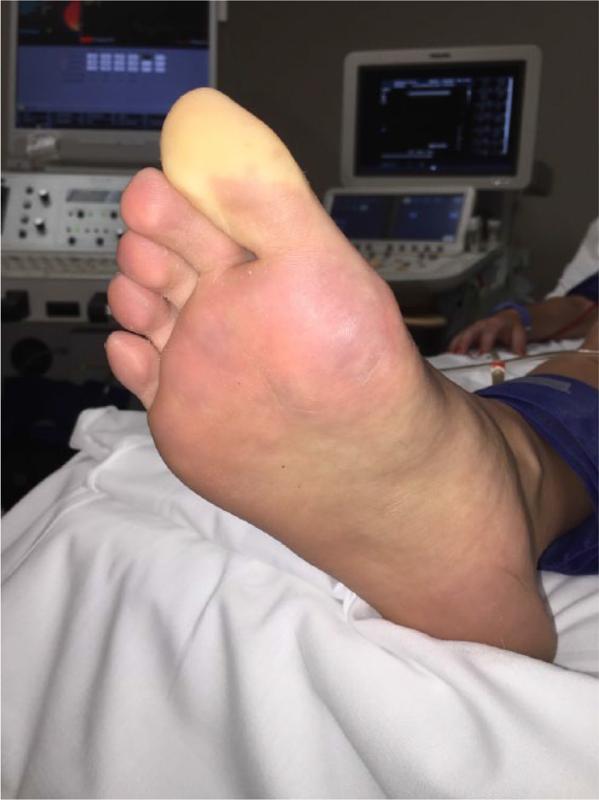
A non-smoking, 19-year-old, college volleyball athlete presented with exertional discomfort, pallor, and coldness of her right hallux, second, and third toes (1). Profound pallor of the hallux was her most alarming manifestation. She reported no symptoms at rest, or with normal activities, only with vigorous workouts. Her symptoms resolved after 30 to 60 minutes upon treatment with hot water and massaging.
Neurogenic vasodysregulation, manifest as Raynaud phenomenon, of her right great toe was provoked with a 6-minute treadmill exercise test (Panel A). Additionally, her resting ankle–brachial index (ABI) was 1.02, which dropped to 0.74 following 6 minutes of treadmill walking.
A computed tomographic (CT) angiogram with active flexion maneuvers of the abdominal aorta and lower extremities demonstrated almost complete occlusion of the right supra-geniculate popliteal artery by the medial head of the gastrocnemius muscle (Panel B). There was also nearly 50% narrowing of the left supra-geniculate popliteal artery, again by the medial head of the gastrocnemius. Upon diagnosis of popliteal artery entrapment syndrome (PAES) of the right lower extremity, the patient was referred for surgical repair.

Surgical exploration did confirm compression of the popliteal artery by the medial head of the gastrocnemius muscle (Panel C), which was successfully resected. Tibial nerve compression could not be demonstrated intraoperatively as the patient was paralyzed.

The clinical presentation of PAES is usually that of exertional calf pain (2). This case was unusual, as a prominent feature was neurogenic vasodysregulation, manifest as Raynaud phenomenon, of the right great toe (3). We theorize that the tibial nerve was intermittently compressed due to its close proximity to the anterior aspect of the medial head of the gastrocnemius, which elicited classic Raynaud phenomenon. Clinicians should consider PAES when a patient presents with exertional Raynaud phenomenon of the toes yet no calf pain. Our patient had an uncomplicated postoperative course, and is now symptom free.
‘Images in vascular medicine’ is a regular feature of Vascular Medicine. Readers may submit original, unpublished images related to clinical vascular medicine. Submissions may be sent to: Heather Gornik, Editor in Chief, Vascular Medicine, via the web-based submission system at http://mc.manuscriptcentral.com/vascular-medicine
Footnotes
Declaration of conflicting interests
The authors declared no potential conflicts of interest with respect to the research, authorship, and/or publication of this article.
Funding
The authors received no financial support for the research, authorship, and/or publication of this article.