
Abstract

A 45-year-old woman presented to our outpatient clinic complaining about positional skin lesions in her lower limbs. Her past medical history was significant for CREST syndrome (CREST: calcinosis, Raynaud phenomenon, esophageal dysmotility, sclerodactyly, and telangiectasia) with skin thickening limited to areas distal to the elbows and knees, severe Raynaud’s phenomenon, calcinosis, sclerodactyly and positive anti-centromere antibodies. Although the calves were unremarkable when examined in a recumbent position (Panel A), leg raising elicited distinct longitudinal venous furrowing consistent with a ‘groove sign’ (Panel B and Video (available as supplementary material)). Laboratory and immunologic analysis showed no evidence of disease activity. Cutaneous biopsy documented an unspecific lymphocytic infiltrate, with no presence of eosinophilic cells.
Panels A and B.
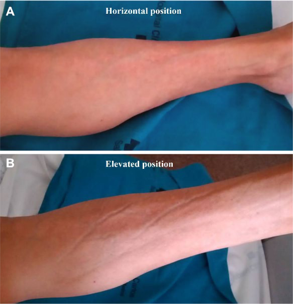
The groove sign presents as skin indentations that follow the pathway of superficial veins. It can be induced by maneuvers that increase venous return, such as leg raising. This sign is probably due to a relative split of skin layers secondary to a fibrotic process between dermis and epidermis. Despite being primarily associated with eosinophilic fasciitis, 1 it can be observed in other diseases of sclerodermal-spectrum disorders.
‘Images in vascular medicine’ is a regular feature of Vascular Medicine. Readers may submit original, unpublished images related to clinical vascular medicine. Submissions may be sent to: Heather Gornik, Editor in Chief, Vascular Medicine, via the web-based submission system at http://mc.manuscriptcentral.com/vascular-medicine
Footnotes
Declaration of conflicting interests
The authors declared no potential conflicts of interest with respect to the research, authorship, and/or publication of this article.
Funding
The authors received no financial support for the research, authorship, and/or publication of this article.
References
Supplementary Material
Please find the following supplemental material available below.
For Open Access articles published under a Creative Commons License, all supplemental material carries the same license as the article it is associated with.
For non-Open Access articles published, all supplemental material carries a non-exclusive license, and permission requests for re-use of supplemental material or any part of supplemental material shall be sent directly to the copyright owner as specified in the copyright notice associated with the article.