



Abstract

A 54-year-old woman with longstanding limited cutaneous systemic sclerosis presented with a several year history of progressively enlarging painful finger nodules and recurrent wintertime fingertip ulcerations. Periodically, the nodules would either ulcerate and/or emit a chalky white discharge.
An examination of her hands and fingers was remarkable for diffusely sclerotic skin, acro-osteolysis, small distal ischemic crusts, punctate telangiectasias, and distally located tumor-like papules and nodules. Yellow-white calcific sediment extruded from the larger nodules, which were especially prominent on the right long finger (Panel A). Plain film radiography of the right hand displayed prominent confluent calcific deposits within the skin and subcutaneous tissues that were most severe within the distribution of the nodules (Panel B). Serum calcium, phosphorous, and parathyroid hormone were normal. Consequently, a diagnosis of severe dystrophic calcinosis cutis was rendered.
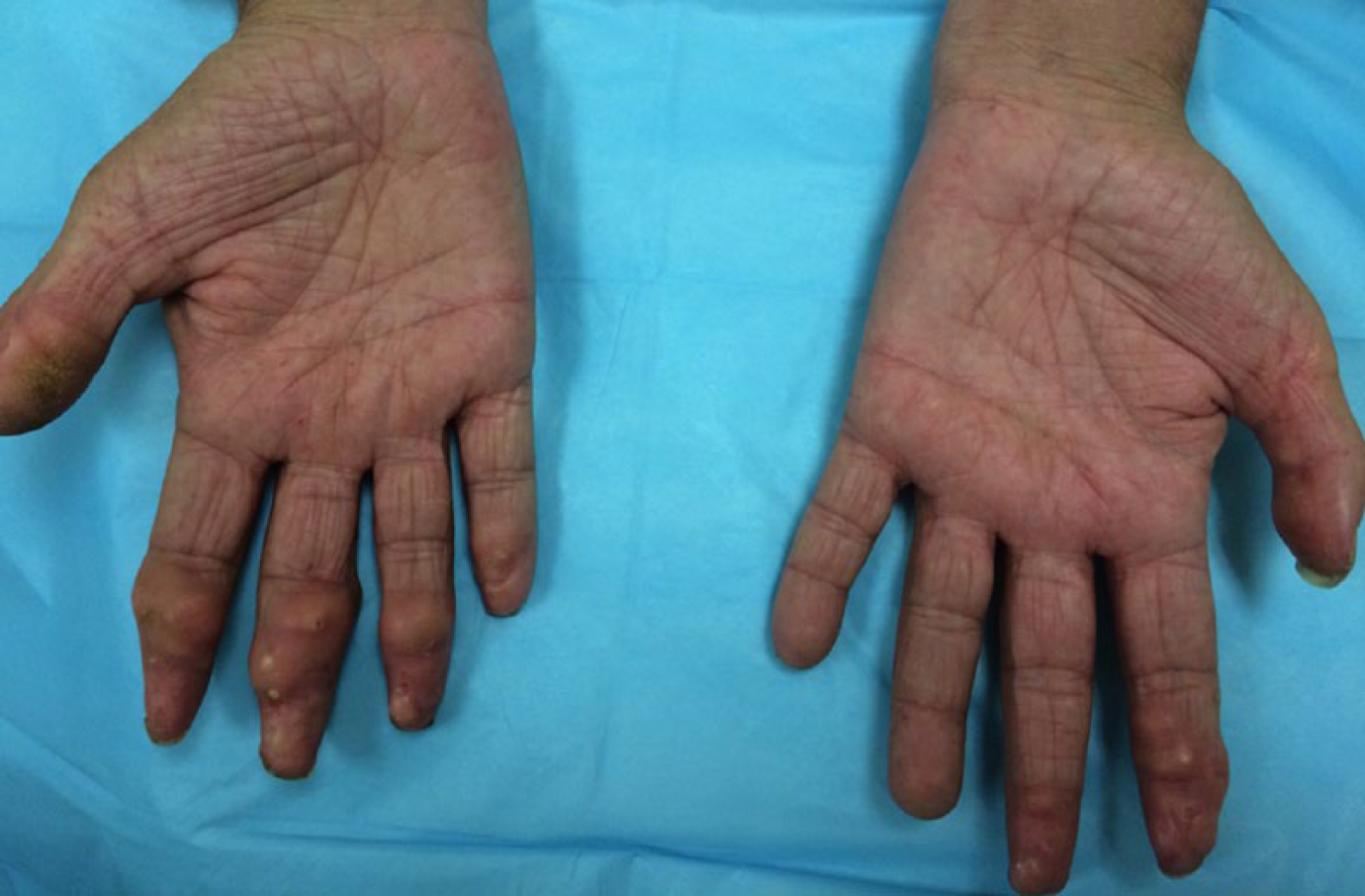
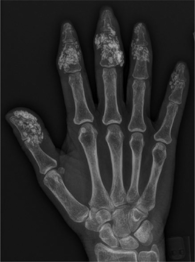
Calcinosis cutis results from the deposition of insoluble calcium within the skin and subcutaneous tissues. Four subtypes of calcinosis cutis have been described: dystrophic, metastatic, idiopathic, and iatrogenic. 1 The dystrophic subtype represents the most common cause of calcinosis cutis and occurs within chronically damaged tissue in the setting of normal serum calcium and phosphorous. Additionally, it is responsible for the calcific deposition that complicates autoimmune disease. Dystrophic calcinosis cutis of variable severity affects approximately 25% of scleroderma patients and is more likely to occur in the limited variant or CREST syndrome. The calcific pathogenesis remains unclear but is potentially related to local trauma, chronic inflammation, and tissue hypoxia. 2 Although calcinosis cutis may be an incidental radiographic finding, it can be debilitating with attendant pain, inflammation, and significant functional impairment. The diagnosis is typically clinically evident (papules/nodules/chalk-like discharge) and supported by plain film radiography. Histology can confirm the diagnosis, yet is rarely required. Concurrent ischemic complications often complicate dystrophic calcinosis cutis and include acro-osteolysis, digital pits and ulcerations.
Therapeutic options are limited and response rates are variable. Vasodilators are recommended in order to reduce tissue ischemia. Specific pharmacological options include diltiazem, minocycline, bisphosphonates, biological agents (infliximab, rituximab), and colchicine. 3 Surgical excision is reserved for severely painful and/or ulcerative nodules yet recurrence is common and intervention may stimulate additional calcification. The patient could not tolerate various medications but was treated with intermittent surgical debridement of the calcific nodules with modest clinical response.
‘Images in vascular medicine’ is a regular feature of Vascular Medicine. Readers may submit original, unpublished images related to clinical vascular medicine. Submissions may be sent to: Heather Gornik, Editor in Chief, Vascular Medicine, via the web-based submission system at http://mc.manuscriptcentral.com/vascular-medicine
Footnotes
Heather L. Gornik served as guest editor for this manuscript.
Declaration of conflicting interests
The author declared no potential conflicts of interest with respect to the research, authorship, and/or publication of this article.
Funding
The author received no financial support for the research, authorship, and/or publication of this article.