



Abstract

We present the case of an 85-year-old woman with acute diverticulosis who underwent abdominal computed tomography (CT). The CT incidentally revealed a giant splenic aneurysm (127 × 125 mm) (Panel A). Selective angiography was performed to demonstrate aneurysm permeability, which confirmed active inflow (Panel B). Consequently, a 10 × 7 mm Amplatzer Vascular Plug was placed into the splenic artery to embolize the aneurysm (Panel C) with no intra or postoperative complications. Abdominal CT performed 3 weeks after the intervention documented complete thrombosis of the aneurysm (Panel D).
Panel A.

Panel B.
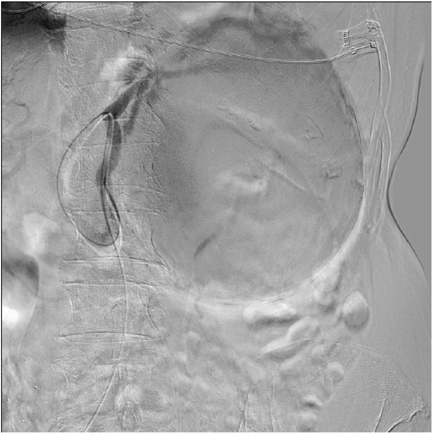
Panel C.

Panel D.

Splenic artery aneurysms are the third most common after aortic and iliac artery aneurysms. They are more commonly reported in female patients (ratio 4:1); however, rupture risk is greater in men. Usually they appear within the distal splenic artery and their diameter is no larger than 3 cm. Portal hypertension, cirrhosis, pregnancy, and hepatic transplant are the most important etiological risk factors. Usually asymptomatic, most of them are incidentally diagnosed. Endovascular treatment has better short-term results and lower perioperative morbidity and mortality; thus it is the treatment of choice if the splenic artery has a suitable anatomy. Remarkably, fewer than 25 giant splenic aneurysm cases have been published in literature.
‘Images in vascular medicine’ is a regular feature of Vascular Medicine. Readers may submit original, unpublished images related to clinical vascular medicine. Submissions may be sent to: Heather Gornik, Editor in Chief, Vascular Medicine, via the web-based submission system at http://mc.manuscriptcentral.com/vascular-medicine
Footnotes
Conflict of interest
The authors declare that there is no conflict of interest.
Funding
This research received no specific grant from any funding agency in the public, commercial, or not-for-profit sectors.