
Abstract

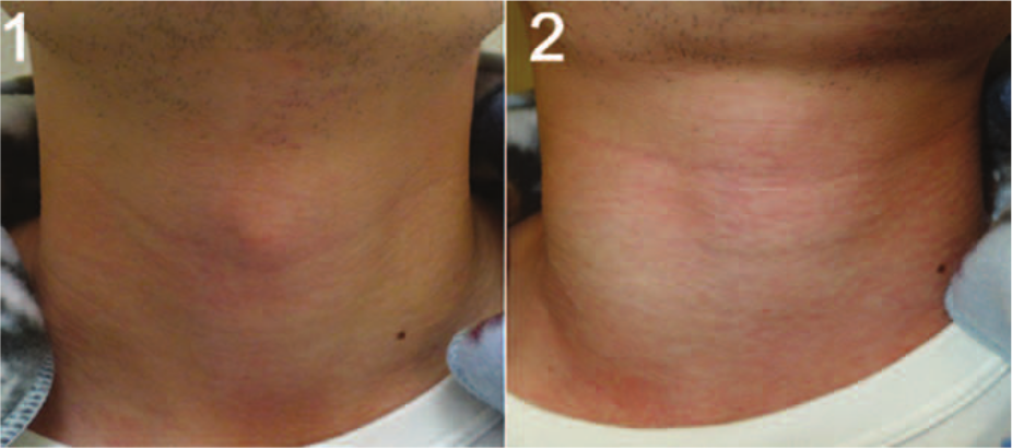
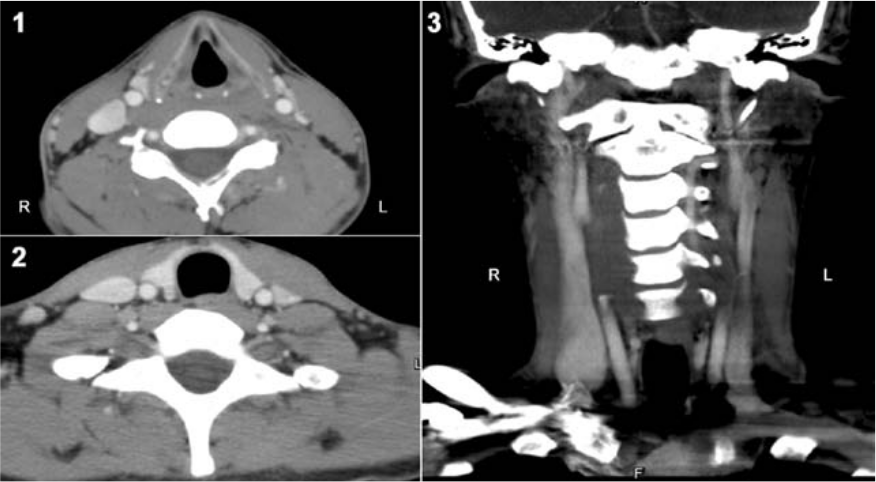
We report the case of a 24-year-old patient who presented with right-sided neck swelling that increased on straining. He had noticed this since his childhood. Physical examination with the patient in the resting state showed no palpable or visible mass in his neck (Panel A1). However, on performance of the Valsalva maneuver, a dynamic swelling over his right neck was noted (Panel A2). Laryngoscopy was performed and showed no visible laryngocele or other lesions. Contrast-enhanced computed tomographic angiography of the neck revealed a dilated lumen of the right internal jugular vein (Panel B1–3) with no mass or diverticulum in the neck or upper mediastinum. A clinical diagnosis of internal jugular vein phlebectasia (IJVP) was made.
IJVP refers to fusiform dilatation of the vein without tortuosity and usually presents as a neck mass in children. 1 Right-sided IJVP is more common than left. Although several theories have been proposed regarding the pathogenesis of IJVP, including certain anatomical and physical factors, its etiologies remain unknown.2,3 The evaluation of suspected IJVP includes medical history, physical examination (the Valsalva maneuver is most important for establishing the diagnosis), and imaging techniques including venography, computed tomographic or magnetic resonance angiography, and color flow Doppler imaging.2,4 The differential diagnosis of dynamic swelling in the neck on straining includes tumors or cysts of the upper mediastinum, external laryngeal diverticula or laryngoceles, and inflation of the apex of the lung. 2
Surgery is recommended in selected cases of IJVP given the potential to rupture, the possibility of formation of intramural thrombosis with subsequent pulmonary embolism, or in consideration of cosmetic and psychological factors. 4 The technique of constriction suture venoplasty plus encapsulation has been preferred for cases of phlebectasia of the internal jugular vein, while ligation or resection of the involved vein may also be considered, particularly in cases of external jugular involvement. 4 Because the patient had no bothersome symptoms, complications or cosmetic requirements, conservative management was recommended. This case illustrates the importance of including IJVP in the differential diagnosis of neck swelling and the importance of performing neck examination with the Valsalva maneuver in such cases.
‘Images in vascular medicine’ is a regular feature of Vascular Medicine. Readers may submit original, unpublished images related to clinical vascular medicine. Submissions may be sent to: Mark A Creager, Editor in Chief, Vascular Medicine, via the web-based submission system at http://mc.manuscriptcentral.com/vascular-medicine
Footnotes
Declaration of conflicting interest
The authors declare that there is no conflict of interest.
Funding
This research received no specific grant from any funding agency in the public, commercial, or not-for-profit sectors.