



Abstract

Renal vascular malformation is an abnormal communication between the intrarenal arterial and venous system. These malformations include arteriovenous malformation (AVM) and arteriovenous fistula (AVF). 1 Renal AVM is a congenital developmental abnormality in which the renal artery and renal vein communicate through a network of abnormal vessels. By contrast, renal AVF is a direct communication between an artery and a vein. The etiology of congenital AVM is unknown, while the cause of acquired renal AVF is usually found to be percutaneous renal biopsy, other procedures, and incidental renal trauma.
A 57-year-old woman visited our hospital with intermittent left flank discomfort of several years’ duration. The symptom had worsened with position change and had become particularly bothersome during the preceding few months. She had no past medical history including trauma or procedures. On physical examination, a continuous bruit was audible over the left flank area. Urinalysis and blood tests showed normal findings. A contrast-enhanced computed tomography scan of the abdomen demonstrated aneurysmally dilated enhancing vessels with attenuation similar to the arteries in the left kidney (Panel A). Digital subtraction arteriography showed a feeding artery to a large AVM with abnormal dilation of the nidus and early opacification of venous flow (Panel B1). In this patient, the large AVM was treated with interlock coils (Panel B2). After super-selective catheterization, coil-embolization of all feeders was performed successfully (Panel B3). Magnetic resonance imaging (MRI) before treatment presented aneurysmally dilated vessels with high flow and suggested an underlying vascular malformation (Panel C1). A follow-up MRI taken 6 months post-embolization showed successful occlusion of the AVM with coils (Panel C2).


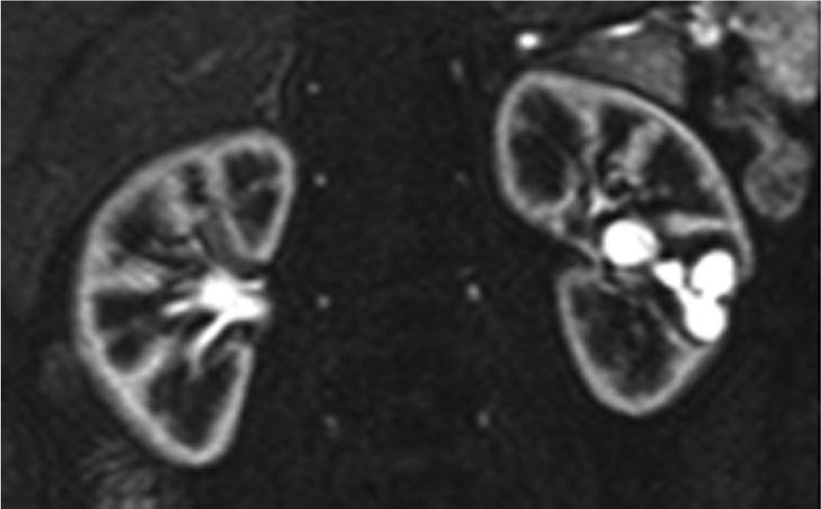

In this case, AVM was managed with deployment of coils into the vascular malformation, which promoted thrombosis. 2 Complications can arise from the rapid shunting of the coils into the venous circulation, leading to emboli entering the pulmonary circulation or the left side of the heart if interarterial communication exists. Blocking the venous output of the AVM can help in preventing this complication. It is also important for the angiographer to be familiar with retrieval techniques if such a complication arises. 3
‘Images in vascular medicine’ is a regular feature of Vascular Medicine. Readers may submit original, unpublished images related to clinical vascular medicine. Submissions may be sent to: Mark A Creager, Editor in Chief, Vascular Medicine, via the web-based submission system at http://mc.manuscriptcentral.com/vascular-medicine
Footnotes
Declaration of conflicting interest
The authors report no conflicts of interest. The authors alone are responsible for the content and writing of the paper.
Funding
This research received no specific grant from any funding agency in the public, commercial, or not-for-profit sectors.