
Abstract

A 72-year-old man, with no medical history of interest, was referred to our clinic for evaluation of tiredness and heaviness in the upper limbs, which was somewhat more pronounced on the left side and was of approximately 6 months in evolution and slow in onset. He denied any association with effort, but reported a certain worsening during the summer and improvement with the local application of cold compresses. Physical examination revealed the presence of multiple, superficial, soft tumors along the venous pathways of bilateral arms, which appeared to be partially thrombosed venous dilatations. Arterial examination of the upper limbs was completely normal, and there were no significant findings on vascular examination of the lower limbs. Magnetic resonance angiography showed ectasia of the deep and superficial veins of the upper limbs, later confirmed by computer tomography. The images correspond to the upper left limb, with diffuse, partially thrombosed ectasia (Panel A) of the deep (Panel B) and superficial (Panel C) venous system, with permeability and without ectasia of the arterial axis (Panel D). The main deep veins, innominate trunk and superior vena cava were patent without any ectasia.

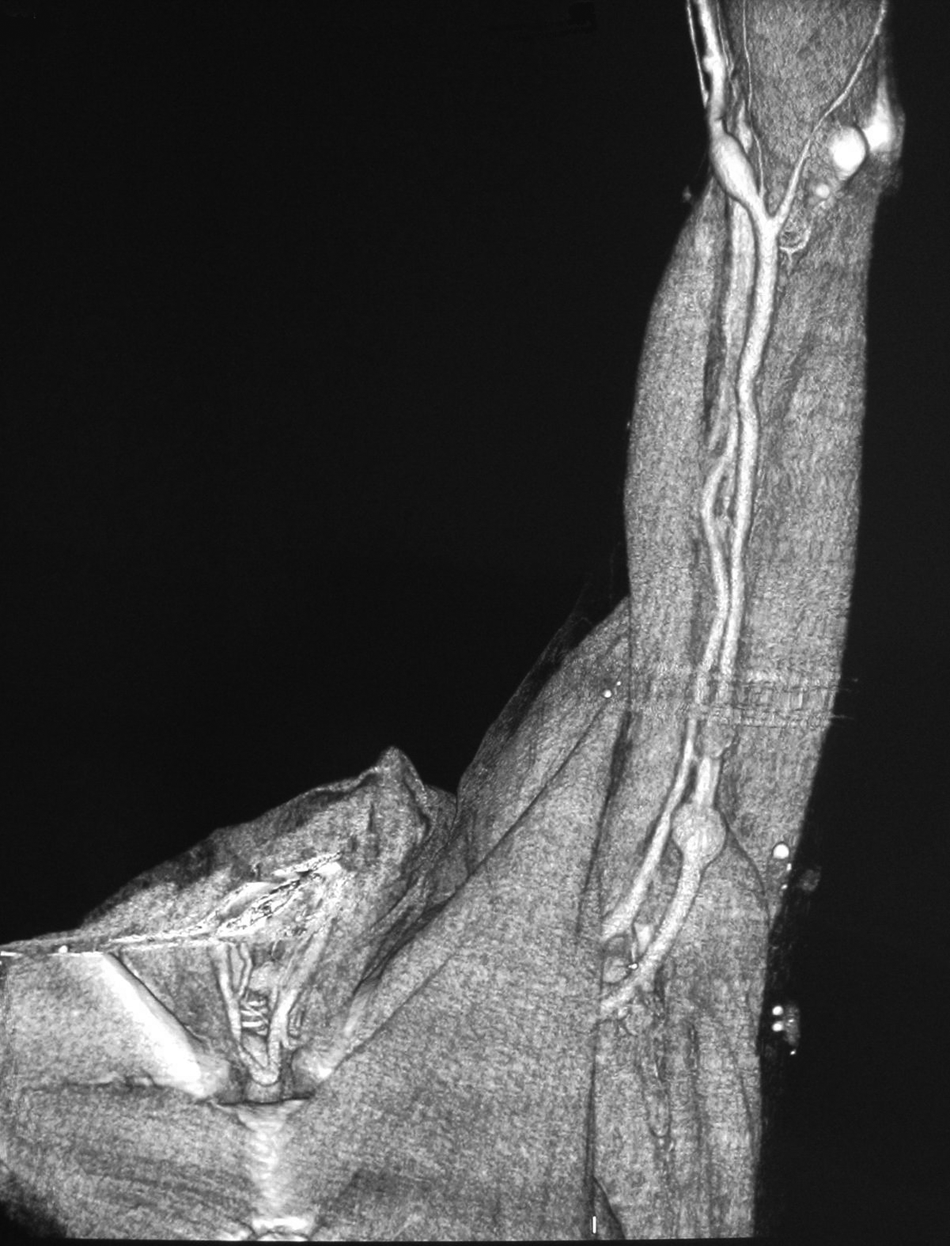


Philipp Bockenheimer described Bockenheimer’s syndrome, or genuine diffuse phlebectasia, for the first time in 1907 1 in a 52-year-old patient with clinical symptoms similar to those of our patient. Later reported cases mainly corresponded to pediatric patients. 2 The clinical signs vary according to the patient, and the severity of symptoms is conditioned by the size of the phlebectasia, generally in the upper limbs as in the original case, although it has also been described in the lower limbs and genitals. 3 A histological examination shows veins with reduced smooth muscle and elastin in the walls, with thrombosis and intermittent phleboliths. 4 The main difference between phlebectasia compared with other venous malformation syndromes (e.g. Parkes Weber, Klippel-Trenaunay) is the absence of port-wine stains. Treatment includes elastic support measures. In general, complete surgical resection is practically impossible and embolization increases the risk of skin necrosis. 2
‘Images in vascular medicine’ is a regular feature of Vascular Medicine. Readers may submit original, unpublished images related to clinical vascular medicine. Submissions may be sent to: Mark A Creager, Editor in Chief, Vascular Medicine, via the web-based submission system at http://mc.manuscriptcentral.com/vascular-medicine
Footnotes
This research received no specific grant from any funding agency in the public, commercial, or not-for-profit sectors.
The authors declare that there is no conflict of interest.