
Abstract
Background:
Autoimmune disorders including nephropathies have been reported more frequently in alemtuzumab-treated multiple sclerosis (MS) patients than in the general population.
Objective:
Describe instances of autoimmune nephropathy in alemtuzumab-treated MS patients.
Methods:
Cases were identified from safety monitoring within the alemtuzumab relapsing-remitting multiple sclerosis (RRMS) clinical development program (CDP) or post-marketing, or following off-label use.
Results:
As of 16 June 2017, 16 autoimmune nephropathies have occurred following alemtuzumab treatment for MS. The incidence of autoimmune nephropathies was 0.34% within the CDP (5/1485 patients). The five CDP cases (one of anti-glomerular basement membrane (anti-GBM) disease, two of membranous glomerulonephropathy, and two of serum anti-GBM antibody without typical anti-GBM disease) were identified early, responded to conventional therapy (where needed), and had favorable outcomes. Three of 11 cases outside the CDP occurred following off-label alemtuzumab use prior to approval for RRMS and were all anti-GBM disease. Diagnosis was delayed in one of these three cases and another did not receive appropriate treatment; all three cases resulted in end-stage renal failure. All anti-GBM disease cases with documented urinalysis demonstrated prior microscopic hematuria.
Conclusion:
Close monitoring of alemtuzumab-treated MS patients facilitates diagnosis and treatment early in the nephropathy course when preservation of renal function is more likely.
Keywords
Introduction
Anti-glomerular basement membrane (anti-GBM) disease following alemtuzumab therapy in multiple sclerosis (MS) patients was first reported in 2006. 1 Subsequently, there has been intense renal surveillance during clinical trial2–6 and non-trial treatments.
Although drug-related nephropathies are well described with several mechanisms recognized, 7 alemtuzumab-related nephropathies occur in the context of an increased risk of various de novo autoimmune disorders following alemtuzumab treatment for MS, most commonly thyroid autoimmune conditions (5-year incidences: all thyroid disorders 40%–45%; serious thyroid adverse events 4%–5%)8,9 and immune thrombocytopenia (1%–3%).8–10
Alemtuzumab was initially approved (Campath®/MabCampath®) for treatment of B-cell chronic lymphocytic leukemia; 11 it has also been used in off-label applications, including MS (prior to approval for this indication), 1 refractory vasculitis, 12 solid organ transplantation, 13 and as a conditioning regimen for hematopoietic stem cell transplantation. 14 Alemtuzumab (LEMTRADA®) has been approved in more than 70 countries for use in adults with relapsing forms of MS, including countries of the European Union for patients with active relapsing-remitting multiple sclerosis (RRMS) defined by clinical and imaging features, and in the US for patients who generally have had an inadequate response to two or more prior therapies.15,16 For RRMS, alemtuzumab is dosed as two initial treatment courses at baseline and 12 months later, and additional courses (up to two in Europe; no limit in the United States) as needed, administered ⩾12 months after the previous dose.15,16 The drug is detectable in serum for ~30 days after administration. 15
De novo autoimmunity may relate to the proposed mechanism of action of alemtuzumab. This humanized monoclonal antibody targets B and T lymphocytes (both CD4+ and CD8+ T cells) for depletion,17,18 followed by a distinctive pattern of B- and T-cell repopulation. B cells return to baseline levels within approximately 6 months; T cells recover more slowly, with levels generally reaching lower limits of normal by 12 months. 19 Relative increases in memory and regulatory T-cell levels occur during immune reconstitution, resulting in a shift from pro- to anti-inflammatory cytokine profiles. 20 Decreases in circulating B and T cells and their altered behavior upon subsequent repopulation may explain the efficacy of alemtuzumab in reducing the risk of relapses, delaying disease progression, and providing efficacy in the absence of continuous treatment.19,21,22 Additional mechanistic studies are required to establish this hypothesis.
Clinical trials have established alemtuzumab as a highly efficacious disease-modifying therapy in patients with active RRMS.2–4 Effects on disease activity include improvements in relapse and disability outcomes, reduction in the occurrence of new brain magnetic resonance imaging (MRI) lesions, and reduction of brain volume loss to levels similar to those in people without MS.8,9,23–27
The purpose of this article is to review the cases of de novo renal autoimmunity in MS patients treated with alemtuzumab within or outside of the clinical development program (CDP) to better understand their natural history, and to assess implications for monitoring and treatment.
Patients and methods
Cases of autoimmune nephropathy arising in alemtuzumab-treated MS patients are reported as of 16 June 2017. Cases occurring within the CDP were identified under a safety monitoring program run by the manufacturer that included renal monitoring (Table 1). Nephropathy cases are described alphabetically based on overall chronology (Figure 1). Five cases occurred within the CDP (patients C–F and J), comprising the phase 2 CAMMS223 (NCT00050778) trial, 2 phase 3 CARE-MS I (NCT00530348) and CARE-MS II (NCT00548405) trials,3,4 4-year open-label CAMMS03409 extension study (NCT00930553),8,9 and ongoing TOPAZ extension study (NCT02255656). 29 Patients provided written informed consent for the analyses described in this study. Surveillance in these trials included measurements of serum creatinine, urine dipstick tests for proteinuria and hematuria, and urine microscopy (monitoring frequency was initially quarterly, but increased to monthly after the first CDP case (patient C) was reported). Additional nephropathy cases have occurred post-marketing, and outside of either the CDP or post-marketing setting (patients A, B, and G) following off-label use of alemtuzumab (Campath®/MabCampath®). A Medline search revealed no additional reported cases.
Summary of routine renal safety monitoring within the alemtuzumab CDP and post-marketing.
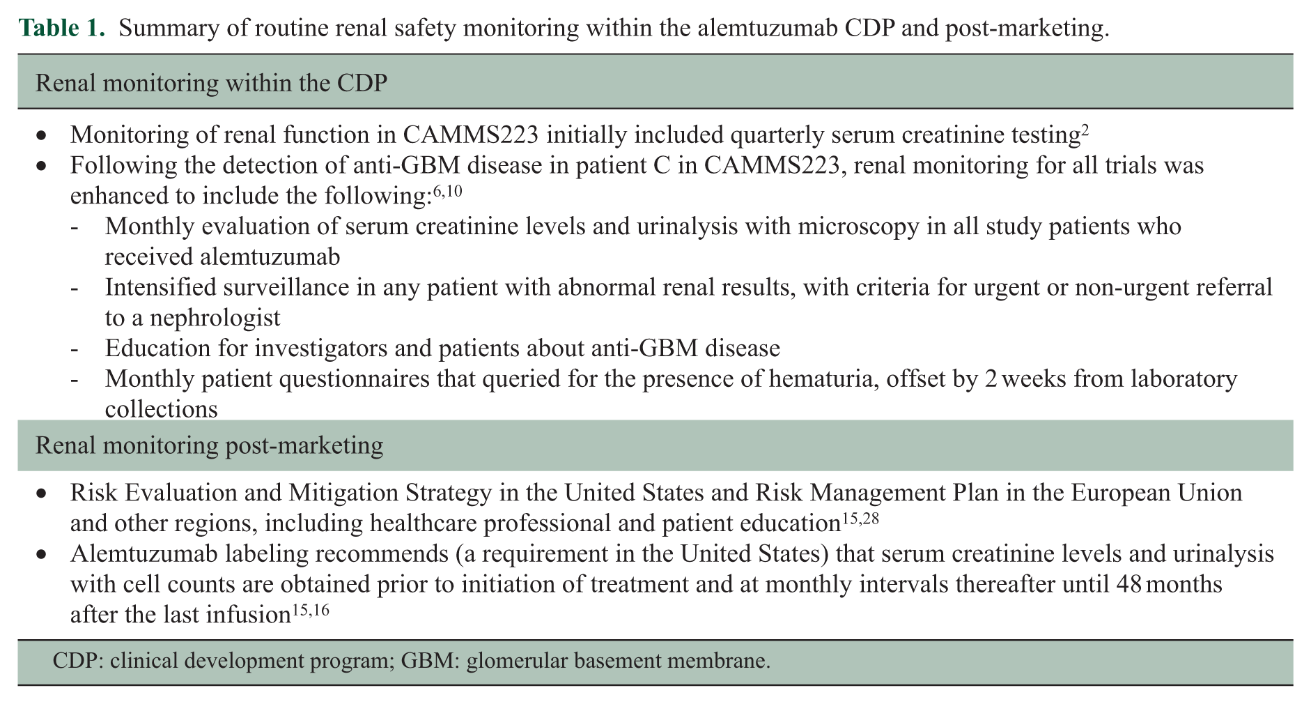
CDP: clinical development program; GBM: glomerular basement membrane.
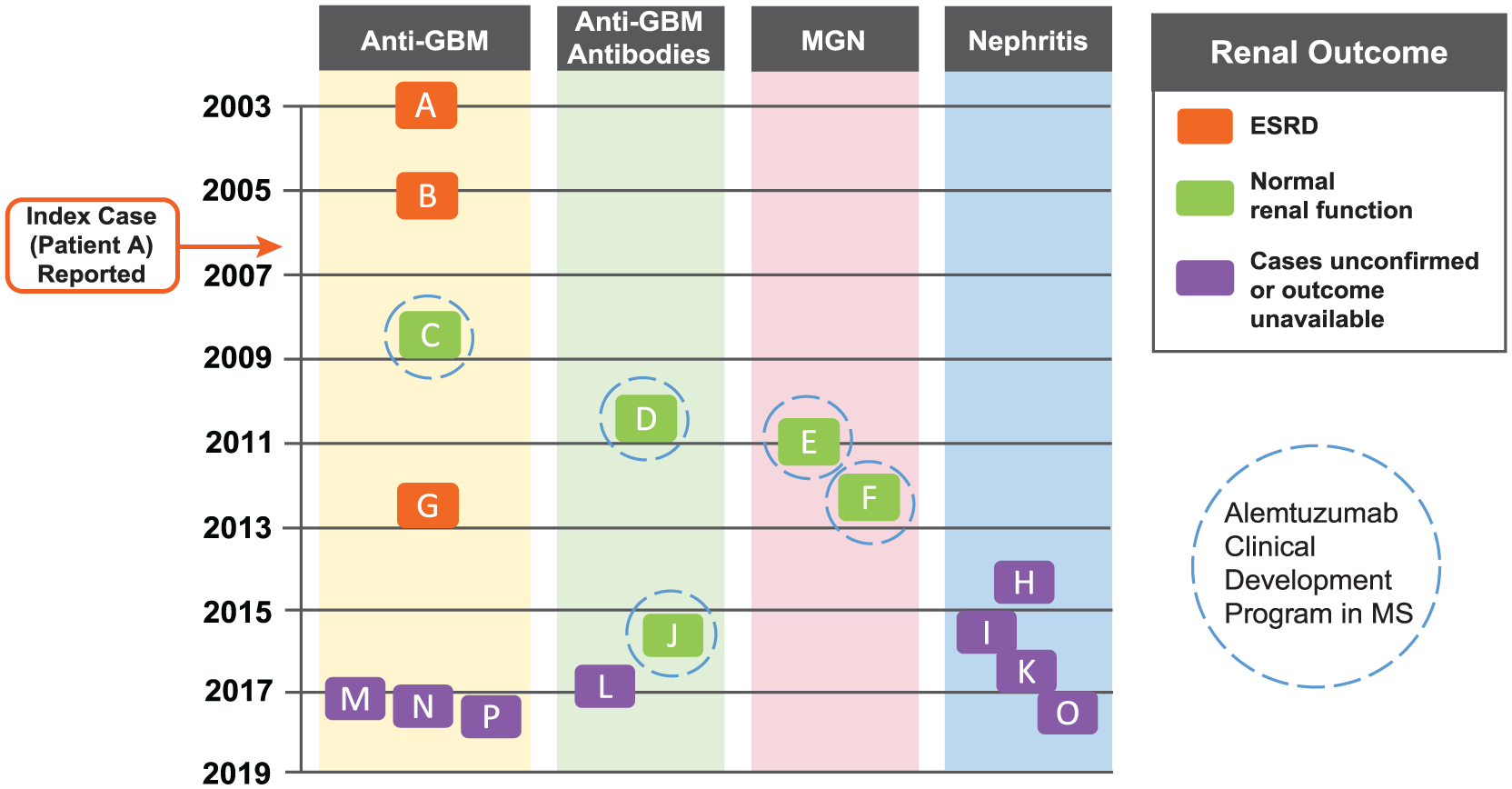
Cases of nephropathy associated with alemtuzumab treatment in MS patients. The nephropathy cases are shown chronologically based on date of diagnosis, and divided according to disease type (anti-GBM disease, anti-GBM antibodies, MGN, and nephritis) and renal outcome. The five cases occurring within the CDP resolved with normal renal function (patients C–F and J). The three cases of anti-GBM disease occurring during off-label use (patients A, B, and G) resulted in end-stage renal disease (ESRD; highlighted in orange). The eight post-marketing cases (patients H, I, and K–P) require further follow-up to determine outcomes; four of these eight cases (patients H, I, K, and O) involved possible immunological renal disease where full information could not be obtained; patients I and O had probable anti-GBM disease, but insufficient information was available to confirm this diagnosis. Patient A (index case) was diagnosed in April 2003 and reported in 2006. 1 Dates of diagnosis for the other cases were: patient B, March 2005; patient C, September 2008; patient D, October 2010; patient E, January 2011; patient F, April 2012; patient G, July 2012; patient H, July 2014; patient I, during 2015; patient J, August 2015; patient K, September 2016; patient L, December 2016; patient M, March 2017; patient N, April 2017; patient O, date unknown (reported May 2017); patient P, date unknown (reported June 2017).
Details of each case were obtained from MedWatch reports and the treating physicians. When available, these included clinical histories, laboratory analyses, and pathology data. Anti-GBM disease cases were classified as those in which a renal biopsy demonstrated crescentic glomerulonephritis with linear deposition of immunoglobulin G (IgG), or there was severe nephritis without interpretable immunofluorescence imaging, but high levels of circulating anti-GBM antibodies. Biopsies from patients C, E, and F were available for review by a renal pathologist (G.S.M.). The status of patients’ MS was also evaluated for cases occurring in the CDP.
Results
Sixteen cases of autoimmune nephropathy have been reported. The first two occurred in an investigator-sponsored pilot study in MS patients 1 before commencement of the alemtuzumab CDP in RRMS. Five cases occurred within the CDP, and one case occurred during independent, physician off-label use of alemtuzumab before its approval for RRMS. Eight further cases have been reported post-marketing. The CDP and post-marketing settings included close monitoring of renal function as part of the trial protocols 6 and Risk Management Plan/Risk Evaluation and Mitigation Strategy,15,28 respectively (Table 1). All cases of autoimmune nephropathy in MS patients treated with alemtuzumab occurred (where known) in patients aged 25–58 years, and with onset within 39 months of the last alemtuzumab administration in all but one patient. The exception was a male patient who, 60 months after treatment, developed a transitory and self-remitting elevation in serum creatinine with detectable serum anti-GBM antibodies, possibly in relation to a mild infectious illness (patient J).
The incidence of autoimmune nephropathies (including anti-GBM disease) with alemtuzumab within the CDP was 0.34%, with a total of 1485 patients at risk and 8632 patient-years of follow-up (median follow-up 6.1 years (range, 0.7–12.7 years)). Through 16 June 2017, approximately 15,000 patients were treated worldwide for MS with alemtuzumab (LEMTRADA®). Based on the nephropathy cases reported so far, this corresponds to an estimated, point in time, post-marketing frequency rate of approximately 0.05%.
Anti-GBM disease cases
Seven patients developed anti-GBM disease, verified in most cases by biopsy showing the presence of crescentic glomerulonephritis and linear deposition of immunoglobulin along the GBM (Table 2). Two of these (patients A (index case) and B) were treated with alemtuzumab at doses higher than those currently approved for treatment of RRMS and with a different treatment regimen. Six cases exhibited rapid loss of renal function between 9 and 39 months after last receiving alemtuzumab (time to event onset not available for patient P). In five cases, there was a period of at least 1 month during which hematuria and/or elevated serum creatinine was detected before accelerating loss of renal function (pre-onset urinalysis not available for patients A, N, and P).
Cases of anti-GBM disease.

F: female; GBM: glomerular basement membrane; GFR: glomerular filtration rate; IF: immunofluorescence; IgG: immunoglobulin G; M: male; N/A: not available.
Cases were classified as anti-GBM disease where a renal biopsy demonstrated crescentic glomerulonephritis with linear deposition of IgG, or severe nephritis without interpretable IF imaging but high levels of circulating anti-GBM antibodies.
Serum creatinine measurement at, or closest to, time of nephropathy event.
Patient B was treated in the month following diagnosis.
Value based on unscheduled lab measurement not reflected in Figure 2.
Anti-GBM antibodies were detected at the time of recognition of acute kidney failure in 6 patients (patient C tested negative). Analysis of serum taken before recognition of acute kidney failure revealed that patient A had no detectable serum anti-GBM antibodies 1 month prior. 1 Patient G had detectable anti-GBM antibodies and microscopic hematuria when serum creatinine was still normal. HLA genotyping data were only available for patient A, who was found to carry a HLA DRB1*15 allele. 30 Alleles of the major histocompatibility complex defined as HLA DRB1*15 are associated with an increased risk for anti-GBM disease and MS.31,32 Kidney biopsy data available for 5 patients revealed crescentic glomerulonephritis with linear IgG deposition (where measured) consistent with anti-GBM disease, including the single case (patient C) with histopathology images available for review (Figure 4(a) and (b)). The percentage of glomeruli with crescents ranged from 15%–98% (Table 2).
All 7 patients received conventional treatment, typically with combinations of plasmapheresis, dialysis, cyclophosphamide, and steroids; however, there was variation in the regimens employed and timing of therapy in relation to the onset of renal failure. Patients A and B developed disease outside of a close monitoring program. Despite being diagnosed and treated (including plasmapheresis and steroids) within 3 days of first symptoms, and within the month after diagnosis, respectively, neither patient recovered renal function; however, both survived and underwent successful renal transplantation without recurrence of anti-GBM disease. Patient C was diagnosed through the CDP monitoring program and was treated with plasmapheresis, cyclophosphamide, and steroids within 6 weeks of first signs of elevated serum creatinine (Figure 2); the patient recovered and retained independent near-normal renal function. Patient G was treated within 2 months of first symptoms of hematuria when she had intact renal function and only 15% crescents on renal biopsy. The patient was treated solely with plasmapheresis (she declined cyclophosphamide), and withdrew from plasmapheresis for 1 week due to personal reasons approximately 1 month after diagnosis. She subsequently experienced a rapid deterioration in renal function. Full conventional treatment with plasmapheresis, cyclophosphamide, and steroids was introduced when renal failure was advanced (serum creatinine 530 µmol/L (6 mg/dL)). After 6 months on dialysis and disappearance of circulating anti-GBM autoantibodies, the patient underwent successful renal transplantation without recurrence of anti-GBM disease. All 3 post-marketing cases (patients M, N, and P) were dialysis-dependent at most recent follow-up based on the information available.
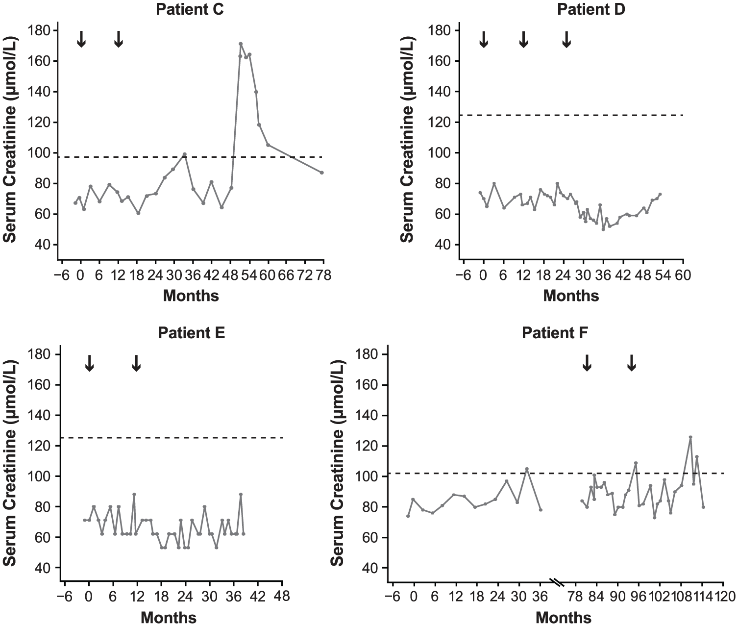
Serum creatinine over time for nephropathy cases occurring during the CDP. Black arrows indicate alemtuzumab treatment courses. Horizontal dashed line indicates upper limit of normal established in each testing laboratory using differently calibrated control levels. Months shown are from first alemtuzumab treatment. Scale of horizontal axes varies due to differing timing of treatment and event onset in each patient. Patient F received subcutaneous interferon beta-1a (44 µg 3× weekly) in the CAMMS223 trial (Month 0 to Month 36) before receiving alemtuzumab.
Cases in which anti-GBM antibodies occurred without typical anti-GBM disease
Three cases are reported in which anti-GBM antibodies were detected without classical anti-GBM disease (Table 3). Patient D presented as nephrotic syndrome with normal glomerular filtration rate (GFR), but the patient’s serum contained a very low titer (just above the laboratory’s reference range) of anti-GBM antibodies. The renal biopsy was reported as membranous nephropathy, but subsequent small rises in serum anti-GBM antibody titer prompted initiation of plasmapheresis, cyclophosphamide, and steroids, resulting in remission of nephrosis and disappearance of anti-GBM antibodies. At 39 months after last treatment for nephropathy, the patient was in remission requiring no medication, anti-GBM antibodies were not detectable, serum creatinine was within the normal range (Figure 2), and there was no detectable proteinuria (Figure 3). Patient J developed a transitory serum anti-GBM antibody titer in the context of a brief self-limiting disease, and patient L was diagnosed with pauci-immune vasculitis and managed accordingly.
Cases in which anti-GBM antibodies occurred without typical anti-GBM disease.
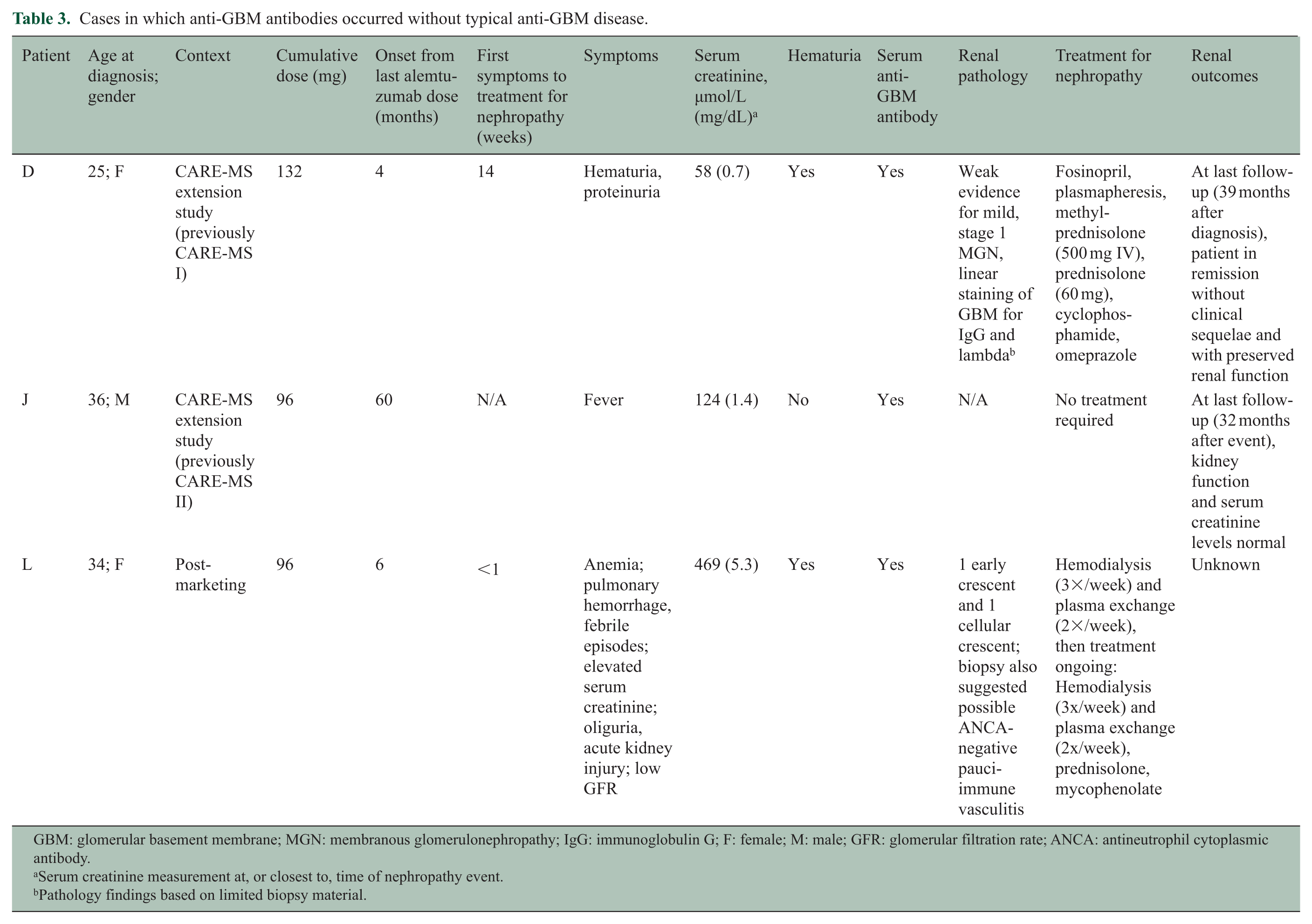
GBM: glomerular basement membrane; MGN: membranous glomerulonephropathy; IgG: immunoglobulin G; F: female; M: male; GFR: glomerular filtration rate; ANCA: antineutrophil cytoplasmic antibody.
Serum creatinine measurement at, or closest to, time of nephropathy event.
Pathology findings based on limited biopsy material.
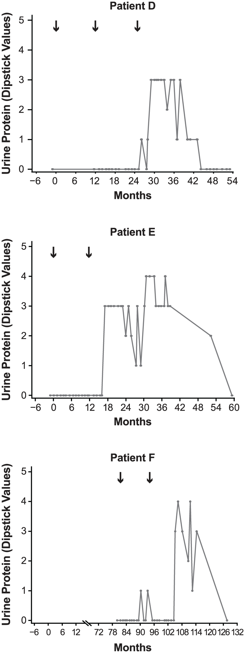
Urine protein (dipstick test) over time for nephropathy cases occurring during the CDP. Urine protein was not available for patient C (routine monitoring was modified to include urinalysis following this case). Black arrows indicate alemtuzumab treatment courses. Numbering on vertical axes refers to number of pluses from dipstick score. Months shown are from first alemtuzumab treatment. Scale of horizontal axes varies due to differing timing of treatment and event onset in each patient. Patient F received subcutaneous interferon beta-1a (44 µg 3× weekly) in the CAMMS223 trial (Month 0 to Month 36) before receiving alemtuzumab.
Membranous glomerulonephropathy cases
Two cases (patients E and F) presented with nephrotic syndrome and normal serum creatinine 5 and 13 months after alemtuzumab treatment, respectively; both were diagnosed through the monitoring program established for the CDP. In both patients, renal biopsy revealed membranous glomerulonephropathy (MGN), stage 1, which was confirmed on review (Table 4 and Figure 4). Immunofluorescence staining in both biopsies was positive for IgG, complement component 3, and kappa and lambda light chains, and electron microscopy revealed global small subepithelial electron dense deposits, without significant intervening GBM spikes. In patient E, mesangial electron dense deposits were also noted. In patient F, who was considerably older than patient E, MGN was superimposed on changes of subcapsular scarring, arterionephrosclerosis, and focal renal atheroemboli.
Cases of MGN.
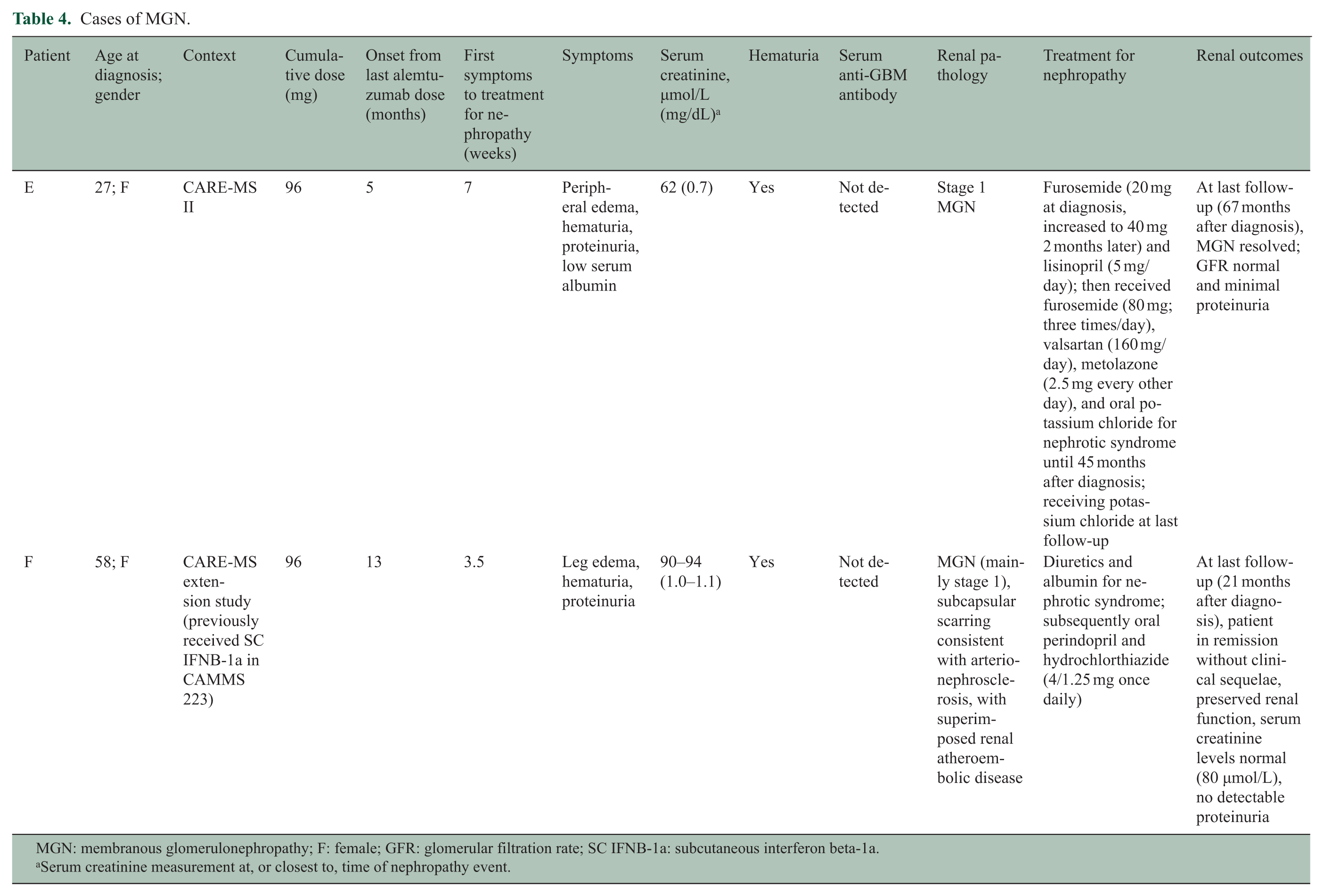
MGN: membranous glomerulonephropathy; F: female; GFR: glomerular filtration rate; SC IFNB-1a: subcutaneous interferon beta-1a.
Serum creatinine measurement at, or closest to, time of nephropathy event.
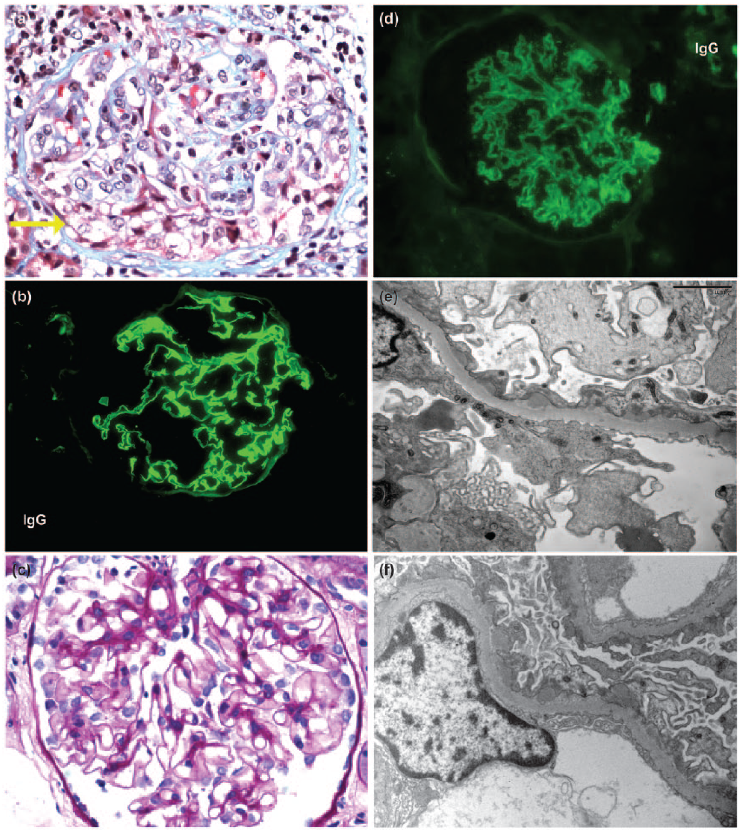
Renal biopsy findings. (a) Patient C: by light microscopy, a glomerulus exhibits a segmental cellular crescent (yellow arrow), characteristic of anti-GBM disease (trichrome, 400× magnification). (b) Patient C: immunofluorescence reveals linear staining of the GBM for IgG, diagnostic of anti-GBM disease (300× magnification). (c) Patient E: by light microscopy, a glomerulus appears unremarkable in this patient with stage 1 MGN (400× magnification). (d) Patient E: immunofluorescence reveals granular global staining of the GBM (300× magnification). (e) Patient E: electron microscopy reveals global small subepithelial electron dense deposits, without significant intervening GBM spikes, diagnostic of stage 1 MGN (5000× magnification). (f) Patient F: similar to patient E, stage 1 changes of MGN are seen on ultrastructural evaluation of the biopsy on patient F (5000× magnification). GBM: glomerular basement membrane; MGN: membranous glomerulonephropathy.
Both patients were treated with angiotensin-converting enzyme inhibitors and diuretics. Patient E was in spontaneous remission 45 months after diagnosis (62 months after initiating alemtuzumab treatment) and, at most recent follow-up (67 months after diagnosis), her GFR was normal with minimal proteinuria (Figure 3). Patient F was in complete remission at last follow-up (21 months following diagnosis) with no detectable proteinuria (Figure 3). Both patients had serum creatinine levels within normal range (Figure 2).
Cases of possible immunological renal disease when full information could not be obtained
In addition to the nephropathy cases mentioned above, four further cases (patients H, I, K, and O) have been reported to the sponsor in the post-marketing setting (Table 5); however, they cannot be independently confirmed due to limitations of pharmacovigilance data collection. These include a case of focal segmental glomerulonephritis with proteinuria (patient H) and two cases of severe nephritis treated as anti-GBM disease (patients I and O). Information on renal outcomes is unavailable in all four cases and further follow-up is awaited.
Cases of possible immunological renal disease where full information could not be obtained.
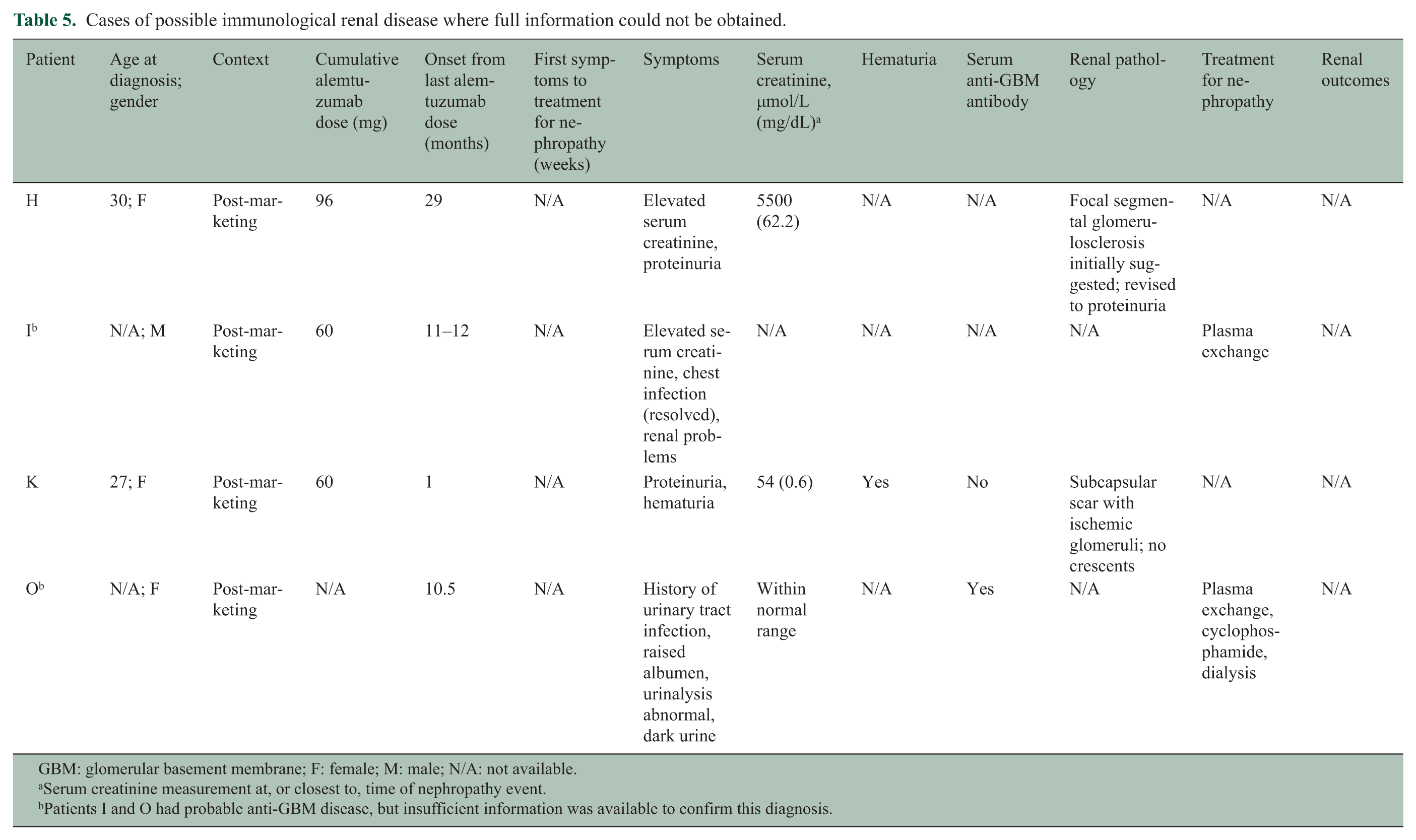
GBM: glomerular basement membrane; F: female; M: male; N/A: not available.
Serum creatinine measurement at, or closest to, time of nephropathy event.
Patients I and O had probable anti-GBM disease, but insufficient information was available to confirm this diagnosis.
MS status of patients within the CDP
Evaluation of the MS status of the five cases (patients C‒F and J) occurring within the CDP suggested that the onset of nephropathy was not associated with exacerbation of MS. Patients C and E experienced a 2-point increase in Expanded Disability Status Scale (EDSS) score from core study baseline to most recent visit, and annualized relapse rate (ARR) remained low (Table 6). Patients D and J showed a decreased EDSS score from baseline to most recent visit and did not relapse over the same period. Patient F experienced an increased EDSS score from 3.5 at baseline to 6.0 at most recent visit and a relatively high ARR (0.45) at most recent visit; however, the patient’s EDSS score was already 6.0 over a 5-month period before the nephropathy event.
MS status of patients within the CDP.
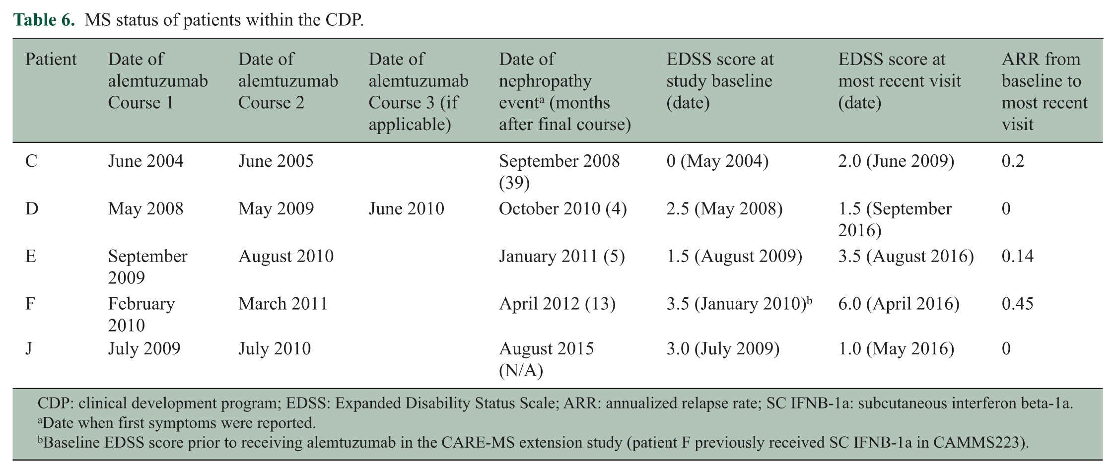
CDP: clinical development program; EDSS: Expanded Disability Status Scale; ARR: annualized relapse rate; SC IFNB-1a: subcutaneous interferon beta-1a.
Date when first symptoms were reported.
Baseline EDSS score prior to receiving alemtuzumab in the CARE-MS extension study (patient F previously received SC IFNB-1a in CAMMS223).
Discussion
The observation of up to 16 cases of de novo renal autoimmunity to date in MS patients treated with alemtuzumab suggests an elevated relative risk for nephropathies. The overall incidence of nephropathies as of 16 June 2017 was 0.34% within the alemtuzumab CDP for RRMS, but so far a lower (0.05%) incidence has been observed in the post-marketing setting. Post-marketing event frequencies are not directly comparable with clinical trial incidences because of differences in ascertainment methodology and follow-up duration, and the limitations of post-marketing reporting, including possible under-reporting, limited availability of clinical details, and potential for inaccurate diagnoses. 33 A case of anti-GBM disease was also previously reported in a patient who received off-label alemtuzumab treatment for systemic vasculitis. 1
Anti-GBM disease occurs rarely in the normal population (up to 1 case/million people/year), with a slight male predominance. 34 It has been reported in MS patients 35 and, although the available data do not enable estimation of the incidence rate, it may be slightly higher than in the normal population, as MS patients have an increased risk for other autoimmune diseases. 36 MS and anti-GBM disease are associated with inheritance of the same HLA class II allele, HLA DRB1*15:01.31,32 In MS, the association with this allele has been reported to be female-specific, 37 and it is interesting to note that the majority of cases of anti-GBM disease following alemtuzumab treatment to date have occurred in female patients, with one patient (patient A) a confirmed carrier of HLA DRB1*15. 30
MGN is a common etiology of nephrotic syndrome in adults, with an incidence in the normal population of 4–10 cases/million people/year. 38 MGN may be idiopathic and associated with phospholipase A2 receptor autoantibodies, or may occur secondary to other diseases such as systemic lupus erythematosus or hepatitis B and C infections. 39 Like anti-GBM disease, very few cases of MGN have been reported in MS patients. 40
Mechanisms underlying the increased risk of autoimmune-mediated conditions with alemtuzumab are not fully understood. However, homeostatic expansion of lymphocytes may be linked to lymphopenia-associated autoimmunity41,42 and represents a potential explanation for increased nephropathy risk in alemtuzumab-treated patients.
Evaluation of the MS status of five autoimmune nephropathy cases occurring within the CDP suggested that nephropathy onset was not associated with exacerbation of MS. Although based on a small number of patients, this finding is consistent with a study showing that another autoimmune disorder, thyroid dysfunction, did not adversely affect MS outcomes in alemtuzumab-treated patients. 43
A number of preliminary conclusions may be drawn regarding the nature of the risk of renal disease with alemtuzumab and the potential for its mitigation. First, with regard to the natural history of anti-GBM disease, five of the seven patients with the disease exhibited a prodrome of at least a month with indications of renal disease in the form of microscopic hematuria and/or elevated serum creatinine, detected while asymptomatic by renal surveillance laboratory testing. Idiopathic anti-GBM disease is usually viewed as fulminant, as it was in the other two alemtuzumab cases, but an asymptomatic prodrome could easily be missed in clinical practice. There have been reports of idiopathic anti-GBM cases with such a prodromal period, notably in patients also presenting with lung hemorrhage and/or hemoptysis and hematuria. 44 Although it is possible that a prodrome is a distinctive feature of anti-GBM disease related to alemtuzumab treatment for MS, more likely, anti-GBM disease (with or without lung involvement) should be reconceived as a condition that takes at least a few weeks to build through minor renal injury to the well-described fulminant presentation.
Second, at least in the alemtuzumab group, serum anti-GBM antibodies were not always detectable by conventional assays during the prodrome and hence cannot be used reliably to rule out a diagnosis of anti-GBM disease. Of particular note, in three of the reported nephropathy cases, serum anti-GBM antibodies were detected without classical anti-GBM disease. False-positives may occur due to non-specific binding in patients with inflammatory diseases. 44 However, two of these three cases occurred in the presence of MGN and pauci-immune vasculitis, respectively, which is consistent with previous reports of anti-GBM antibodies occurring in combination with these diseases. 34
Third, outcomes and response to treatment are not obviously different from those in patients with these conditions occurring outside the context of alemtuzumab treatment. In particular, conventional treatment of anti-GBM disease in alemtuzumab-treated patients was effective when started before substantial renal damage had occurred (patient C, within the CDP with close monitoring), and failed to preserve native renal function when treatment was interrupted (patient G) or when renal injury was very advanced (patients A, B, N, and P, all of whom were reported to have severe injury on biopsy with >90% glomerular crescents).
Finally, close observation of alemtuzumab patients seems to enable, in at least some patients, recognition of anti-GBM disease early, when treatment is most likely to preserve kidney function. Unfortunately, the earliest indication of the disease is microscopic hematuria, which has a low positive predictive value for anti-GBM disease, particularly in younger women who comprise the majority of the at-risk group. 45 The Risk Management Plan/Risk Evaluation and Mitigation Strategy for alemtuzumab adopted for use in clinical practice, including monthly laboratory monitoring required for 48 months following the last infusion, and healthcare professional and patient education on signs and symptoms of autoimmune events, may serve to mitigate against significant renal damage.15,28
Conclusion
Autoimmune nephropathies occur at increased rates in MS patients treated with alemtuzumab. Close monitoring can identify new cases early in the evolution of the nephropathy when treatment is most likely to be effective in preserving renal function. These findings are of critical importance, as the treatment of RRMS with alemtuzumab is expanding, having been judged to have a favorable benefit:risk profile in the majority of patients.
Footnotes
Acknowledgements
The authors and Sanofi thank the patients for their participation in the CAMMS223, CARE-MS I, CARE-MS II, CAMMS03409, and TOPAZ studies, as well as the Steering Committees and the investigators. Critical review of the manuscript was provided by Isabel Firmino, MD, and Colin Mitchell, PhD, of Sanofi. Editorial support was provided by David R Thomas, PhD, and Richard J Hogan, PhD, of Eloquent Scientific Solutions.
Declaration of Conflicting Interests
The author(s) declared the following potential conflicts of interest with respect to the research, authorship, and/or publication of this article: R.P. has received consulting fees from Sanofi. J.A.W. has received personal fees from Gilead Sciences and Sanofi. D.W. has received research support and/or consulting fees from Acorda Therapeutics, Avanir Pharmaceuticals, EMD Serono, GlaxoSmithKline, Roche/Genentech, Novartis, Ono Pharmaceutical, Opexa Therapeutics, Osmotica, Pfizer, Questcor, Receptos, Sanofi, Teva, and XenoPort. M.H. has participated as clinical investigator and/or speaker for Actelion, Alexion, Alvogen, Bayer, Biogen, Merck, Novartis, Roche, Sanofi, and Teva. H.-P.H. has received honoraria for consulting, serving on steering and data monitoring committees, and speaking at symposia from Bayer Healthcare Pharmaceuticals, Biogen, CSL Behring, Merck Serono, Novartis, Octapharma, Roche, Sanofi, and Teva, with approval by the Rector of Heinrich-Heine University. E.K.H. has received honoraria and grant support from Actelion, Biogen, Merck Serono, Novartis, Receptos, Roche, Sanofi, and Teva. G.S.M. reviewed clinical histories, laboratory analyses, and pathology data for nephropathy patients C–F, and provided post hoc definitive diagnoses as a paid consultant to Sanofi; he did not receive compensation for contributing to writing and critical review of the manuscript, or for approval of the final submission draft. D.H.M., C.E.R., and D.P.B. received compensation as employees of Sanofi. A.J.C. has received consulting fees, lecture fees, and institutional grant support from Sanofi.
Funding
The author(s) disclosed receipt of the following financial support for the research, authorship, and/or publication of this article: Prof. Eva Kubala Havrdová was supported by the Czech Ministry of Education, Project PROGRES Q27/LF1. Editorial support in the development of this paper was funded by Sanofi. The CAMMS223, CARE-MS, CAMMS03409, and TOPAZ studies were funded by Sanofi and Bayer HealthCare Pharmaceuticals.