
Abstract
Background:
Lentigo maligna (LM), traditionally observed in older adults, is increasingly reported among younger patients, in whom diagnostic suspicion is lower.
Objective:
To characterize clinical, dermoscopic, and reflectance confocal microscopy (RCM) features of LM in patients under and over 50 years of age.
Methods:
This retrospective, single-centre study analyzed 200 patients with 229 biopsy-confirmed LM lesions diagnosed between 2018 and 2023. Patients were stratified by age (<50 vs ≥50 years). Clinical presentation, dermoscopic features, and RCM findings were analyzed using chi-square, Mann-Whitney U tests, and linear regression (P < .05).
Results:
Of 229 lesions, 33 occurred in patients <50 years. Younger patients, predominantly female (57% vs 40%, P = .067), showed less chronic actinic damage (41% vs 83%, P < .001) and more frequent trunk (36%) or lower limb (31%) lesions compared to older patients (face/neck 26.7%, trunk 40%, P = .004). Lesions in younger patients tended to be more clinically noticeable and solitary, but showed fewer dermoscopic features (mean 3.26 vs 6.92, P < .001). The 4 most common dermoscopic patterns—central featureless areas, irregular pigmentation foci, asymmetrical follicular pigmentation, and angular lines—though less frequent in younger patients, remain useful for early LM diagnosis. All lesions displayed at least 1 atypical colour, with dark brown being the most frequent (87%). RCM (23 younger, 145 older patients) revealed cytoarchitectural atypia. Invasion rates (21.2% vs 22.6%, P = .86) were similar.
Conclusion:
LM in younger patients presents as clinically prominent but dermoscopically subtle lesions, necessitating heightened suspicion. The use of RCM may be of value to ensure accurate diagnosis and avoid inappropriate treatments.
This is a visual representation of the abstract.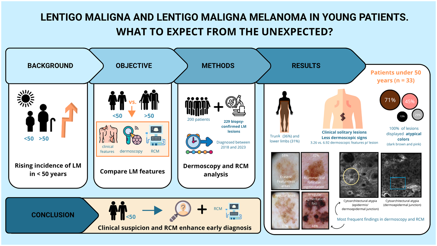
Get full access to this article
View all access options for this article.