
Abstract
Introduction:
Vascular closure devices are commonly used to achieve rapid haemostasis and early ambulation following arterial puncture for endovascular procedures. Although device failure rates are low, the consequences of arterial occlusion include severe limb ischaemia. We describe a novel endovascular technique for the treatment of Angio-Seal arterial closure device (Terumo, Europe NV) failure causing femoral artery occlusion.
Materials and methods:
We describe 2 cases of lower limb angioplasty performed for critical limb ischaemia where the access site was closed using an Angio-Seal according to the manufacturer instructions for use (IFU). In both cases, however, ultrasound could not be used during deployment of the Angio-Seal due to body habitus and small subcutaneous haematoma. In both cases, the device failed and occluded the femoral artery.
Results:
Access was achieved via a retrograde approach from the contralateral limb in one case and a retrograde approach from the ipsilateral profunda artery in the other case. Angiography confirmed that the footplate of the Angio-Seal had occluded the femoral artery. Subsequently, the occlusion was crossed and a short balloon-mounted bare metal stent placed to push the footplate against the arterial wall that resulted in resolution of the occlusion and haemorrhage control.
Conclusions:
Crossing the occlusion caused by failure of the Angio-Seal closure device and subsequent stenting resulted in satisfactory relief of the femoral artery occlusion and haemostasis without the added risks of open surgical revascularisation and general anaesthetic.
Introduction
Arterial closure devices, following arterial puncture for endovascular procedures, are frequently used because of their ability to secure reliable haemostasis allowing for early mobilisation and shorter hospital stays.1-4 The Angio-Seal device (Terumo, Europe NV) is one of the most commonly used devices for closure of the femoral artery and incorporates a collagen plug and footplate to seal the puncture site.2,4
Failure of the device can lead to uncontrolled haemorrhage and/or occlusion of the femoral artery. The incidence of femoral artery occlusion secondary to device failure is quoted as being in the order of 0.06%.4-6 When this complication occurs, revascularisation is required to salvage the limb.
Open surgical exploration and repair is often thought to be the only treatment option.5,6 We describe the safe and rapid endovascular management of 2 patients with a femoral artery occlusion using a balloon-mounted bare metal stent.
Case 1
A 77-year-old obese diabetic man presented with rest pain and non-healing ulceration of both lower limbs that was worse on the right. A duplex ultrasound had demonstrated a heavily calcified and stenosed right popliteal artery and stenoses of the superficial femoral artery (SFA) and popliteal artery on the left.
Angioplasty was first attempted on the right limb which was the most symptomatic. The right common femoral artery was punctured and a 6F sheath inserted. The SFA and popliteal artery were stented with excellent angiographic results. A 6F Angio-Seal closure device was used to attempt to achieve haemostasis; however, after releasing the footplate and pulling it back, the device failed and there was continued haemorrhage.
The left common femoral artery was punctured with a retrograde approach and a 5F sheath inserted. Angiogram demonstrated that the right SFA was totally occluded by the footplate that had got caught in the SFA origin presumably onto plaque (Image 1). The occlusion was crossed with a 0.018-in guidewire, the catheter advanced, and angiographically confirmed to be within the luminal mid-common femoral artery (CFA). A 6 mm × 40 mm (EverFlex; ev3) and 5 mm × 15 mm (Express Vascular SD; Boston Scientific) balloon-mounted bare metal nitinol stents were deployed flush to the SFA origin with excellent angiographic result, maintained haemostasis, and no residual stenosis (Image 2). Haemostasis on the left was achieved with manual compression. The patient returned 3 months later for left leg SFA angioplasty and a duplex study confirmed a patent right femoral artery.
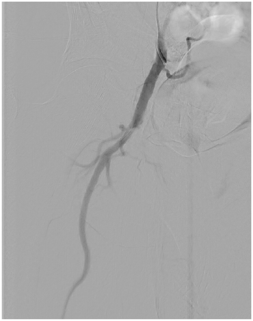
Angiogram of the footplate occluding the femoral artery.
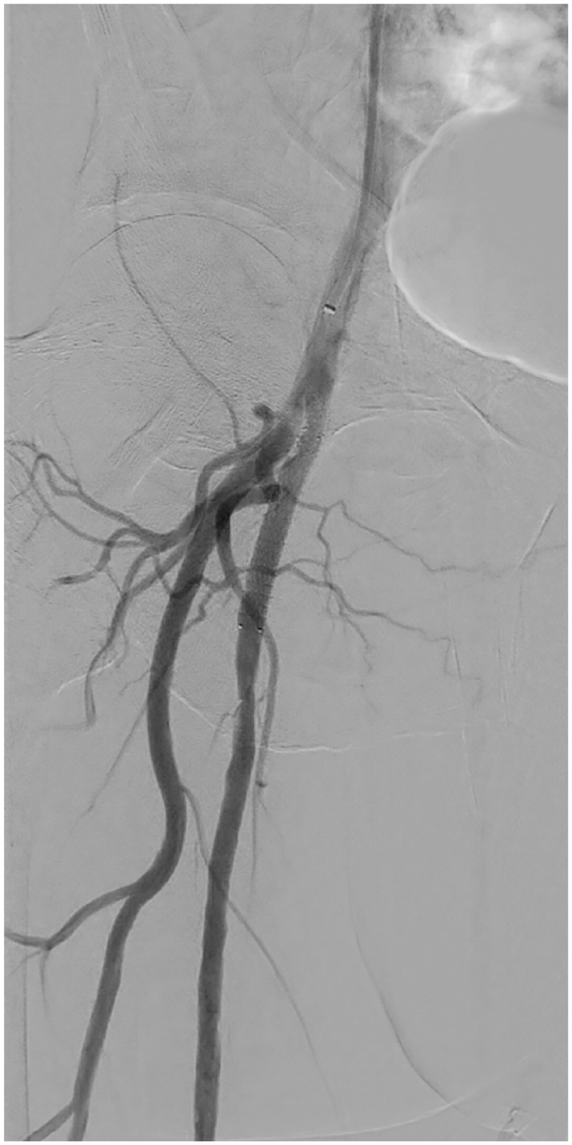
Post-treatment angiogram.
Case 2
A 72-year-old diabetic man had a non-healing surgical wound from his right transmetatarsal amputation. A magnetic resonance angiography demonstrated significant iliofemoral disease and occluded right SFA. He underwent bilateral kissing stents of the common iliac arteries and the 7F right SFA puncture site was closed using an Angio-Seal. This was complicated, however, by the failure of Angio-Seal and continuous bleeding. The heavily calcified right profunda was punctured under fluoroscopic guidance and a 5F sheath inserted. Angiogram confirmed that the footplate had become lodged in the proximal CFA (Image 3).

Angiogram demonstrating footplate occlusion.
The occlusion was crossed with catheter and wire and a 5 mm × 19 mm (Express Vascular SD; Boston Scientific) balloon-mounted bare metal stent deployed that re-established blood flow to the profunda (the SFA was chronically occluded; Image 4). Haemostasis was then achieved with prolonged manual haemostasis.

Angiogram post-treatment.
Discussion
The Angio-Seal closure device is intended for use in the femoral arteries and is contraindicated for use in heavily diseased or small calibre access sites.7,8 The device comprises 3 bioabsorbable components: a suture connecting the collagen plug and a polymer anchor; the suture forms the third absorbable component.9,10 The footplate is designed to be deployed within the arterial lumen and is pulled back against the anterior wall of the artery at the puncture site. The collagen plug is then pushed down over a suture and placed in the perivascular space above the arteriotomy where it expands allowing for haemostasis.7,8,10 The Angio-Seal can fail and occlude the vessel by 2 primary methods. Either the footplate is deployed in a vessel lumen of insufficient diameter to permit arterial flow past the footplate or the footplate pulls against a calcified atheroma on the posterior vessel wall causing luminal occlusion. Alternatively, inadvertent deployment of the collagen plug into the intravascular space can occlude the vessel.
Historically, femoral artery occlusion and stenosis have been treated by open surgical revascularisation to prevent severe limb ischaemia. 6 Open surgery remains the gold standard for treating CFA lesions; however, this usually requires a general anaesthetic which may not be suitable for the comorbid vasculopathy. In elderly patients in particular, careful selection for open surgery is mandated as the risk of death and wound complication is not insignificant with mortality and wound-related complications quoted as being 3.4% and 8%, respectively, and return to theatre being as high as 10%. 11 The prompt availability of a vascular surgeon and theatre space for the acutely occluded CFA is required in addition.
The concern of endovascular stenting in the CFA centres on the perceived risk of stent fracture from external compression in what is a mobile artery over the hip joint. More recent studies, however, have demonstrated comparable primary patency and freedom of target lesion revascularisation in short and medium term.12-14 In a trial comparing stenting and surgery for CFA stenosis, Goueffic and colleagues reported significantly reduced perioperative morbidity and mortality in patients having endovascular repair over open surgical repair but with comparable clinical improvement and primary patency with a mean follow-up of 2 years. 15 Stent selection, however, seems important. A study by Linni and colleagues compared a bioabsorbable stent to open surgery and found higher rates of redo procedures in those patients stented which outweighed any reduction in surgical site infection. 16 In our reported cases, both patients were comorbid and relatively elderly with associated risks of a general anaesthetic. We believe that in this subset of patients, an endovascular management strategy is preferable as it can be performed quickly, safely, and effectively under local anaesthetic.
To our knowledge, this is the first described case of stenting following femoral occlusion caused by a closure device. Other endovascular techniques described include the use of a directional atherectomy device to cut both the plaque and Angio-Seal footplate, followed by balloon angioplasty as described by Babaev and colleagues 17 in a case series of 13 patients. The authors experienced good midterm outcomes and no complications. Although the use of an atherectomy device may avoid the complications associated with proximal SFA stenting such as re-occlusion and stent fracture, atherectomy may not be readily available in all institutions and stenting provides quick and simple way to regain flow and maintain haemostasis.
Conclusions
Angio-Seal–related complications can result in vessel occlusion and/or haemorrhage with the associated risks of threatened limb viability. An open surgical approach for revascularisation may not be appropriate in the severely comorbid patient and a fast and safe endovascular treatment method may be preferable. We believe that crossing the occlusion and stenting offers a safe and effective method for achieving revascularisation.
Footnotes
Funding:
The author(s) received no financial support for the research, authorship, and/or publication of this article.
Declaration of conflicting interests:
The author(s) declared no potential conflicts of interest with respect to the research, authorship, and/or publication of this article.
Author Contributions
TCH and SH contributed equally to the manuscript preparation and checking.
Ethical Approval
All procedures performed in studies involving human participants were in accordance with the ethical standards of the institutional and/or national research committee and with the 1964 Helsinki Declaration and its later amendments or comparable ethical standards.
Informed Consent
Informed consent was obtained from all individual participants included in the study.