
Abstract
Chronic strongyloidiasis is an infection of the tropical regions, caused by the nematode Strongyloides stercoralis. In the United States, patients are typically immigrants. The very long asymptomatic phase followed by the clinical presentation of the disease mimics asthma, chronic obstructive pulmonary disease (COPD), or inflammatory bowel disease (IBD) as in this case report. The inconsistency of eosinophilia and the low sensitivity of microscopic stool examination make chronic strongyloidiasis a disease that is frequently misdiagnosed in the United States. The use of corticosteroids in these misdiagnosed cases transforms chronic strongyloidiasis into disseminated strongyloidiasis or hyperacute syndrome, which leads to high mortality. Iatrogenic errors represent the essential cause of mortality due to chronic strongyloidiasis in the United States. We recommend a high index of suspicion of chronic strongyloidiasis when a physician approaches an immigrant presenting with symptoms mimicking asthma, COPD, and IBD with subcutaneous masses.
Introduction
Chronic strongyloidiasis is a lifelong 1 parasitic infection caused by the nematode Strongyloides stercoralis, which has the unique ability, among all helminths, to silently establish a cycle of autoinfection in humans and can remain undetected for many decades. 1 Chronic strongyloidiasis is an endemic infection occurring in tropical regions which include Africa, Asia, and South America. However, in the United States, chronic strongyloidiasis is rare but more prevalent in immigrant populations from tropical regions. 2 It is often misdiagnosed or diagnosed with significant delay. 2 The diagnosis of chronic strongyloidiasis should always be considered in immigrants who present with persistent respiratory, cutaneous, or gastrointestinal complaints, regardless of the number of years they have spent away from tropical regions. In addition, immigrants from tropical regions should be treated empirically for possible chronic strongyloidiasis before they undergo immunosuppressive therapy, 2 whether they are symptomatic or not. When the immune system is compromised, a patient with chronic strongyloidiasis may develop two life-threatening and highly mortal complications: disseminated strongyloidiasis and hyperinfection syndrome.3,4
Case Description
A 53-year-old African man who emigrated from Mali 25 years ago complained of skin nodules in the periumbilical area and left lower quadrant which were present for the past 5 years. He also presented with a maculopapular rash over the legs, intermittent abdominal pain, nausea, and diarrhea, alternating with constipation, which were all present for the past 14 years. During this time, the patient had seen many physicians without a definitive diagnosis and without effective treatment. The patient admitted to occasionally relying on herbal medicine sent from Mali, without relief of his symptoms.
The physical examination was positive for two 0.5 cm diameter subcutaneous nodules in the left lower quadrant and periumbilical area and one in the gluteal region, freely movable under the skin. The overlying skin appeared normal. The patient presented a picture of a 3 cm × 14 cm longitudinal desquamative maculopapular rash in the midline of his back, as shown in Figure 1. This rash appeared 2 months prior to consultation and completely cleared on its own after 2 weeks. An examination of his legs revealed scattered areas of a maculopapular rash. The rest of the physical examination was normal.
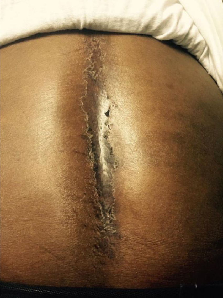
Back of the patient: self-limited desquamative rash.
The complete blood count did not indicate eosinophilia. However, the patient’s serology for anti-strongyloidiasis IgG was positive. The diagnosis of chronic strongyloidiasis was made and the patient was started on ivermectin 200 µg/kg/day for 2 days and repeated after 2 weeks. Two months after initial treatment, the patient became asymptomatic and the skin was completely clear of any rash and nodule. Eight months after the initial diagnosis, the patient remained asymptomatic and the skin was still clear of any rash and nodule.
Discussion
In this case, the patient presented with chronic symptoms, which included intermittent abdominal pain and diarrhea, alternating with constipation, maculopapular rash, and subcutaneous masses. These symptoms were consistent with inflammatory bowel diseases.5,3 Patient symptoms had a characteristic pattern that led to looking for another diagnosis: Symptoms occurred from different systems. Each system was affected distinctly in time. When the symptoms manifested, they were limited in time (transient rash, intermittent abdominal pain). These characteristics were better explained by a parasitic cycle: an infectious agent that affects the skin and gastrointestinal tract and elicits an inflammatory response during its passage (causing symptoms limited in time). Inflammation, because of a parasite cycle as the origin of symptoms, was more inclusive than inflammatory bowel diseases because it provided an explanation for the presence of masses: Although they were freely mobile under the skin, they were not lipomas 6 ; rather they were inflammatory masses around the parasite in subcutaneous tissues. Strongyloides stercoralis, a parasite that infects the skin and the gastrointestinal tract, was more likely the cause. The review of the patient’s remote immigration history was the key element that solidified the postulation by providing a possible explanation for the origin of S stercoralis: Mali 25 years ago. The inflammatory origin of the subcutaneous masses was reinforced later when they were completely cleared after ivermectin treatment.
Chronic strongyloidiasis results from the unique ability of S stercoralis, among all helminths, to establish a cycle of autoinfection in humans that can be lifelong. 7 People become infected through direct skin contact with soil contaminated with the infective filariform larvae. This parasite goes from the skin, into the then pass through the blood to the lungs climb the respiratory tract and and then are swallowed and go in the GI tract.. 8 The high concentration of S stercoralis in the lymphatic system suggests that it uses this manner of circulation to reach the lungs. 9 Once swallowed, in the jejunum, the infective filariform becomes an adult worm that excretes eggs in the feces. 7 When the eggs hatch internally, the rhabditiform larvae may be excreted in the feces or be transformed into the infective filariform larvae that may penetrate the walls of the gut or perianal skin and restart the cycle anew. This autoinfection cycle is the pathogenesis underlying a chronic strongyloidiasis. 10 It is often asymptomatic; however, when symptomatic, symptoms involved include the skin (recurrent maculopapular rash), the respiratory tract (cough, wheezing, tracheal irritation), or the gastrointestinal tract (abdominal pain, diarrhea 11 ).
There is a delay in the diagnosis of chronic strongyloidiasis, an average of 5 years in the United States. 2 Our patient was initially asymptomatic for at least 9 years. Then, the patient experienced gastrointestinal and cutaneous symptoms for 16 years, during which the different physicians he saw did not evaluate him for possible chronic strongyloidiasis. At least 3 factors contribute to the delay in the diagnosis of chronic strongyloidiasis. First is the rarity of chronic strongyloidiasis cases. Second is the ability of S stercoralis to infect a patient outside the United States and to remain undetected and asymptomatic for many years after the patient has moved to the United States. Third is the high variability in the clinical presentation of infection when S stercoralis becomes symptomatic. As a result, the clinician may not have the presence of mind to correlate the patient’s symptoms with possible exposure to S stercoralis, although it may have occurred many years earlier, outside the United States. Therefore, a very high clinical index of suspicion is necessary to include chronic strongyloidiasis among the differential diagnoses, when the clinician is confronted with an immigrant of tropical origin with symptoms involving the lung, skin, or gastrointestinal tract.
Strongyloides stercoralis is the leading cause of mortality due to helminth infections in the United States. 12 This is attributed to the fact that most fatalities are iatrogenic with the use of corticosteroids. 2 Immunosuppression causes the transformation of chronic strongyloidiasis into disseminated strongyloidiasis or hyperinfection syndrome. These are two highly fatal complications.3,13 As a result of immunosuppression, rhabditiform larvae load may increase in the organs involved in the normal cycle (gastrointestinal tract, lungs, skin) 14 and cause hyperinfection syndrome. Immunosuppression may also cause the rhabditiform larvae to migrate to the organs that are not involved in its natural cycle, such as the brain, and cause disseminated strongloidiasis. 13 In the development of either disseminated strongyloidiasis or hyperinfection syndrome, the rhabditiform larvae penetrate the wall of the gastrointestinal tract, causing secondary gram-negative sepsis and meningitis. Acute respiratory distress from enteric bacteria may occur, 14 with a mortality rate of 73% for disseminated strongyloidiasis 15 and 50% for hyperinfection syndrome. 2 Unfortunately, the clinical presentation of chronic strongyloidiasis can mimic chronic obstructive pulmonary disease (COPD) exacerbation, 16 acute new-onset asthma, 17 and ulcerative colitis, 5 leading to the use of empiric corticosteroids, which will be fatal in these misdiagnoses. As a matter of fact, only 9% of US physicians consider further parasitology evaluation in an immigrant who presents with an onset of wheezing and abnormal eosinophilia instead of empiric corticotherapy. 2
Chronic strongyloidiasis is easy to treat, 18 but difficult to diagnose. Some laboratory findings are strongly suggestive of an infection with S stercoralis. However, they are not always consistent. Eosinophilia, which is a common finding in helminth infections, shows a sensitivity of 82.6% 5 in infections with S stercoralis. However, the Centers for Disease Control and Prevention (CDC) enzyme immunoassay (EIA) Strongyloides serology proves to be highly sensitive at 94.6%. 18 In our case, the patient’s blood count did not show eosinophilia. However, the patient was positive for IgG anti-Strongyloides. Therefore, the lack of eosinophilia in the initial diagnostic workup should not prevent the clinician from asking further investigative tests if clinical suspicion exists. The serology test can be difficult to interpret because it may show a cross-reaction with other helminths such as Loa loa and Ascaris lumbricoides. 19 Unlike S stercoralis, none of these helminths can establish a cycle in humans and persist for decades 7 after exposure before causing symptoms. Helminths other than S stercoralis decline progressively 8 in a patient who has left endemic areas, and new symptoms are almost exclusively related to S stercoralis infection. The definitive diagnosis involves visualization of the parasite. Microscopic examination of a stool sample detects the parasite in 25% of cases. Multiple stool microscopic sample examinations using the Baermann technique may increase the sensitivity to 85%. 3 Stool examination was not performed for this patient. Regarding the relative lack of sensitivity of different tests, a situation such as the initiation of an immunosuppressive treatment, which requires ruling out an infection of S stercoralis, may become a challenge for a physician. The recommended strategy for high-risk patients from southeast Asia or agriculture worker is to obtain serology and initiate empiric treatment with ivermectin 200 µg/kg before immunosuppressive therapy. 2
Conclusions
Chronic infection with S stercoralis is easily treatable. However, its diagnosis presents several challenges, including the delayed onset of symptoms after exposure to S stercoralis, the variable clinical presentations, and the lack of sensitivity of the different diagnostic tests available. These challenges directly contribute to the high rate of misdiagnoses and delayed diagnosis. As a result, the mortality of S stercoralis infection is essentially iatrogenically induced by the use of immunosuppressive therapy in a patient who had an unknown S stercoralis infection. A high index of clinical suspicion is necessary to establish the diagnosis. Specifically, physicians should also consider the possibility of chronic strongyloidiasis when they are confronted with patients presenting with conditions such as asthma, COPD exacerbation, or ulcerative colitis if they have once been in tropical regions. The presence of subcutaneous masses in the context of chronic strongyloidiasis suspicion should not lead to excision because they may disappear with treatment. Finally, immigrants from tropical regions should be offered an empiric treatment for chronic strongyloidiasis before they receive any immunosuppressive therapy to prevent the development of hyperinfection syndrome or disseminated strongyloidiasis.
Footnotes
Peer Review:
Six peer reviewers contributed to the peer review report. Reviewers’ reports totaled 982 words, excluding any confidential comments to the academic editor.
Funding:
The author(s) received no financial support for the research, authorship, and/or publication of this article.
Declaration of Conflicting Interests:
The author(s) declared no potential conflicts of interest with respect to the research, authorship, and/or publication of this article.