
Abstract
Orbiviruses are double-stranded RNA viruses that have profound economic and veterinary significance, 3 of the most important being African horse sickness virus (AHSV), bluetongue virus (BTV), and epizootic hemorrhagic disease virus (EHDV). Currently, vaccination and vector control are used as preventative measures; however, there are several problems with the current vaccines. Comparing viral amino acid sequences, we obtained an AHSV-BTV-EHDV consensus sequence for VP5 (viral protein 5) and for VP7 (viral protein 7) and generated homology models for these proteins. The structures and sequences were analyzed for amino acid sequence conservation, entropy, surface accessibility, and epitope propensity, to computationally determine whether consensus sequences still possess potential epitope regions. In total, 5 potential linear epitope regions on VP5 and 11 on VP7, as well as potential discontinuous B-cell epitopes, were identified and mapped onto the homology models created. Regions identified for VP5 and VP7 could be important in vaccine design against orbiviruses.
Keywords
Introduction
Bluetongue virus (BTV), African horse sickness virus (AHSV), and epizootic hemorrhagic disease virus (EHDV) all belong to the Reoviridae family under the genus Orbivirus. Orbiviruses generally have icosahedral virions with a genome of 10-11 double-stranded RNA segments. 1 All 3 viruses are noncontagious and are transmitted by biting midges (Culicoides).2,3 All 3 viruses are recognized globally and have been isolated on every continent except Antarctica, but due to orbiviruses being transmitted predominantly by arthropod vectors, the global and seasonal distribution of each individual virus varies depending on which specific vector is responsible for its transmission and what conditions it requires to thrive. 4
Many of the Reoviridae viruses have profound economic and veterinary significance. These viruses are economically important due to the direct loss of livestock and indirect loss because of restricted movement of livestock as there is the potential to spread the infection into previously unaffected regions.4–6 Both BTV and EHDV infect wild and domestic ruminants, causing varying severity of disease depending on different species.7–11 Epizootic hemorrhagic disease virus affects white-tailed deer the most severely. 12 Bluetongue virus is symptomatically seen most often in sheep.9,13 African horse sickness affects not only horses, ponies, and the European donkey most severely but also mules, African donkeys, and zebra to a much lesser extent.14,15 African horse sickness has also been discovered in camels, goats, and buffalo and other wild and domestic ruminants. 16 Both BTV and AHS have been found in carnivores such as dogs, which contracted the disease from consuming infected meat.4,17,18 Duration of infection varies greatly for all 3 viruses depending on which species is infected.4,9,13,14,18
The 3 viruses are known to cause similar symptoms, including fever, anorexia, dysphagia, edema, weakness and hemorrhage, which can become fatal.19–22 There is no treatment available other than symptomatic treatment; therefore, the main defense against these viruses is proactive measures taken such as vaccines and vector control.15,23 Currently, there are various live-attenuated monovalent and/or polyvalent vaccines available for the different viruses.6,15,24 However, there are several problems with the vaccines currently available for BTV, EHDV, and AHS including the following: (1) the vaccines can cause viremia, (2) there is currently no way to distinguish infected animals from vaccinated ones, (3) use of polyvalent vaccines leads to potential genetic reassortment between serotypes, and (4) current vaccines are also serotype specific, and although they are closely related, cross-protection is rarely seen.24–26 Orbiviruses have numerous serotypes, which are different strains of the same virus that are distinguishable from one another by the immune system. Each serologic strain shares specific antigens and will illicit the same immune reaction. Currently, 27 serotypes of BTV,24–27 8 serotypes of EHDV, 6 and 9 serotypes of AHSV 28 have been identified to date.
The BTV, EHDV, and AHSV virions consist of an outer capsid that surrounds an inner capsid core that is composed of 2 protein layers (a core and subcore).29–32 For each virus, the outer capsid consists of 2 viral proteins, namely, VP2 and VP5, which are involved in host cell attachment, and are the most variable, and have antigenic properties, causing them to be the main serotype-determining proteins. The core in made up of VP7, and the subcore consists of VP3. VP7 is the group-specific antigen that distinguishes between each Orbivirus virus regardless of the serotype. 6 Three other minor proteins (VP1, VP4, and VP6) as well as 4 nonstructural proteins (NS1, NS2, NS3, and NS4) have also been identified which play a role in several processes including virus-host interaction, inclusion body formation, viral protein translation, virion assembly, and virus budding in each virus.33–35
Bluetongue virus is the most studied of the 3 viruses, but the mechanism of replication is thought to be very similar in all 3 viruses. The virus enters the bloodstream via the Culicoides saliva during its blood meal,13,36,37 adheres itself to host cell membranes by VP2 and VP5 binding to surface glycoproteins, and then enters the host cells by endocytosis.38–40 The outer capsid proteins are shed and the core moves into the cytoplasm. 40 The core contains all the transcription machinery needed to transcribe the 10 genome segments.41–43 The core particles assemble within inclusion bodies,44,45 and the outer capsid proteins are subsequently added after the core particles are released from the viral inclusion body surfaces. 46 The complete BTV particles then exit the host cell either by exocytosis or by direct penetration of the cell membrane, which in turn damages the membrane and causes lysis of the cell.47,48 Diagnostic tools used vary widely and include agar immunodiffusion, enzyme-linked immunosorbent assay, and reverse transcription-polymerase chain reaction, next-generation sequencing, and DNA microarrays, to name a few, and VP7 is the predominant protein used for identification.6,49
In this study, we aimed to analyze the sequence and structure of VP5 and VP7 of 3 orbiviruses, namely, BTV, AHSV, and EHDV, to specifically identify functionally important regions as well as potential epitopes that are conserved across all 3 of the viruses. This provides insight into the design of potential subunit vaccine components that could be used to protect animals against infection of orbiviruses. To do this, we first derived consensus sequences for VP5 and VP7 within each virus individually and then one for all 3 viruses combined, which we called AHSV-BTV-EHDV VP5 consensus and AHSV-BTV-EHDV VP7 consensus. Homology models were constructed for the viruses that do not have structural data currently available for the full-length protein, namely, AHSV VP5 and VP7 and EHDV VP5 and VP7. To view the structural implications of regions in the consensus sequences, homology models were created to view the sequence information in structural space for AHSV-BTV-EHDV VP5 consensus and AHSV-BTV-EHDV VP7 consensus. Variability, entropy, and conservation within the consensus sequence as well as accessible surface area of individual residues and their propensity to be part of an epitope were determined for both proteins. The combined analysis provided estimates of 5 potential epitope sites on VP5 that are conserved across the 3 viruses and 11 potential epitope sites on VP7 as well as numerous potential discontinuous B-cell epitope residues. The epitope regions were visualized on the protein structure to display position in relation to the viral particle and emphasize the regions with higher potential.
Materials and Methods
Sequence retrieval
Amino acid sequences of BTV, AHSV, and EHDV VP5 and VP7 proteins were retrieved from the National Center for Biotechnology Information database in FASTA format using the search string “‘Name of virus’ AND VPx AND (amino acid full length)[sequence length].”
Consensus sequence deduction
The consensus sequence and conservation score for each protein from each virus were determined using CLC Genomics Workbench software. The FASTA format for each virus and capsid protein was imported into CLC Genomics Workbench 3.6.1 software (QIAGEN, Arhus, Denmark) (http://www.clcbio.com). The consensus sequences for VP5 and VP7 were generated for individual viruses using the “create alignment” tool. A consensus sequence of all 3 viruses was created using a FASTA file containing consensus sequence of each virus generated above and using the “create alignment” tool. This sequence was then named “AHSV-BTV-EHDV consensus.”
Homology model generation
Homology models of each of the proteins were constructed using the homology builder in the Accelrys Discovery Studio 2017 suite of tools using Protein Data Bank (PDB) codes: 3J9E (VP5) 50 and 1BVP (VP7) 51 as the model templates. Structure renderings were performed using PyMOL (1.3rl Edu). 52
Conservation and entropy calculation
Scop3D 53 was used to calculate the sequence variation and entropy of both viral proteins for each individual virus as well as the consensus. Homology models created were used to visualize the sequence conservation and entropy. Structures generated to visualize conservation were created with PyMOL (1.3rl Edu). 52
Calculate relative solvent accessibility
Relative solvent accessibility (RSA) analysis is used to give an indication of buried and accessible residues in a given protein. The RSA of VP7 and VP5 proteins of each virus was calculated by the “Calculate Residue Solvent Accessibility” tool in Discovery Studio 2017 software with probe radius of 1.4 Ǻ. Exposed residues were calculated to have a maximum RSA value greater than 25% and buried residues were calculated to have a maximum RSA of less than 10%.54,55
Epitope prediction
ElliPro epitope prediction software 56 was used to obtain predicted linear epitopes, predicted discontinuous epitopes, and residue values for the appropriate proteins. Homology model PDB files were used as input to obtain a structure-based prediction.
Results
Consensus sequences
There is nearly double the number of protein sequences available for BTV as compared with AHSV and very few sequences available for EHDV. Table 1 shows the number of sequences retrieved for each protein within each virus. Sequences retrieved for each protein were used to determine the consensus sequence of BTV VP5 and VP7, AHSV VP5 and VP7, EHDV VP5 and VP7, as well as consensus of VP5 and VP7 of all 3 viruses together named AHSV-BTV-EHDV consensus. To avoid bias in the consensus sequence for all 3 viruses toward viruses that have more sequence data, the consensus of each virus was used during the calculation of the consensus sequence of all 3 viruses. Consensus sequences generated for VP5 and VP7 are displayed in Figure 1A and B, respectively.
Number of protein sequences used and conservation score.

Abbreviations: AHSV, African horse sickness virus; BTV, bluetongue virus; EHDV, epizootic hemorrhagic disease virus; VP, viral protein.
The value represents the % of the sequence that is fully conserved.

(A) Consensus sequences obtained for VP5 from all 3 viruses as well as the combined AHSV-BTV-EHDV VP5 consensus using CLC Workbench version 3.6.1 (QIAGEN) (http://www.clcbio.com). (B) Consensus sequences obtained for VP7 from all 3 viruses as well as the AHSV-BTV-EHDV V7 consensus using CLC Workbench version 3.6.1 (QIAGEN) (http://www.clcbio.com). AHSV indicates African horse sickness virus; BTV, bluetongue virus; EHDV, epizootic hemorrhagic disease virus; VP, viral protein.
Homology modeling
The high sequence similarity between BTV, AHSV, and EHDV made it possible to generate homology models for the consensus sequences to visualize the sequence information in structural space. The models were generated using PDB codes: 3J9E (VP5 BTV) 50 and 1BVP (VP7 BTV), 52 as the model templates. Recently, Bekker et al 57 generated a VP7 monomer and trimer homology model for AHSV using 1BVP and 1AHS. 58 Twenty homology models were generated for each protein using the standard method in Discovery Studio (BLAST search, alignment of template protein sequence to structure, and using “Build Homology Model” tool), and stereochemical quality of the 3-dimensional (3D) models was assessed by looking at DOPE score, PDF Total Energy, PDF Physical Energy value and by obtaining Verify Scores, Verify-3D score and drawing Ramachandran plots. A model with the most appropriate values was chosen for each protein. Values obtained for the chosen models can be seen in Table 2, and the Ramachandran plots generated for individual viruses’ proteins can be found in Figure 2A, with the models chosen displaying outlier patterns similar to the template sequences.
Values obtained to assess the stereochemical quality of the 3-dimensional (3D) models.

Abbreviations: AHSV, African horse sickness virus; BTV, bluetongue virus; EHDV, epizootic hemorrhagic disease virus; VP, viral protein.
Table shows values obtained for each of the final chosen homology models. Values demonstrate validity of the homology models generated and used in further studies in this article.

(A) Ramachandran plots in indicating validity of homology models generated. Top models chosen had least amount of outliers, indicated by red triangles, and when plots were compared with template sequence, similar outlier patterns were seen. Homology models generated for (B) VP5 and (C) VP7. Models of AHSV (green), BTV (pink), and EHDV (blue) have been superimposed on one another to show structural similarity. Images rendered using PyMOL (1.3rl Edu). 52 AHSV indicates African horse sickness virus; BTV, bluetongue virus; EHDV, epizootic hemorrhagic disease virus; VP, viral protein.
The DOPE scores for the VP5 models varied between −37 716.5 and −55 933 and for VP7 between −28 575.9 and −40 573.3. Regions on VP5 where the most variability is seen are hinge-like regions which would be presumed to be more flexible, which could explain the model flexibility seen here. Root-mean-square deviation (RMSD) value for 1BVP is 0.229 and the homology models generated for the viral VP7’s RMSD varied from 0.229 to 0.468, and the RMSD value for 3J9E is 0.8 and VP5 homology models’ RMSD values varied from 0.616 to 1.068. The RMSD values show that there is no marked difference between the models and original structures. Verify scores were calculated, for validation of packing quality of 3D homology models using the Accelrys Discovery Studio 2017 “Verify protein” tool. Three scores are reported: Verify Score, Verify Expected High Score, and Verify Expected Low Score. If the Verify Score is higher than the Verify Expected Low Score, the model is considered of acceptable quality. These scores are shown as DS Verify scores in Table 2. Verify-3D scores were calculated for sequence-structure compatibility (https://services.mbi.ucla.edu/SAVES/). It scores between −1.0 (bad) and +1.0 (good). The model must have a positive value to be acceptable. All homology models generated had at least 83% of the residues at ≥0.2.59,60 The homology models generated for the AHSV, BTV, and EHDV VP5 and VP7 can be found in Figure 2B and C, respectively, which demonstrates the structural similarity generated for all 3 viruses’ proteins for both VP5 and VP7.
Conservation and entropy
Scop3D software produces 2 values, namely, a “conservation” value and an “entropy” value based on the sequence and structure data for proteins. In Scop3D, conservation is the percentage variation at each amino acid position and it gives an idea of the distribution of sequence conservation on a protein structure. 53 Entropy, however, provides information on the random spread of the observed variation across all 20 possible amino acids in each position. 53 Low entropy values therefore indicate that only a few amino acids are possible in a said position, whereas a high entropy value means that many different amino acids are found in a said position. Conservation scores for each individual viral capsid protein can be found in Table 1. A significant percentage conservation is seen within and between viruses which is consistent with the findings in a previous study. 61 The spread of conservation and entropy values for the AHSV-BTV-EHDV consensus can be found in Figures 3 and 4 for VP5 and VP7, respectively. VP5 is seen to be less conserved than VP7 which is consistent with findings of by Iwata et al. 61 Residues that are conserved for these capsid proteins more than likely play key structural or functional roles during the viral life cycle, and if these regions are recognized as epitopes, the protection provided by the consensus sequence proteins would be longer lasting and effective potentially across viruses.

A comparative representation of conservation, entropy, RSA, and ElliPro residue score for AHSV-BTV-EHDV VP5 consensus. The scale for the conservation and entropy scores ranges from highly conserved, low entropy (red) to poorly conserved, high entropy (green). The scale for the RSA values ranges from buried (blue) to accessible (dark blue). The ElliPro residue score has a scale of low epitope potential (white) to high epitope potential (black). Potential linear epitope regions are indicated by roman numerals (boxed in light blue). AHSV indicates African horse sickness virus; BTV, bluetongue virus; EHDV, epizootic hemorrhagic disease virus; RSA, relative solvent accessibility; VP, viral protein.
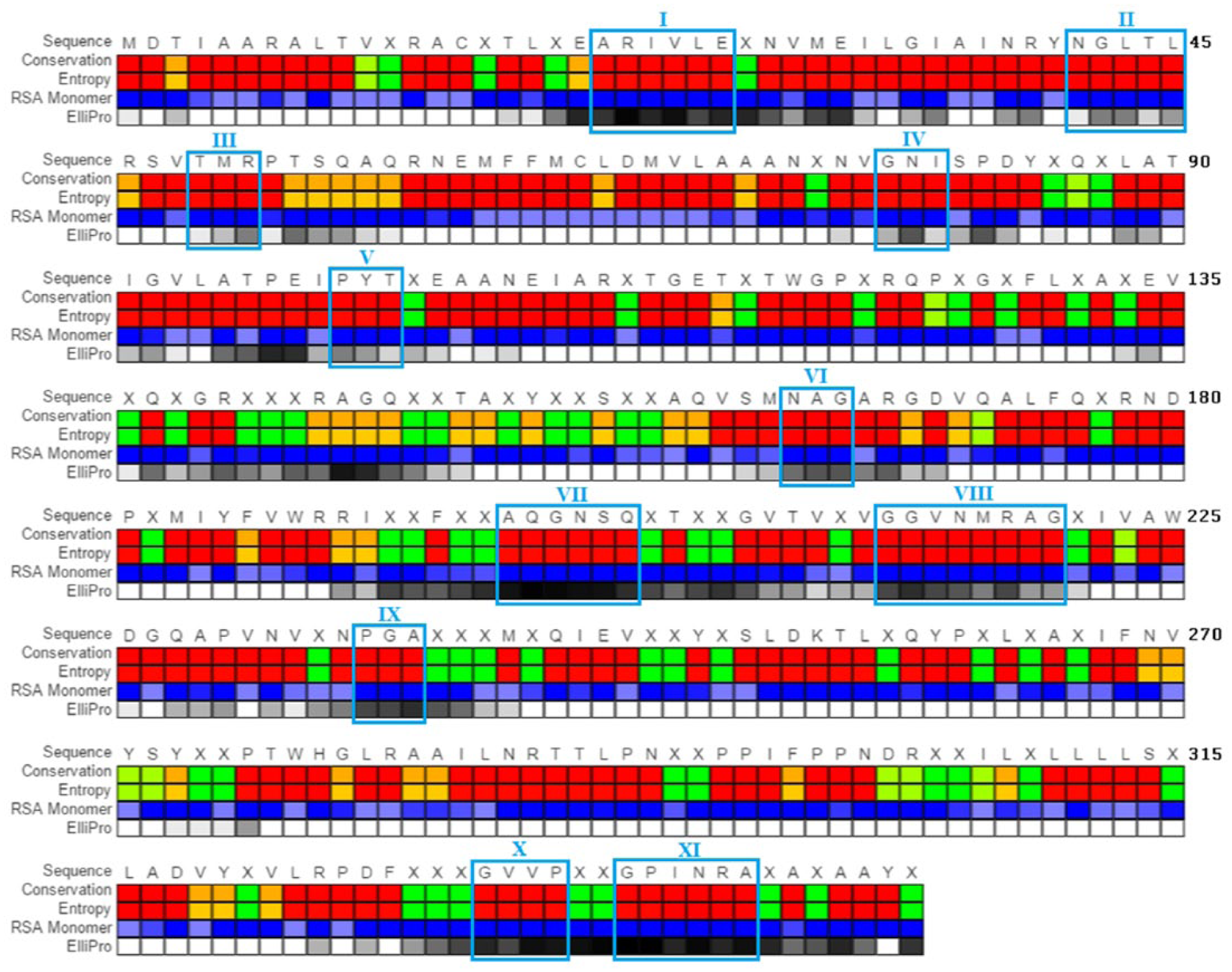
A comparative representation of conservation, entropy, RSA, and ElliPro residue score for AHSV-BTV-EHDV VP7 consensus. The scale for the conservation and entropy scores ranges from highly conserved, low entropy (red) to poorly conserved, high entropy (green). The scale for the RSA values ranges from buried (blue) to accessible (dark blue). The ElliPro residue score has a scale of low epitope potential (white) to high epitope potential (black). Potential linear epitope regions are indicated by roman numerals (boxed in light blue). AHSV indicates African horse sickness virus; BTV, bluetongue virus; EHDV, epizootic hemorrhagic disease virus; RSA, relative solvent accessibility; VP, viral protein.
Relative solvent accessibility (percentage residue solvent accessibility)
Amino acid residues that are surface exposed in the virion or the protein during the viral life cycle could potentially act as epitopes. Discovery studio’s “Calculate Residue Solvent Accessibility” tool was used to obtain RSA values for residues found in the monomer AHSV-BTV-EHDV consensus sequences for VP5 and VP7. Discovery studio calculates the RSA with respect to an extended Ala-X-Ala tripeptide (where X is residue of interest). Residues with a value above 25 are considered to be exposed and those with a value below 10 are buried within the molecule. Relative solvent accessibility values were determined for the AHSV-BTV-EHDV consensus sequences for VP5 and VP7 and are displayed in Figures 3 and 4 for VP5 and VP7, respectively. A high percentage of residues were found to have RSA values above the 25% threshold and this can be explained by the RSA value having been calculated for the monomeric protein structure of each protein.
Epitope prediction
Not all surface-exposed residues will be recognized as epitopes during infection or vaccination. ElliPro software 56 was used to generate predicted linear epitopes, predicted discontinuous epitopes, and residue epitope propensity values for the AHSV-BTV-EHDV VP5 and VP7 consensus sequences. Epitope regions can be either continuous (linear) or discontinuous (conformational). 62 B-cell epitopes can be either continuous or discontinuous but are predominantly discontinuous and T-cell epitopes are only linear. In this study, we investigated predicted B-cell epitopes. Potential regions identified in ElliPro were then further verified for epitope potential by incorporating all the data collected, including conservation and RSA. In Figures 3 and 4, a comparison of conservation, entropy, RSA, and ElliPro residue scores were used to highlight new regions of potential conserved epitopes for VP5 and VP7, respectively. A conservation value of 0.5 and below was acceptable and entropy value of 1 and below was acceptable, 53 a RSA value greater than 25%54,55 was acceptable, and an ElliPro residue value greater than 0.5 56 was used when identifying potential epitope regions in the AHSV-BTV-EHDV consensus sequences. Studies have shown that amino acid sequences as short as 3 amino acids are able to act as both B-cell and T-cell epitope regions and are in fact capable of eliciting a response even more potent than there longer counterparts,63,64 and therefore for this study, 3 amino acids is the minimal length of amino acids for linear epitopes predicted.
Five potential linear epitope regions were found in VP5: (I) 18 to 22, (II) 45 to 50, (III) 238 to 241, (IV) 370 to 374, and (V) 458 to 460 (Figure 3). The computed potential linear epitopes predicted were analyzed visually to determine the possible impact when presented within the virion (Figure 5). Of the 5 computed epitopes, II and III are exposed on the surface of the virion and would be readily accessible to antibodies. Eleven potential linear epitope regions were found in VP7: (I) 21 to 26, (II) 41 to 45, (III) 49 to 51, (IV) 78 to 80, (V) 100 to 102, (VI) 164 to 166, (VII) 197 to 202, (VIII) 213 to 220, (IX) 236 to 238, (X) 331 to 334, and (XI) 337 to 342 (Figure 4). Mapping these epitopes onto the protein structure with surface representation of VP7 (Figure 6) indicates that computed epitopes VI, VII, VIII, and IX are all present in the virion just below the VP5/VP2 outer layer. It has been shown that antibodies can interact with VP7 65 and hence it is possible that these regions on VP7 would be accessible to the immune system to elicit a protective response.

Surface representation of monomeric VP5 of the various orientations with potential linear epitope regions (blue) and numbered to correspond to regions found on Figure 3 and potential discontinuous B-cell epitope residues are highlighted (green). The cube is placed as a visualizing aid for the various orientations of the viral protein, with shaded side depicting the surface being viewed, if the center of the cube is the center of the trimeric unit. Images rendered using PyMOL (1.3rl Edu). 52 VP indicates viral protein.

Surface representation of monomeric VP7 of the various orientations with potential linear epitope regions (blue) and numbered to correspond to regions found in Figure 4 and potential discontinuous B-cell epitope residues are highlighted (green). The cube is placed as a visualizing aid for the various orientations of the viral protein, with shaded side depicting the surface being viewed, if the center of the cube is the center of the trimeric unit. Images rendered using PyMOL (1.3rl Edu). 52 VP indicates viral protein.
Discontinuous B-cell epitopes are epitope residues that are separated in linear sequence, but when the protein takes on its 3D conformation, the residues come in contact and interact to form an epitope region recognizable by the immune system. The potential linear epitope regions could also pair with discontinuous B-cell epitope residues to form larger B-cell epitopes. Based on the conservation, entropy, RSA, and ElliPro residue score, potential discontinuous epitope residues on VP5 include 1; 3; 12; 16; 24; 28; 32; 33; 40; 41; 53; 54; 58; 60; 62; 63; 66; 117; 121; 126; 143; 153; 177; 179; 183; 188; 190, 193; 195; 200; 206; 214; 215; 243; 337; 338; 341; 355; 412; 418; 423; 428; 429; 431; 432; 436; 439; 447; 449; 453; 454; 494; 497; 498 (shown in green in Figure 5). The outer surface of VP5 has more discontinuous computed epitope regions and only 2 computed linear epitope regions. Potential discontinuous epitope residues on VP7 include 1; 17; 28; 30; 31; 34; 35; 38; 39; 76; 82; 83; 88; 89; 91; 95; 104; 106; 107; 140; 168; 170; 204; 207; 209; 230; 232; 276; 324; 326; 346; 347 (show in green in Figure 6). The discontinuous regions of high epitope propensity cover a large portion of the surface of VP7 that is in contact with the outer protein layer of the virion.
Discussion
In orbiviruses, VP2 and VP5 form the outermost layer of the virion that covers VP7 which forms the middle layer. VP2 is known to be the major antigenic protein and a major target for neutralizing antibodies 66 ; however, VP2 has the highest degree of sequence variability 61 which makes it a poor candidate when looking for potential cross-neutralization proteins between AHSV, BTV, and EHDV that will have long-lasting effects.
VP5 has a higher conservation than VP2, although it still shows a degree of variability within and between viruses. VP5’s sequence flexibility may be vital to enable it to mutate and evade the immune system as it forms part of the outer layer which is exposed to potential antibodies. On VP5, the linear B-cell epitope 265-LSGID-269 67 is highly conserved across all 3 viruses; however, the 266-S amino acid was not conserved in AHS. Other B-cell epitopes have previously been identified for VP568–70 but have been found to not be effective at providing protection across serotypes; this could potentially be due to the lack of amino acid sequence conservation not only between the different orbiviruses but even also between the serotypes.
When we obtained the AHSV-BTV-EHDV VP5 consensus, there were still at least 2 predicted linear regions (II and III) that are conserved across the 3 viruses, which are exposed to the surface in the viral structure and have a good probability of being an epitope for an orbivirus vaccine (Figure 5). The other potential epitope regions, although not exposed on the outer surface of the viron (I, IV, V) (Figure 5), may still be important as recently it has been discovered that the VP5 BTV undergoes structural rearrangements 50 during the infection cycle. There are also numerous potential discontinuous B-cell epitope regions which are shown in green in Figure 5. Evidence of VP5’s role in the immune response is seen in studies that show that a vaccine combining VP2 and VP5 resulted in better protection than when VP2 is administrated alone in a vaccine. It has also been seen that the amount of VP2 within a vaccine dose can be more than halved when administered with VP5 and still produce the same level of protection.71,72 VP5 alone has not been shown to provide protection. When multiple viral proteins are combined to produce a subunit vaccine, it enables an enhanced immune response and produces a greater protection against infection. This has been seen with the combination of all 4 major proteins into a vaccine (VP2, VP3, VP5, and VP7) which reduced the amount of VP2 required in the vaccine 50-fold. 72
Our data for VP7 show a higher degree of conservation than is seen for VP5 (Table 1); this is likely due to role this protein plays in the viral structure as it acts as the bridge between the outer layer VP5/VP2 and the inner layer VP3, which protects the genome and plays many roles during viral life cycle. The structural constraints on VP7 are important to allow it to undergo the required conformational changes throughout the viral life cycle 73 and therefore might explain a higher degree of conservation as compared with VP5. This potentially makes it a very valuable candidate as a component for a cross-protective vaccine. Interestingly, the RGD motif (169-170) is conserved across all 3 viruses and it is thought to be key for the double-layered virion to bind to Culicoides cells. 74 It therefore appears that this mechanism is conserved in the 3 viruses. All 3 viruses need to be able to infect the Culicoides but proteases found in the saliva of Culicoides stimulate the release of VP2 and VP5 by cleaving 75 and therefore infection must occur via the double-layered virion. The RGD motif must be key in the double-layered particle infection of Culicoides cells across the orbiviruses. To date, little investigation has been done into the potential of VP7 to play a role in B-cell activation. Our data indicates that there are numerous potential discontinuous B-cell epitope residues marked in green in Figure 6 as well as at least 4 potential epitope regions (VI, VII, VIII, and IX) that are conserved for all 3 viruses and are found on the surface of VP7 and are exposed when the virus loses the outer capsid layer and potentially exposed in the full virion (Figure 6). Investigations into BTV VP7 have shown that VP7 has regions that are exposed on the virion surface and are able to act as epitopes, 65 and vectors presenting VP7 are capable to produce an immune response that is at least partially protective.76,77 This, along with evidence that VP6, the middle layer protein in Rotavirus, which is also a member of the Reoviridae family, has shown to be involved in B-cell immunity which suggests that VP7 of the orbiviruses is a good candidate for B-cell activation as numerous papers point out the similarity of BTV VP7 and Rotavirus VP6. 78 , 79 , 80 In addition to this, in BTV, VP7 is seen to be a major group reactive antigen that has been seen to contain CD8+ and CD4+ T-cell epitopes that are conserved among different serotypes. 81 Most significantly BTV VP7 has been seen to induce a strong T-cell response when used in vaccination trails, and more specifically VP7 is vital to the activation of BTV-specific CD8+ and CD4+ cells. 77 In this study, certain residues within the previously identified T-cell epitopes on VP781–83 are highly conserved across all 3 viruses suggesting the possibility that VP7 might be capable of activating a T-cell response across the 3 orbiviruses. This is significant as high promise has been seen in the development of T–cell-inducing vaccines that are able to induce CD4 and/or CD8 cells to be involved in the response to the pathogen and activate cell-mediated effector mechanisms. T cells are responsible for the activation of B-cell preparation by the germinal centers (GCs), and therefore antigens that fail to activate the GCs produce a much weaker and shorter antibody response and little to no immune memory is generated.84,85 VP7 is known to play a large role in T-cell activation, and therefore the linear epitopes (I, II, III, IV, V, X, XI) and ones found in previous studies that are found throughout the viral protein structure may play key roles in developing immune protection.
A vaccine containing at least VP5 and VP7 possessing a sequence equivalent to our consensus sequences could provide protection against these 3 viruses (BTV, AHSV, and EHDV) over a long period due to sequence stability seen in the high conservation and the ability to activate both B and T cells. To confirm the immune response and protection that the identified epitope regions of the proteins would provide, further experimental studies would need to be undertaken. These include the development of protein expression systems that are cost-effective and purification protocols that can be scaled up for mass production of these 2 proteins. The protein produced would also need to be fully characterized to ensure that the structure under various conditions represents that of the virus during key viral life cycle stages. A lot of research is currently being done into developing new vaccines, and newer techniques including reverse genetic systems for BTV and AHSV have broadened the possibilities in current vaccine devlopement. 86 However, while current vaccines have been focused on inducing neutralizing antibodies to structural components found on the virion, studies have shown that this approach can be greatly improved by the inclusion of immunogens to induce T-cell responses as this helps in the prompt recognition and destruction of infected cells and activation of B cells via the GCs and therefore an effective elimination of the virus from the host. 87 It must also be noted that current vaccines which target VP2 are serotype specific and give short-lived protection. 88
Conclusions
After analysis of the available amino acid sequences for VP5 and VP7 of AHSV, BTV, and EHDV, we propose a consensus sequence for VP5 and VP7 across 3 viruses (AHSV, BTV, and EHDV). The proteins produced form the consensus sequence should fold to produce similar structures to those found in the viral particles based on the homology models created. We found that the proteins have several regions that are conserved, surface exposed, and have a high propensity to be epitope regions. The high sequence conservation for VP7 and to a lesser extent for VP5 across viruses indicates that a number of the residues within these protein structures facilitate their function. This is advantageous because these highly conserved regions could be exploited in the development of preventative applications. Several potential epitopes of both B and T cells were computed which could provide long-lasting protection against all 3 viruses and even potentially for other orbiviruses as well.
Footnotes
Acknowledgements
The authors thank Mr Jason S Evans for formulating the graphical representations for Figures 3 and 4.
Funding:
The author(s) disclosed receipt of the following financial support for the research, authorship, and/or publication of this article: This research as supported by UNISA (Woman in Research grant and High Performance Computing Center of UNISA) and by the NRF for the availability of necessary software and time.
Declaration of conflicting interests:
The author(s) declared no potential conflicts of interest with respect to the research, authorship, and/or publication of this article.
Author Contributions
The generation of data and its analysis was conducted by BLR, NP, and SG. The draft manuscript was prepared by BLR. BLR, NP, and SG have critically reviewed the manuscript and participated in the study design and choice of methods. SG conceived the idea and reviewed the draft and final manuscripts and interpretation of results. All authors read and approved the final manuscript for submission.