
Abstract
SARS-CoV-2 has been highly susceptible to mutations since its emergence in Wuhan, China, and its subsequent propagation due to containing an RNA as its genome. The emergence of variants with improved transmissibility still poses a grave threat to global health. The spike protein mutation is mainly responsible for higher transmissibility and risk severity. This study retrieved SARS-CoV-2 variants structural and nonstructural proteins (NSPs) sequences from several geographic locations, including Africa, Asia, Europe, Oceania, and North and South America. First, multiple sequence alignments with BioEdit and protein homology modeling were performed using the SWISS Model. Then the structure visualization and structural analysis were performed by superimposing against the Wuhan sequence by Pymol to retrieve the RMSD values. Sequence alignment revealed familiar, uncommon regional among variants and, interestingly, a few unique mutations in Beta, Delta, and Omicron. Structural analysis of such unique mutations revealed that they caused structural deviations in Beta, Delta, and Omicron spike proteins. In addition, these variants were more severe in terms of hospitalization, sickness, and higher mortality, which have a substantial relationship with the structural deviations because of those unique mutations. Such evidence provides insight into the SARS-CoV-2 spike protein vulnerability toward mutation and their structural and functional deviations, particularly in Beta, Delta, and Omicron, which might be the cause of their broader coverage. This knowledge can help us with regional vaccine strain selection, virus pathogenicity testing, diagnosis, and treatment with more specific vaccines.
Introduction
The severe acute respiratory syndrome coronavirus 2 (SARS-CoV-2) has already reached pandemic proportions and still threatens our way of life. It is an enclosed, single-stranded, positive-sense RNA virus with a 29.7 kb genome. 1 There are 6 open reading frames in the genomic RNA (ORFs). The ORF1ab gene of SARS-CoV-2 encodes a polypeptide broken into 16 nonstructural proteins. 2 Among them are 3 nonstructural proteins: Papain Like-protease (PLpro) or NSP3, RNA-dependent RNA polymerase (RdRp) or NSP12, and Helicase or NSP13, 2 and a structural protein, for example, the spike is among crucial.
Spike glycoprotein is the principal structural protein of SARS-CoV-2 and is highly conserved across all human coronaviruses. The spike protein of SARS-CoV-2 plays a vital role in the entry to host cell 3 as it participates in receptor recognition and viral attachment. It is one of the most critical targets for the COVID-19 vaccine and therapeutic because of its crucial significance. 4
PLpro is a protein with 1945 amino acid residues that have multiple functions. The papain-like domain cleaves nonstructural protein peptide bonds5,6 and inhibits the host cell’s type 1 interferon (IFN) antiviral actions. 7 Because PLpro cleaves the interferon-stimulating gene-15 (ISG-15) protein, host cells cannot effectively respond to antiviral signals.8,9 RdRp is a 932 amino acid RNA polymerase that copies viral RNA. 2 RdRp is essential for viral replication. It is the primary surface point for antiviral drugs to bind and inhibit or reduce virus replication via remdesivir binding. 10 Helicase with 601 amino acids has multiple enzymatic activities, for example, helicase, 2 RNA 5′-triphosphatase, 11 and NTPase 12 activities. All 3 nonstructural proteins play a vital role in genome replication and propagation in the host cell and can be ideal targets for antiviral drugs. 13
However, the subsequent advent of SARS-CoV-2 strains that are more transmissible and, in some cases, more severe than the Wuhan, China-origin strain can occasionally dash hope for new vaccinations. 14 The World Health Organization (WHO) has identified 5 SARS-CoV-2 variants as variations of concern (VOC): the Alpha, Beta, Gamma, Delta, and Omicron. These were crucial in increasing their virulence and transmissibility or decreasing the effectiveness of therapies and immunizations.
According to the worldometer, 6 549 724 deaths (till October 1, 2022) have been attributed to SARS-CoV-2. Numerous investigations have been conducted evaluating the severity of this respiratory disease among different variants, particularly Beta, Delta, and Omicron. The Beta and Delta variations were associated with a higher risk than the Alpha, and Gamma variants, as shown by a higher hospitalization rate, severity of illness, and mortality. 15 The Beta and Delta genetic profiles are distinguished by a distinct combination of some unique mutations that may result in structural and functional abnormalities. Despite being different from earlier successful variants, Delta has spread globally and become the sole VOC since around September 2021. 16
In addition to these 4 VOCs, a new variant of concern emerged at the end of November 2021, named Omicron. 17 In the early days, Omicron was found to have less severity in sickness than infection than Delta. But now, Omicron is also as severe a threat as Delta. A comparative analysis of the disease severity between the Omicron and Delta variants in the USA found that the cumulative number of deaths in the Omicron wave was very similar to that seen in the Delta wave. Similarly, it was shown that during the peak outbreak of the Omicron variant in the UK, US, and Australia, daily hospitalization and ICU cases were about one-time greater than during the peak outbreak of the Delta variant. 18 These results imply that structural variations of the spike protein caused by mutational changes may increase the severity of illness and the risk of death in the Beta, Delta, and Omicron types.
Understanding the SARS-CoV-2 proteome is essential for developing proteomic-based COVID-19 control strategies. 3 That’s why the investigation of structural variations is required to connect the genomic patterns of coronaviruses to the phenotypic attributes to show the binding discrepancies and antibody response for variants of concern. 19 Such structural analysis data can then be used to determine binding affinity with receptor binding sites and design potential therapeutics against the new variants. 20
In this study, we aimed to describe mutations in all 5 VOCs from various nations, catalog them, and then determine whether these structural variations in nonstructural and structural proteins significantly influenced disease severity and increased transmissibility. In addition, our research centered on the relationship between these 5 variants’ mutational and structural changes and their associated mortality rate.
Materials and Methods
Sequence collection
In our study, we selected 3 main nonstructural proteins (NSPs), Papain-like proteinase (NSP3), RdRp (NSP12), and Helicase (NSP13), and one structural protein, spike protein, from 5 different types of SARS-CoV-2 variants, Alpha, Beta, Gamma, Delta, and Omicron. Based on the following criteria, we retrieved sequences from the NCBI SARS-CoV-2 database 21 sequence lengths of 25 000 and 35 000 as the minimum and maximum, respectively. The pangolin lineage B.1.1.7, B.1.351, P.1, B.1.617.2, and BA.1, BA.2, and BA.3 were given for Alpha, Beta, Gamma, Delta, and Omicron accordingly, while the human was chosen as the host. Fasta format of the sequences was collected. In addition, the completeness of nucleotide was selected as a criterion. At first, we picked and downloaded 50 sequences of each of the 4 proteins from each of the 5 variants from the NCBI SARS-CoV-2 database. In this case, 50% of sequences were from the origin country of the variant and the rest from different geographic regions such as Africa, Asia, Europe, Oceania, North America, and South America, depending on NCBI availability. However, the sequences of NSP13 were not directly found in the NCBI database. Therefore, we downloaded the whole genome sequence of SARS-CoV-2 and retrieved the NSP13 sequence. The reference sequences of SARS-CoV-2 and Wuhan strain (YP_009724389 for NSPs and YP_009724390 for the spike protein) were also recovered from the NCBI SARS-CoV-2 database.
Mutational analysis
Multiple sequence alignments of these 3 NSPs and spike proteins of 5 variants were analyzed by the Clustal Omega application using BioEdit software. We identified some common mutations in almost all the sequences and named them “common mutations.” Interestingly, we also discovered a few mutations classified as “uncommon regional mutations,” which were only seen in specific regions in our analysis. Only mutations in more than 10% of the total sequences were considered unusual regional mutations.
Protein modeling and quality assessment
Homology modeling of PLpro, RdRp, Helicase, and spike proteins of all 5 variants (NCBI ID UFA39486.1, UAL50113.1, QXF22993.1, UHK30205.1, UJU86736.1 for Alpha, Beta, Gamma, Delta, and Omicron respectively) was performed using the SWISS-MODEL database. 22 In addition, 6wuu, 5rl6, and 7krn were used as PLpro, Helicase, and RdRp template proteins, respectively, for each variant, and 7n1u, 7n1q, 7sbt, 7sbo, and 7cn4 were used as template spike proteins for Alpha, Beta, Gamma, Delta, and Omicron respectively to determine protein structure. Swiss-Model QMEAN (Quality model energy analysis) quality assessment was used to assess the quality of the modeled protein structures using QMEAN Z-score, 23 GMQE (Global Model Quality Estimation), 24 and Ramachandran Plot. 25
Structural analysis
Pymol was used for structural visualization by which protein models were superimposed individually with wild RdRp, Helicase, and spike protein of SARS-COV 2 (NCBI ID: YP 009724390.1; Wuhan, China) to determine the structural changes where mutations occurred, and RMSD values provided by pymol during superimposing were noted. The root-mean-square-deviation (RMSD) in their best-superimposed atomic coordinates is used to analyze the structural similarity of 2 proteins. 26 In fact, the root means square deviation (RMSD) between corresponding atoms of 2 protein chains is a commonly used measure of similarity between 2 protein structures. 27 A lower RMSD between 2 structures suggests a closer resemblance, whereas a higher RMSD indicates a more significant structural difference. RMSD Value less than 2 Å is considered an insignificant or minor structural deviation. 28 In the case of PLpro, the superimposition with the wild structure was not possible due to the unavailability of the wild structure of this protein in the protein database.
Results
Common mutations found worldwide
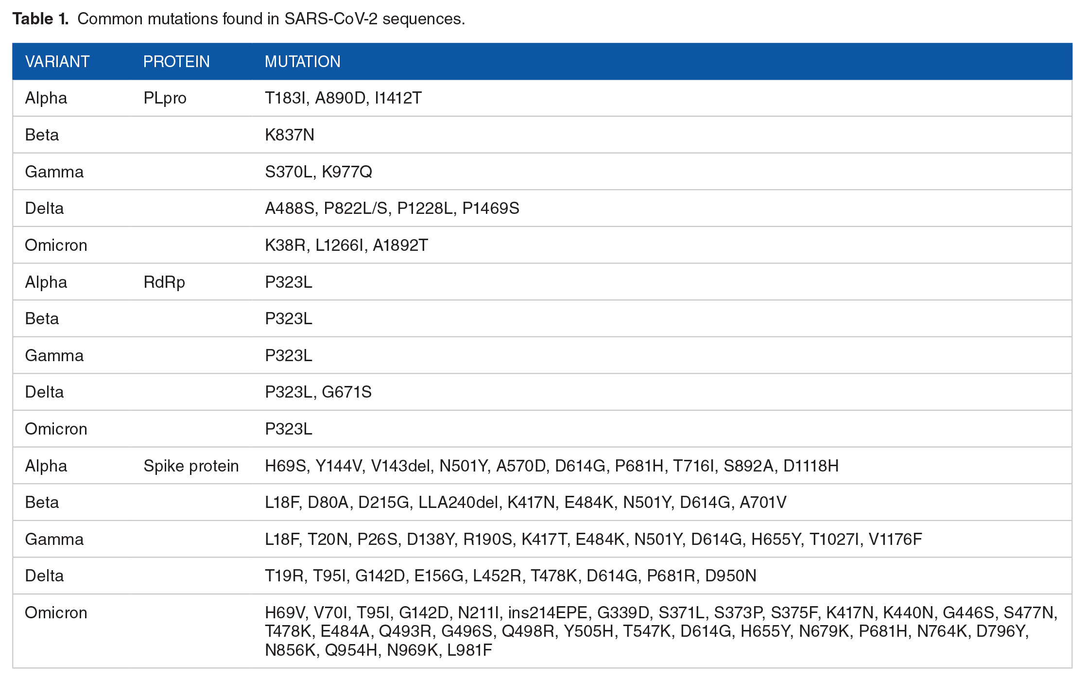
We mainly worked with 3 nonstructural proteins (NSPs), for example, PLpro, RdRp, Helicase, and 1 structural protein, for example, the spike protein of the 5 concern variants from different countries. All variants showed some mutations found in almost all (50) sequences analyzed. In the RdRp case, we found P323L mutation in all 5 variants of all regions, and G671S was found only in the Delta variant. In the PLpro, T183I, A890D, and I1412T mutations were found for the Alpha variant, K837N for Beta, S370L, K977Q for Gamma, A488S, P822L/S, P1228L, P1469S for Delta, and K38R, L1266I, A1892T for Omicron variant. However, the structural protein spike protein has the most mutational changes among the proteins concerned. In Omicron, there is an insertion mutation, ins214EPE. In sequence alignment figures, BioEdit shows the omicron spike mutations 3 positions later than those mentioned because of adding those insertion mutations. Among them, only the D614G mutation was common for the spike protein of all 5 variants. All the mutations of these 5 variants are listed in Table 1.
Common mutations found in SARS-CoV-2 sequences.
Regional uncommon mutations of the nonstructural and structural proteins
Some uncommon mutations were found only in the sequences of some specific regions, not all over the world, which we named uncommon or unique regional mutations listed in Table 2. Mutational analysis revealed that these regional mutations were found mainly in the USA-based sequences (Figure 1). For example, in the Alpha variant, P227L mutation was found in the RdRp protein, K1191N mutation was found in the spike protein, and K159R mutation was found in the Helicase in the sequences retrieved from different states of the USA. In the case of the Beta variant, the T293I mutation was found in the RdRp protein from Djibouti, whereas E37D and V416A mutations were found in the PLpro from the USA. A688V and T1303I mutations were found in the sequences of spike proteins and PLpro, respectively, of the Gamma variant from the USA. T95I mutation was found in the spike of both the Delta variants of the USA and Indian sequences. P822L and K159R mutations were found in the Indian Delta sequences of PLpro and Helicase, respectively. However, the Omicron variant has only one notable mutation, T1004I, which was found in the PLpro of the Indian sequences.
Uncommon regional mutations found in SARS-CoV-2 sequences.


Uncommon regional mutational analysis of non-structural proteins (nsps): (A) P227L mutation in the RdRp protein of Alpha variant in the USA, (B) K159R mutation in the Helicase protein of Alpha variant in the USA, (C) T293I mutation in the RdRp protein of Beta variant in Djibouti, (D) E37D mutation in the PLpro protein of Beta variant in the USA, (E) A416V mutation in the PLpro protein of Beta variant in the USA, (F) T1303I mutation in the PLpro protein of Gamma variant in the USA, (G) P822L mutation in the PLpro protein of Delta variant in India, (H) K159R mutation in the Helicase protein of Delta variant in India, and (I) T1004I mutation in the PLpro protein of Omicron variant in India.
Homology modeling quality assessment
For quality assessment of homology models sequence identity, QMEAN Z-Score, GMQE values, and Ramachandran Favoured Score for each variant spike protein are noted (Table 3). Models were also visualized by Ramachandra plot (Figure 2).
Homology modeling quality assessment.


Spike protein models were also visualized through Ramachandran plot. Most of the amino acids are in the favored regions.
Analysis of structural deviation by measuring RMSD value of RdRp and Helicase
We determined the structural changes of proteins by comparing them with reference structures (Wuhan). For this purpose, we measured the root mean square deviation (RMSD) of RdRp and Helicase proteins by superimposing them on reference structures (NCBI ID: YP 009724390.1). RdRp and Helicase of all variants had RMSD values of 1.094 and 0.85 Å. So, there were minor changes in analyzing structural variation.
Analysis of structural deviation by measuring RMSD value of spike proteins
We superimposed spike proteins of variants with reference protein (NCBI ID: YP 009724390.1). Spike of Delta showed the highest RMSD value (11.067 Å), whereas Beta and Omicron showed RMSD values of 10.055 and 10.083 Å, respectively. On the other hand, Alpha (4.354 Å) and Gamma (2.840 Å) had the lowest RMSD value (Table 4). Furthermore, we found the Delta spike showed a higher and notable structural deviation from Wuhan, followed by Beta and Omicron. And Alpha and Gamma contained minimal structural variation.
Impact of common mutations with structural deviation of structural proteins.

Bold indicating higher or significant RMSD values.
Impact of common mutations and uncommon regional mutations within the structure of spike proteins
Due to A688V and T95I mutations, structural changes were found in the Gamma (NCBI ID UEF38454.1) and Delta spike proteins (NCBI ID UBU60165.1) of the USA after comparing them with the reference structure. Changes occurred at amino acid number 688 of the structure due to the removal of one methyl group in the first case. Whereas polar to nonpolar amino acid change caused the difference in the structure at position 95 due to the removal of the hydroxyl group and the addition of a few methyl groups in the second case (Figure 3). These might affect the binding affinity with receptors. Similarly, structural changes were found in the Indian Delta spike protein compared to the reference structure at position 95 (Figure 3).

Uncommon regional mutational and structural analysis of structural proteins (spike proteins): A(a) A688V mutation in the USA’s spike protein of Gamma variant, A(b) structural deviations of A688V mutation in Gamma spike A688V, and A(c) change in amino acid Alanine to Valine, B(a) T95I Mutation in the Delta spike protein in the USA, B(b) structural deviations of Delta spike T95I, and B(c) change in amino acid Threonine to Isoleucine.
Mutational and structural analysis of the spike proteins of beta variants
Mutational analysis of the Beta spike revealed some novel mutations which had structural alteration. We found a structural change in Beta spike protein at position 80. Aspartic acid (negatively charged) found in the Wuhan spike was changed to Alanine (nonpolar) in the Beta spike protein (Figure 4). Leucine-Leucine-Alanine found in the Wuhan spike was deleted in the Beta spike protein. The deletion mutation caused changes in Leucine (nonpolar) to Histidine (positively charged) at atom position 242 and of Alanine (nonpolar) to Arginine (positively charged). Those changes caused structural differences due to the extra addition of the amine group (Figure 4). Apart from this, structural change was also found at atom position 215 (Supplemental Figure 1). The mutation occurred of Aspartic Acid (negatively charged) to Glycine (Nonpolar), which caused a change in structure due to the removal of a carboxyl group and the addition of hydrogen. Structural change was also found at atom position 701. The mutation replaced Alanine (nonpolar) with Valine (nonpolar), which caused the addition of methyl groups, and a negligible change in the structure occurred at that position (Supplemental Figure 1).

Mutational and structural analysis of the D80A and LLA242del mutations of the spike proteins of beta variant. A(a) D80A mutation in the spike protein of the Beta variant, A(b) structural change in Beta spike D80A, and A(c) amino acid alteration of Aspartic Acid to Alanine, B(a) LLA242del mutation in the spike protein of the Beta variant, B(b) structural change in Beta spike LLA242del, and B(c) amino acid alteration of Arginine to Alanine.
Mutational analysis and structural analysis of the spike proteins of delta variants
Similarly, T19R, E156G, L452R, T478K, P681R, and D950N were common mutations found in the spike protein of the delta variant by mutational analysis. Structural change was found in the Delta spike protein at position 19. The mutation took place from Threonine (polar) to Arginine (positively charged), which caused a significant change in structure due to the removal of the hydroxyl group and the addition of amine groups (Supplemental Figure 2). At position 156, the mutation took place from Glutamic Acid (negatively charged) to Glycine (nonpolar), which caused structural change due to removing a carboxyl group (Supplemental Figure 2). At position 452, the mutation took place from Leucine (nonpolar) to Arginine (positively charged), which caused structural change due to the addition of amine groups (Figure 5). At position 478, the mutation took place from Threonine (polar) to Lysine (positively charged), which caused structural change because of the removal of the hydroxyl group and the addition of amine groups (Figure 5). At position 681, the mutation occurred from Proline (nonpolar) to Arginine (positively charged), which caused structural change due to adding amine groups (Supplemental Figure 3). Structural change was also found in Delta spike protein at position 950. The mutation caused changing of the amino acid from Aspartic Acid (negatively charged) to Valine (Nonpolar) which caused a change in structure at the position due to the removal of a carboxyl group and the addition of a methyl group (Supplemental Figure 3).

Mutational and structural analysis of the L452R and T478K mutations of the spike proteins of delta variant. A(a) L452R mutation in the spike protein of the Delta variant, A(b) structural change in Delta spike L452R, and A(c) amino acid alteration of Leucine to Arginine. B(a) T478K mutation in the spike protein of the Delta variant, B(b) structural change in Delta spike T478K, and B(c) amino acid alteration of Threonine to Lysine.
Mutational analysis and structural analysis of the spike proteins of omicron variants
Spike protein of the omicron variant has a significant number of shared mutations found by mutational analysis in this study. H69V, V70I, N211I, G339D, S371L, S373P, S375F, N440K, G446S, S477N, E484A, Q493R, G496S, Q498R, T547K, N679K, N764K, D796Y, N856K, N969K were the common mutations found in omicron spike. A notable structural change was found at position 69, where Valine replaced Histidine due to the removal of amine groups (Supplemental Figure 4). Structural change was found at position 211 because of the amino acid change from polar to nonpolar with the removal of amine groups (Supplemental Figure 5).
Due to the addition of a carboxylic group at position 339, Structural change occurred (Figure 6) when Glycine (nonpolar) was replaced by Aspartic Acid (negatively charged). In addition, Polar to nonpolar structural changes was found at positions 371, 373, and 375, where Leucine, Proline, and Phenylalanine replaced Serine (Supplemental Figures 5 and 6). On the contrary, nonpolar to polar structural changes were found at positions 446 and 496, where Glycine was replaced by Serine (Supplemental Figures 7 and 9).

Mutational and structural analysis of the G339D and N440K mutations of the spike proteins of omicron variant: A(a) G339D mutation in the spike protein of the Omicron variant, A(b) structural change in omicron spike G339D, and A(c) amino acid alteration of Glycine to Aspartic acid, B(a) N440K mutation in the spike protein of the omicron variant, B(b) structural change in omicron spike N440K, and B(c) amino acid alteration of Asparagine to Lysine.
Another notable structural change was found at position 484, where the mutation occurred from Glutamic Acid (negatively charged) to Alanine (Nonpolar) which caused a change in structure at that position due to the removal of a carboxyl group and the addition of a methyl group (Supplemental Figure 8). Finally, at positions 493 (Supplemental Figure 9) and 498 (Supplemental Figure 8), the mutation took place from Glutamine (polar) to Arginine (positively charged), which caused structural change due to the removal of the hydroxyl group and the addition of amine groups.
Similarly, at position 547, the mutation took place from Threonine (polar) to Lysine (positively charged), which caused structural change (Supplemental Figure 10) due to the removal of the hydroxyl group and the addition of amine groups. Apart from these, negligible structural change was found at positions 70 (nonpolar to nonpolar) and 477 (polar to polar) (Supplemental Figures 4 and 7). Structural change was found at position 796, where the mutation occurred from Aspartic Acid (negatively charged) to Tyrosine (Polar) which caused a change in structure at that position due to the removal of a carboxyl group and the addition of a phenol group (Supplemental Figure 12).
Structural change was found at positions 440, 764, 856, 679, and 969, where mutation took place from Asparagine (polar) to Lysine (positively charged), which caused a significant change in structure due to the removal of the keto group and the addition of amine groups (Figure 6, Supplemental Figures 10–12).
Discussions
Emerging mutations in SARS-CoV-2 variants affect the structure and functions of nonstructural and structural proteins, making the disease difficult to treat and control. 15 All 5 SARS-CoV-2 VOCs have many common mutations reported in previous studies. In this study, we focused on the mutational analysis of uncommon regional mutations found in 3 NSPs, namely PLpro, RdRp, and Helicase, as well as one structural protein, the spike protein of 5 VOCs of SARS-CoV-2. We further investigated the structural alterations caused by a few unique mutations among these uncommon regional mutations and their relationship with mortality.
PLpro cleaves off interferon-stimulating gene-15 (ISG-15) protein, resulting in improper antiviral signaling by host cells.8,9 In addition, RdRp is a fundamental surface point for the antiviral drug to stop or slow down viral replication. 10 And, Helicase is responsible for DNA strand unwinding and translocation. 29 Mutational analysis of these 3 nonstructural proteins revealed that some uncommon mutations found in a different region might affect the above functions of such proteins. For example, in RdRp, P227L mutation has recently appeared, and its prevalence may have increased during the most recent global peak, 30 and P323L has been associated with the disease severity of COVID-19. 31
The RMSD values of PLpro, RdRp, and Helicase were too low to analyze structural deviations. For this reason, we did not perform the structural analysis of nonstructural proteins. But spike protein, one of the major structural proteins of SARS-CoV-2, had a higher RMSD value, prompting its structural investigation and mutational analysis of 2 uncommon regional mutations.
Structures of spike proteins of each variant were assessed as good quality as QMEAN Z scores were below −2.5. Z-scores close to 0.0 represent a “native-like structure,” As a general rule, a “QMEAN” Z-score below −4.0 denotes a model with low quality. 32 The quality of the models was also validated by GMQE scores which give an overall model quality measurement between 0 and 1, with higher numbers indicating higher expected quality 33 and Ramachandran plot, which is a way to visualize energetically favored regions for backbone dihedral angles against of amino acid residues in protein structure. 34 In our study, the GMQE scores of all models are between 0 and 1, finding maximum amino acids in the favored region by visualizing the Ramachandran plot and favored scores. 35
Our structural analysis of uncommon regional mutations of spike protein explored that structural change caused by T95I mutation of Indian spike protein might be responsible for high viral load. 36 Also, mutational and structural modifications of P688V of Gamma spike protein found in the USA in our study might cause a notable increase in infectious rate.
In addition, the RMSD values for the Beta (10.055 Å), Delta (11.067 Å), and Omicron (10.083 Å) variants of spike proteins were extremely high in comparison to the reference structure. Some unique mutations of Beta, Delta, and Omicron spike protein that induced significant structural changes were further evaluated to determine the association between structural change and risk of severity.
The alteration of amino acids from polar or nonpolar to positively or negatively charged and vice versa may alter protein structure and function. 37 Our study discovered the structural change of D80A mutation found only in the Beta spike that could be responsible for escaping neutralizing antibodies and compromising vaccine efficacy. 38 In this study, we performed a structural analysis of the Delta spike protein of T19R mutation, which revealed that structure change might cause the sidechain conformational change and weakened interactions with antibodies. 39 Similarly, the structural changes found in our study in the delta spike of L452R mutation present in the receptor-binding domain (RBD) of SARS-COV-2, spike protein might cause the reduction of neutralizing activities in many monoclonal antibodies 15 as well as the immune evasion by increasing the spike protein’s affinity for the hACE2 receptor and decreasing the immune system’s detection capability. 40 Our study further investigated the structural change of T478K mutation found in the Delta spike that could be predicted to be responsible for increasing binding affinity between RBD and ACE2 receptor (entry point of SARS-COV-2 into the human cell) and enabling immune escape. 41
Interestingly, we also found that P681R, the most prominent mutation detected only in the delta spike, causes structural alteration and may be responsible for the increased cell-level infectivity. This structural alteration, which occurred right at the furin cleavage site of the delta variation, played a crucial role in boosting S-protein cleavage and was proposed as the primary cause of the Delta variant’s infectivity. 42
We found that the D950N mutation of delta spike protein changed structure, but no functional change was yet to detect. Similarly, LLA242del and D215G mutations in the beta spike protein caused a difference in the structure found in our analysis but are yet to find functional alteration. A701V mutation in the Beta spike protein showed minor structural alteration of amino acids from nonpolar to nonpolar.
Structural changes were also found in G339D, S371L, S375F, K440N, G446S, S477N, E484A, G496S, Q498R, and Y505H mutations in our study of Omicron spikes may lead to increased abilities of immune escape. 43 Besides, Omicron has N679K mutation where structural alteration was detected in this study near the furin cleavage site which incorporates basic amino acids around the furin cleavage site and, perhaps, facilitates the cleavage of the spike protein into S1 and S2, thereby increasing fusion and virus infection. 44 Furthermore, N764K, D796Y, and N856K mutations also caused structural deviations in our study to help attain improved stability in the Omicron variant. 43
Conclusion
Our findings suggested that notable structural changes in the spike protein of Beta, Delta, and Omicron variants may have a significant association with rates of sickness severity and mortality rates. However, until now, no specific database exists that correlates the mortality rates caused by SARS-CoV-2 variants that could be associated with the structural changes found in our study. Since there is a greater possibility of making an effect on mortality by structural changes of SARS-CoV-2 variants, both mathematical and statistical analysis, and a patient database will be needed to establish the relationship between structural changes and mortality rates.
This new information from our study and more detailed information about SARS-CoV-2 infections, mutation, and structural changes will be better equipped to assess the selection of regional vaccine strains and treat them with that more targeted vaccines.
Supplemental Material
sj-pptx-1-evb-10.1177_11769343231182258 – Supplemental material for Beta, Delta, and Omicron, Deadliest Among SARS-CoV-2 Variants: A Computational Repurposing Approach
Supplemental material, sj-pptx-1-evb-10.1177_11769343231182258 for Beta, Delta, and Omicron, Deadliest Among SARS-CoV-2 Variants: A Computational Repurposing Approach by Mohammad Mamun Alam, Sumaiya Binte Hannan, Tanvir Ahmed Saikat, Md Belayet Hasan Limon, Md Raihan Topu, Md Jowel Rana, Asma Salauddin, Sagar Bosu and Mohammed Ziaur Rahman in Evolutionary Bioinformatics
Footnotes
Author Contributions
Funding:
The author(s) disclosed receipt of the following financial support for the research, authorship, and/or publication of this article: This research study was funded by core donors who provide unrestricted support to icddr,b for its operations and research. Current donors providing unrestricted support include the Governments of Bangladesh, Canada, Sweden, and the UK.
Declaration of Conflicting Interests:
The author(s) declared no potential conflicts of interest with respect to the research, authorship, and/or publication of this article.
Supplemental Material
Supplemental material for this article is available online.
References
Supplementary Material
Please find the following supplemental material available below.
For Open Access articles published under a Creative Commons License, all supplemental material carries the same license as the article it is associated with.
For non-Open Access articles published, all supplemental material carries a non-exclusive license, and permission requests for re-use of supplemental material or any part of supplemental material shall be sent directly to the copyright owner as specified in the copyright notice associated with the article.